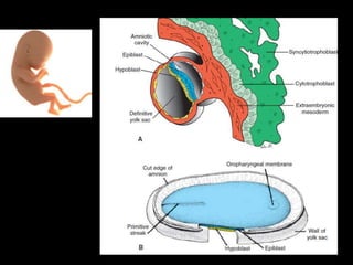
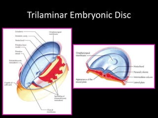
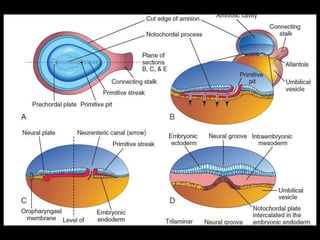
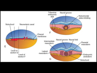
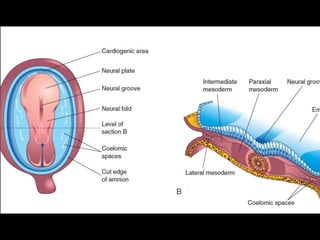
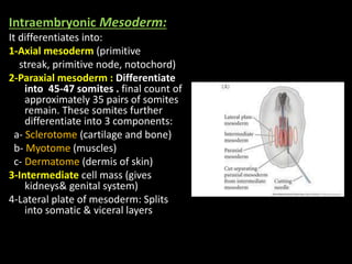
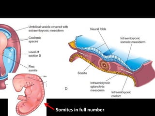
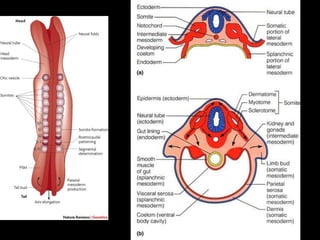
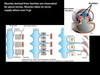
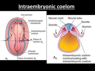
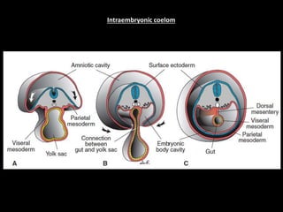
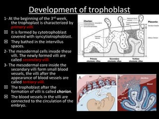
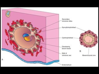
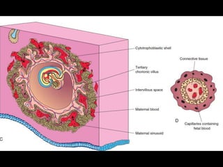
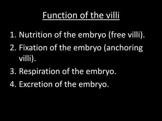
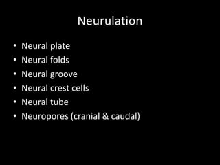
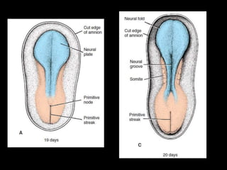
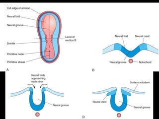
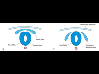
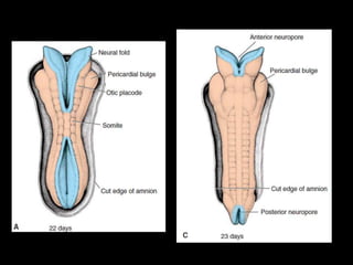
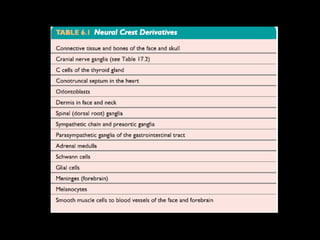
2. Events of 3rd week
• Gastrulation: Formation of Germ Layers
• Primitive Streak
• Fate of Primitive Streak
• Notochordal Process and Notochord
• Allantois
• Neurulation: Formation of Neural Tube
• Neural Plate and Neural Tube &Neural Crest cells
• Development of Somites
• Development of Intraembryonic Coelom
• Further Development of Chorionic Villi
6. Gastrulation:
Is the process of formation of the 3 germ layers (ectoderm,
mesoderm & endoderm).
It begins by the formation of a mid line groove on the
epiblast called primitive streak.
The cephalic end of this streak is called primitive node. In
the center of this node there is primitive pit.
The epiblastic cells migrate & reach the primitive streak.
They slip under the primitive streak (invaginate) forming
endoderm.
Some of invaginated epiblastic cells forms the intra-
embryonic mesoderm.
Thus the epiblast is the source of the 3 germ layers, which
are ectoderm (dorsally), endoderm (ventrally) and in-
between these 2 layers is the mesoderm.
From these 3 germ layers arise all organs & tissues of the
body.
11. Formation of the notochord
• Prenotochordal cells invaginating in the primitive
node move cranially reach the prechordal plate
• Prenotochordal cells become intercalated in the
hypoblast , consists of two cell layers.
• They form the notochordal plate .
• As the hypoblast is replaced by endoderm cells, cells of
the notochordal plate detach from the endoderm.
• They then form a solid cord of cells, the definitive
• notochord
12. Functions of the notochord
1.It forms the
axial skeleton in embryo
2. It induces the overlying
ectoderm to thicken and
form the neural plate( the
primordium of the central
nervous system )
Notochord is the organizer
for nervous system
formation.
13. Functions of the notochord
3. The notochord degenerates
and disappears as the
bodies of the vertebrae
form. Its remnant is the
nucleus pulposus of the
intervertebral discs.
14. Intraembryonic Mesoderm:
It differentiates into:
1-Axial mesoderm (primitive
streak, primitive node, notochord)
2-Paraxial mesoderm : Differentiate
into 45-47 somites . final count of
approximately 35 pairs of somites
remain. These somites further
differentiate into 3 components:
a- Sclerotome (cartilage and bone)
b- Myotome (muscles)
c- Dermatome (dermis of skin)
3-Intermediate cell mass (gives
kidneys& genital system)
4-Lateral plate of mesoderm: Splits
into somatic & viceral layers
20. Development of trophoblast
1- At the beginning of the 3rd week,
the trophoplast is characterized by
primary villi.
It is formed by cytotrophoblast
covered with syncytiotrophoblast.
They bathed in the intervillus
spaces.
2- The mesodermal cells invade these
villi. The newly formed villi are
called secondary villi.
3- The mesodermal core inside the
secondary villi form small blood
vessels, the villi after the
appearance of blood vessels are
called tertiary villi.
The trophoblast after the
formation of villi is called chorion.
The blood vessels in the villi are
connected to the circulation of the
embryo.
21.
22.
23. Clinical application
• Third week of development is a very
sensitive period in fetal
development. Many factors such as
drugs, alcohol or irradiation to the
mother may cause congenital
anomalies to her embryo.
24. Function of the villi
1. Nutrition of the embryo (free villi).
2. Fixation of the embryo (anchoring
villi).
3. Respiration of the embryo.
4. Excretion of the embryo.