CFTR is a chloride channel located on the cell membrane. Under the mediation of cAMP, CFTR is phosphorylated, causing the channel to open and transporting about 10 CIs extracellularly per minute. When the cftr gene is mutated (most commonly, the codon encoding 508 phenylalanine is lost), the defective CFTR cannot be processed normally in the endoplasmic reticulum, and most cannot be transported to the cell membrane;
Structure and function of plasma membrane 2ICHHA PURAK
The presentation consists of 72 slides,describes following heads
DEFINITION : STRUCTURE OF PLASMA MEMBRANE
COMPONENTS OF PLASMA MEMBRANE ( (BIOCHEMICAL PROPERTIES)
LIPID BILAYER
PROTEINS
CARBOHYDRATES
CHOLESTEROL
MODELS EXPLAINING STRUCTURE OF BIO MEMBRANE
FLUID MOSAIC MODEL
MOBILITY OF MEMBRANE
GLYCOCALYX : GLYCOPROTEINS AND GLYCOLIPIDS
TRANSPORT OF IONS AND MOLECULES ACROSS PLASMA MEMBRANE
FUNCTIONS OF PLASMA MEMBRANE
DIVERSITY OF CELL MEMBRANES
SITE OF ATPASE ION CARRIER CHANNELS AND PUMPS-RECEPTORS
Plasma membrane dynamic structure By KK Sahu SirKAUSHAL SAHU
Introduction
History
Definition
Structure of plasma membrane(fluid mosaic model)
Examples to show dynamic nature of plasma membrane
The diffusion of membrane protein after fusion
Restriction on protein and mobility
Control of membrane protein and mobility
Membrane lipid mobility
Membrane domains and cell polarity
Function of plasma membrane
Conclusion.
References.
What is the function of CFTR (details are important) (5 points).pdfrupeshmehta151
What is the function of CFTR? (details are important) (5 points)
Solution
Ans:- Cystic fibrosis transmembrane conductance regulator (CFTR) is a membrane protein and
chloride channel in vertebrates that is encoded by the CFTR gene.CFTR functions as a ATP
gated anion channel increasing the conductance for certain anions to flow down their
electrochemical gradient.ATP driven conformational changes in CFTR open and close a gate to
allow transmembrane flow of anions down their electrochemical gradient. Essentially CFTR is
an ion channel that evolved as a broken ABC transporter that leaks when in open
conformation.The CFTR is found in the epithelial cells of many organs including the lungs,liver
,pancreas,digestive tract ,reproductive tract and skin.Normally the protein moves chloride and
thiocyanate ions out of an epithelial cell covering mucus.Positively chargedsodium ions follow
passively increasing the total electrolyte concentration in the mucus resulting in the movement of
water out of the cell via osmosis.In sweat glands defective CSTR results in reduced transport of
sodium chloride and sodium thiocyanate in the reabsorptive duct and therefore saltier sweat..
The CFTR gene belongs to a family of genes that regulate the energy transfer that allows a cell to open and close its ion channels. It is located on human chromosome 7 and consists of twenty-seven sequences of DNA that encode 1,480 amino acids. The CFTR gene produces the CFTR protein, which regulates the chloride ion content of epithelial cells that line the nasal cavity, lungs, and stomach.
Where is [Cl] normally highest, inside or outside of a cell How do.pdfbarristeressaseren71
Where is [Cl\'] normally highest, inside or outside of a cell? How does this compare to [Na*] in
a cell? If the membrane became permeable to Cl^-, what direction would Cl^- move based on its
chemical gradient? In a healthy person, when the CFTR channel opens, Cl^- moves from the
inside of the cell to membranes, and your response to Question 2 as you answer) What direction
will water move in this system when the CFTR is open? If the CFTR doesn\'t work correctly due
to a mutation, Cl^- movement is blocked and an Inside Cell abnormally thick sticky mucus is
produced on the outside of the cell. This mucus clogs the airways in the lungs, and increases the
risk of infection by bacteria. Figure 2 illustrates a mutant CFTR channel in the epithelial cells
lining the lung.
Solution
1. Mostly Chloride and Sodium are present in high concentrations in the extracellular region and
low concentration in the intra cellular region. This is the reason behind generation of membrane
potential as potassium is present at low concentrations in the extracellular region and higher in
intracellular areas.
2. Since the concentration of Chloride is relatively higher in the extracellular region, if the
membrane becomes permeable, it would lead to movement of chloride ions from extracellular to
intracellular region because of the existing concentration gradient.
3. CFTR channels stand for Cystic fibrosis Transmembrane Conductance Regulator Channels. It
is an ABC transporter class protein which conducts chloride ions and water along with
thiocynate ions across epithelial cell membranes. It is responsible for carrying out active
transport of Chloride ions against the concentration gradient by the use of ATP as well as
phosphorylation. That is why the movement of chloride ions is possible against the concentration
gradient.
4. The ionic concentration of the mucus slowly increases with the functioning of CFTR channel
and this leads to movement of water from the inside to the outside via osmosis..
Cystic Fibrosis and Protein Synthesis Introduction Cystic fi.pdfsunilverma8487
Cystic Fibrosis and Protein Synthesis
Introduction
Cystic fibrosis is an inherited disease that is marked by the buildup of thick, sticky mucus that can
damage the lungs and many other organs. Cystic fibrosis affects the viscosity of the mucus lining
of the lungs. Mucus is a collection of many substances including enzymes, proteins and mucins. In
the lungs, specialized cells called Goblet cells help produce mucus. The lung tissues keep the
mucus hydrated and moving. The mucus also traps bacteria and particulates and flushes them out
of the lungs to avoid infection. In a person with Cystic Fibrosis, mucus becomes dehydrated and
thick. Bacteria and particles get trapped and lead to infection.
These infections cause chronic coughing, wheezing, and inflammation. Over time, mucus buildup
and infections result in permanent lung damage.
CFTR
CFTR gene is found on human chromosome 7 and the gene is 4400 nucleotides in length. CFTR
gene produces CFTR protein. In the human body, it functions as a channel across the membrane
of cells that produce mucus, sweat, saliva, tears, and digestive enzymes. In the lungs, the
membrane channel transports negatively charged chloride ions into and out of cells. The transport
of chloride ions helps control the movement of water in tissues, which is necessary for the
production of thin, freely flowing mucus.
Figure 1: Normal and mutated versions of the CFTR structure and function.
In cells from non-CF (Cystic Fibrosis) individuals, the chloride channels open periodically to allow
the cell to maintain a normal balance of chloride ion between the inside and outside of the cell. In
CF individuals, these chloride channels do not function, and chloride ions build up inside the cell.
Figure 2: Image of the open and closed CFTR transmembrane protein. The NBD region
-
binds ATP in order to transport Cl .
Why might the mucus of CF patients be thicker than that of non-CF individuals?
-
Using your knowledge of osmosis and water potential, explain why a lack of transport of Cl ions
might result in thicker mucus for people with CF.
Using the model below, describe what is happening to the mucus thickness, cilia and airway
surface liquid of the CF person.
-
Figure 3: Model of a normal and a CF airway due to lack of Cl transport..
Describe the structure of CFTR. You must provide a written descripti.pdfarchanadesignfashion
Describe the structure of CFTR. You must provide a written description for full credit. A picture
on its own is not enough. A picture with a description is acceptable. (5 points)
What is the function of CFTR? (details are important) (5 points)
Why is the mucus thicker than normal in CF patients? A good answer will include a description
of how osmosis is affected by CF. (5 points)
The most common mutation causing CF is a deletion of F508. What are the cellular
consequences of the F508 deletion? (5 points)
What are the organismal consequences of the F508 deletion? i.e. how does this mutation affect a
human being? (5 points)
How would you use gene therapy to treat CF? What problems might the plasma membrane cause
for successful gene therapy? (5 points)
Solution
Describe the structure of CFTR
CFTR protein :
CFTR :
Mutation
F508 is a class II CFTR mutation.[
The CFTR protein is to a great extent communicated in cells of the pancreas, intestinal and
respiratory epithelia, and every exocrine organ. At the point when appropriately collapsed, it is
moved to the cell layer, where it turns into a transmembrane protein in charge of opening
channels which discharge chloride particles out of cells; it likewise at the same time hinders the
take-up of sodium particles by another channel protein. Both of these capacities help to keep up a
particle angle that causes osmosis to coax water out of the cells.
The F508 transformation prompts to the misfolding of CFTR and its inevitable debasement in the
ER. In life forms with two supplements of the transformation, the protein is totally truant from
the cell film, and these basic particle transport capacities are not performed.
in this notes we will study and learn about
cell membrane
parts of cell membrane
different formation of cell membrane
lipids present in cell membrane
function of cell membrane
Cell Physiology Spring 2016 Name Page 1 This test is .docxtidwellveronique
Cell Physiology Spring 2016 Name:
Page 1
This test is to be completed on your own, the essay sections should not be the same as anyone else’s
(word for word) If that is what I find you both will receive 0 for this test.
Structure/Function Cell Part
1) Stores material within the cell (General)
2) The sites of protein synthesis
3) Transports materials within the cell
4) Organelle that manages or controls all the cell functions in a
eukaryotic cell
5) Digests excess or worn-out cell parts, food particles and invading
viruses or bacteria
6) Small bumps located on portions of the endoplasmic reticulum
7) Provides temporary storage of food, enzymes and waste products
8) Produces a usable form of energy for the cell
9) Packages proteins for transport out of the cell
10) The membrane surrounding the cell
11) Name for the collection of DNA in the nucleus of eukaryotic cells
12) Consist of hollow tubes which provide support for the cell
13) Small hair-like structures used for movement or sensing things
Cell Physiology Spring 2016 Name:
Page 2
1) In what organelle does cellular respiration take place?
2) Name two storage organelles?
3) What is the list of organelles that take part in protein synthesis?
4) How is the nucleus involved in protein synthesis?
5) What is the difference between rough ER and smooth ER? What is the ER doing that is
different in each case?
6) What are lysosomes? What types of molecules would be found inside a lysosome?
7) Why might a lysosome fuse with or link up with a food vacuole?
8) In what organelle do molecules move from the ER to the Golgi bodies?
Cell Physiology Spring 2016 Name:
Page 3
9) The pH of lysosomes is lower than that of the cytosol because of the action of
A. Na+ and OH− transport proteins in the lysosomal membrane
B. H+ and Cl− transport proteins in the plasma membrane
C. acid-producing enzymes in the lysosomal lumen
D. H+ and Cl− transport proteins in the lysosomal membrane
10) The phenomenon in which a chemical absorbs light at one wavelength and emits it at a specific
and longer wavelength is called
A. Differential interference contrast.
B. fluorescence.
C. deconvolution.
D. shadowing.
11) Which of the following could be used to visualize subcellular structure in living cells?
A. transmission electron microscopy
B. scanning electron microscopy
C. bright field microscopy
D. differential Fluorescence interference light microscopy
12) If a cellular homogenate were subjected to differential centrifugation, which of the following
would be expected to pellet first?
A. the endoplasmic reticulum
B. mitochondria
C. the cytosol
D. nuclei
Cell Physiology Spring 2016 Name:
Page 4
13) The disruption of a cell is necessary to release its organelles and contents for subsequent
isolation. One method, called _________________________________, uses ultrahigh-
...
The sodium channel is a channel present on the membrane that allows a small amount of Na+ to enter the cell along its electrochemical gradient, as discovered by British scientists Hodgkin and Huxley. It can be divided into two types, voltage-gated and ligand-gated. The sodium ion channel is the primary activation bond for electrical signals in all animals, while the electrical signal is the basis for a series of physiological processes such as neural activity and muscle contraction.
Numerous cells are able to ingest foreign materials, but the ability to increase this activity in response to opsonization by antibody and/or complement, so as to acquire antigen specificity, is restricted to cells of the myeloid series, principally polymorphs, monocytes and macrophages; these are sometimes termed ‘professional’ phagocytes.
Neuroscience is characterized by multi-disciplinary and multi-level intersections. It combines behavior, cognition and brain mechanism, and it attempts to elaborate the neural mechanism of human and animal in perceiving objects, forming images, using language, memorizing information, reasoning and decision-making at the micro level of molecule, synapse and neuron and at the macro level of system, whole brain and behavior.
Transient receptor potenital (TRP) is a large family of non-selective cation channels located on the cell membrane. One type of channel can be activated by Vanillic acid compounds, so this type of channel is called the TRPV subfamily. Mutations in TRPV are associated with neurodegenerative diseases, skeletal dysplasia, kidney disease and cancer and TRPV is an important therapeutic target for these diseases.
The organic cation transporter (OCT) is an important drug delivery protein with a broad tissue distribution in the body that mediates the metabolic processes of most drugs. At present, the gene sequence, transport mechanism, substrate structure specificity, regulatory mechanism, gene polymorphism andin vivodistribution characteristics of this transporter have been deeply studied. Based on this knowledge, pharmacologists have successfully delivered many drugs at the transporter molecule level and applied them to clinical practice.
Epigenetics is the study of heritable changes in gene function that do not involve changes in the DNA sequence. A variety of epigenetic mechanisms can be perturbed in different types of cancer. Epigenetic alterations of DNA repair genes or cell cycle control genes are very frequent in sporadic (non-germ line) cancers, being significantly more common than germ line (familial) mutations in these sporadic cancers.
In genetics, genotoxicity describes the property of chemical agents that damage the genetic information within a cell causing mutations(Genotoxicity is often confused with mutagenicity. All mutagens are genotoxic, whereas not all genotoxic substances are mutagenic.). The alteration can have direct or indirect effects on the DNA: the induction of mutations, mistimed event activation, and direct DNA damage resulting in mutations.
The process of cell cycle regulation is the activation or inactivation of various regulatory factors under the surveillance of checkpoints, thereby initiating the process of cell DNA replication and division into two daughter cells.
DNA mismatch repair (MMR) recognizes and repairs erroneous insertion, deletion, and mis-incorporation of bases that can arise during DNA replication and recombination, and repair some forms of DNA damage. It plays an important role in maintaining genomic stability and cellular homeostasis.
Post-translational modifications play an important role in cells, such as DNA recognition, protein-protein interactions, catalytic activity, and protein stability. Protein acetylation/deacetylation is a histone covalent modification that is mainly catalyzed by histone acetylase and histone deacetylase, respectively.
DNA mismatch repair (MMR) recognizes and repairs erroneous insertion, deletion, and mis-incorporation of bases that can arise during DNA replication and recombination, and repair some forms of DNA damage. It plays an important role in maintaining genomic stability and cellular homeostasis.
Mitochondria are a subcellular structure prevalent in eukaryotic cells and the most important source of energy in cells. Most tissue cells in the human body rely on oxidative phosphorylation of mitochondria to obtain the energy needed to maintain their metabolism.
Slide 1: Title Slide
Extrachromosomal Inheritance
Slide 2: Introduction to Extrachromosomal Inheritance
Definition: Extrachromosomal inheritance refers to the transmission of genetic material that is not found within the nucleus.
Key Components: Involves genes located in mitochondria, chloroplasts, and plasmids.
Slide 3: Mitochondrial Inheritance
Mitochondria: Organelles responsible for energy production.
Mitochondrial DNA (mtDNA): Circular DNA molecule found in mitochondria.
Inheritance Pattern: Maternally inherited, meaning it is passed from mothers to all their offspring.
Diseases: Examples include Leber’s hereditary optic neuropathy (LHON) and mitochondrial myopathy.
Slide 4: Chloroplast Inheritance
Chloroplasts: Organelles responsible for photosynthesis in plants.
Chloroplast DNA (cpDNA): Circular DNA molecule found in chloroplasts.
Inheritance Pattern: Often maternally inherited in most plants, but can vary in some species.
Examples: Variegation in plants, where leaf color patterns are determined by chloroplast DNA.
Slide 5: Plasmid Inheritance
Plasmids: Small, circular DNA molecules found in bacteria and some eukaryotes.
Features: Can carry antibiotic resistance genes and can be transferred between cells through processes like conjugation.
Significance: Important in biotechnology for gene cloning and genetic engineering.
Slide 6: Mechanisms of Extrachromosomal Inheritance
Non-Mendelian Patterns: Do not follow Mendel’s laws of inheritance.
Cytoplasmic Segregation: During cell division, organelles like mitochondria and chloroplasts are randomly distributed to daughter cells.
Heteroplasmy: Presence of more than one type of organellar genome within a cell, leading to variation in expression.
Slide 7: Examples of Extrachromosomal Inheritance
Four O’clock Plant (Mirabilis jalapa): Shows variegated leaves due to different cpDNA in leaf cells.
Petite Mutants in Yeast: Result from mutations in mitochondrial DNA affecting respiration.
Slide 8: Importance of Extrachromosomal Inheritance
Evolution: Provides insight into the evolution of eukaryotic cells.
Medicine: Understanding mitochondrial inheritance helps in diagnosing and treating mitochondrial diseases.
Agriculture: Chloroplast inheritance can be used in plant breeding and genetic modification.
Slide 9: Recent Research and Advances
Gene Editing: Techniques like CRISPR-Cas9 are being used to edit mitochondrial and chloroplast DNA.
Therapies: Development of mitochondrial replacement therapy (MRT) for preventing mitochondrial diseases.
Slide 10: Conclusion
Summary: Extrachromosomal inheritance involves the transmission of genetic material outside the nucleus and plays a crucial role in genetics, medicine, and biotechnology.
Future Directions: Continued research and technological advancements hold promise for new treatments and applications.
Slide 11: Questions and Discussion
Invite Audience: Open the floor for any questions or further discussion on the topic.
This pdf is about the Schizophrenia.
For more details visit on YouTube; @SELF-EXPLANATORY;
https://www.youtube.com/channel/UCAiarMZDNhe1A3Rnpr_WkzA/videos
Thanks...!
(May 29th, 2024) Advancements in Intravital Microscopy- Insights for Preclini...Scintica Instrumentation
Intravital microscopy (IVM) is a powerful tool utilized to study cellular behavior over time and space in vivo. Much of our understanding of cell biology has been accomplished using various in vitro and ex vivo methods; however, these studies do not necessarily reflect the natural dynamics of biological processes. Unlike traditional cell culture or fixed tissue imaging, IVM allows for the ultra-fast high-resolution imaging of cellular processes over time and space and were studied in its natural environment. Real-time visualization of biological processes in the context of an intact organism helps maintain physiological relevance and provide insights into the progression of disease, response to treatments or developmental processes.
In this webinar we give an overview of advanced applications of the IVM system in preclinical research. IVIM technology is a provider of all-in-one intravital microscopy systems and solutions optimized for in vivo imaging of live animal models at sub-micron resolution. The system’s unique features and user-friendly software enables researchers to probe fast dynamic biological processes such as immune cell tracking, cell-cell interaction as well as vascularization and tumor metastasis with exceptional detail. This webinar will also give an overview of IVM being utilized in drug development, offering a view into the intricate interaction between drugs/nanoparticles and tissues in vivo and allows for the evaluation of therapeutic intervention in a variety of tissues and organs. This interdisciplinary collaboration continues to drive the advancements of novel therapeutic strategies.
Multi-source connectivity as the driver of solar wind variability in the heli...Sérgio Sacani
The ambient solar wind that flls the heliosphere originates from multiple
sources in the solar corona and is highly structured. It is often described
as high-speed, relatively homogeneous, plasma streams from coronal
holes and slow-speed, highly variable, streams whose source regions are
under debate. A key goal of ESA/NASA’s Solar Orbiter mission is to identify
solar wind sources and understand what drives the complexity seen in the
heliosphere. By combining magnetic feld modelling and spectroscopic
techniques with high-resolution observations and measurements, we show
that the solar wind variability detected in situ by Solar Orbiter in March
2022 is driven by spatio-temporal changes in the magnetic connectivity to
multiple sources in the solar atmosphere. The magnetic feld footpoints
connected to the spacecraft moved from the boundaries of a coronal hole
to one active region (12961) and then across to another region (12957). This
is refected in the in situ measurements, which show the transition from fast
to highly Alfvénic then to slow solar wind that is disrupted by the arrival of
a coronal mass ejection. Our results describe solar wind variability at 0.5 au
but are applicable to near-Earth observatories.
Earliest Galaxies in the JADES Origins Field: Luminosity Function and Cosmic ...Sérgio Sacani
We characterize the earliest galaxy population in the JADES Origins Field (JOF), the deepest
imaging field observed with JWST. We make use of the ancillary Hubble optical images (5 filters
spanning 0.4−0.9µm) and novel JWST images with 14 filters spanning 0.8−5µm, including 7 mediumband filters, and reaching total exposure times of up to 46 hours per filter. We combine all our data
at > 2.3µm to construct an ultradeep image, reaching as deep as ≈ 31.4 AB mag in the stack and
30.3-31.0 AB mag (5σ, r = 0.1” circular aperture) in individual filters. We measure photometric
redshifts and use robust selection criteria to identify a sample of eight galaxy candidates at redshifts
z = 11.5 − 15. These objects show compact half-light radii of R1/2 ∼ 50 − 200pc, stellar masses of
M⋆ ∼ 107−108M⊙, and star-formation rates of SFR ∼ 0.1−1 M⊙ yr−1
. Our search finds no candidates
at 15 < z < 20, placing upper limits at these redshifts. We develop a forward modeling approach to
infer the properties of the evolving luminosity function without binning in redshift or luminosity that
marginalizes over the photometric redshift uncertainty of our candidate galaxies and incorporates the
impact of non-detections. We find a z = 12 luminosity function in good agreement with prior results,
and that the luminosity function normalization and UV luminosity density decline by a factor of ∼ 2.5
from z = 12 to z = 14. We discuss the possible implications of our results in the context of theoretical
models for evolution of the dark matter halo mass function.
Cancer cell metabolism: special Reference to Lactate PathwayAADYARAJPANDEY1
Normal Cell Metabolism:
Cellular respiration describes the series of steps that cells use to break down sugar and other chemicals to get the energy we need to function.
Energy is stored in the bonds of glucose and when glucose is broken down, much of that energy is released.
Cell utilize energy in the form of ATP.
The first step of respiration is called glycolysis. In a series of steps, glycolysis breaks glucose into two smaller molecules - a chemical called pyruvate. A small amount of ATP is formed during this process.
Most healthy cells continue the breakdown in a second process, called the Kreb's cycle. The Kreb's cycle allows cells to “burn” the pyruvates made in glycolysis to get more ATP.
The last step in the breakdown of glucose is called oxidative phosphorylation (Ox-Phos).
It takes place in specialized cell structures called mitochondria. This process produces a large amount of ATP. Importantly, cells need oxygen to complete oxidative phosphorylation.
If a cell completes only glycolysis, only 2 molecules of ATP are made per glucose. However, if the cell completes the entire respiration process (glycolysis - Kreb's - oxidative phosphorylation), about 36 molecules of ATP are created, giving it much more energy to use.
IN CANCER CELL:
Unlike healthy cells that "burn" the entire molecule of sugar to capture a large amount of energy as ATP, cancer cells are wasteful.
Cancer cells only partially break down sugar molecules. They overuse the first step of respiration, glycolysis. They frequently do not complete the second step, oxidative phosphorylation.
This results in only 2 molecules of ATP per each glucose molecule instead of the 36 or so ATPs healthy cells gain. As a result, cancer cells need to use a lot more sugar molecules to get enough energy to survive.
introduction to WARBERG PHENOMENA:
WARBURG EFFECT Usually, cancer cells are highly glycolytic (glucose addiction) and take up more glucose than do normal cells from outside.
Otto Heinrich Warburg (; 8 October 1883 – 1 August 1970) In 1931 was awarded the Nobel Prize in Physiology for his "discovery of the nature and mode of action of the respiratory enzyme.
WARNBURG EFFECT : cancer cells under aerobic (well-oxygenated) conditions to metabolize glucose to lactate (aerobic glycolysis) is known as the Warburg effect. Warburg made the observation that tumor slices consume glucose and secrete lactate at a higher rate than normal tissues.
Richard's entangled aventures in wonderlandRichard Gill
Since the loophole-free Bell experiments of 2020 and the Nobel prizes in physics of 2022, critics of Bell's work have retreated to the fortress of super-determinism. Now, super-determinism is a derogatory word - it just means "determinism". Palmer, Hance and Hossenfelder argue that quantum mechanics and determinism are not incompatible, using a sophisticated mathematical construction based on a subtle thinning of allowed states and measurements in quantum mechanics, such that what is left appears to make Bell's argument fail, without altering the empirical predictions of quantum mechanics. I think however that it is a smoke screen, and the slogan "lost in math" comes to my mind. I will discuss some other recent disproofs of Bell's theorem using the language of causality based on causal graphs. Causal thinking is also central to law and justice. I will mention surprising connections to my work on serial killer nurse cases, in particular the Dutch case of Lucia de Berk and the current UK case of Lucy Letby.
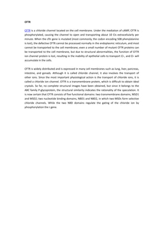
1. CFTR
CFTR is a chloride channel located on the cell membrane. Under the mediation of cAMP, CFTR is
phosphorylated, causing the channel to open and transporting about 10 CIs extracellularly per
minute. When the cftr gene is mutated (most commonly, the codon encoding 508 phenylalanine
is lost), the defective CFTR cannot be processed normally in the endoplasmic reticulum, and most
cannot be transported to the cell membrane; even a small number of mutant CFTR proteins can
be transported to the cell membrane, but due to structural abnormalities, the function of CFTR
ion channel protein is lost, resulting in the inability of epithelial cells to transport CI-, and CI- will
accumulate in the cells.
CFTR is widely distributed and is expressed in many cell membranes such as lung, liver, pancreas,
intestine, and gonads. Although it is called chloride channel, it also involves the transport of
other ions. Since the most important physiological action is the transport of chloride ions, it is
called a chloride ion channel. CFTR is a transmembrane protein, which is difficult to obtain ideal
crystals. So far, no complete structural images have been obtained, but since it belongs to the
ABC family P-glycoprotein, the structural similarity indicates the rationality of the speculation. It
is now certain that CFTR consists of five functional domains: two transmembrane domains, MSD1
and MSD2; two nucleotide binding domains, NBD1 and NBD2, in which two MSDs form selective
chloride channels. While the two NBD domains regulate the gating of the chloride ion by
phosphorylation the r gene.