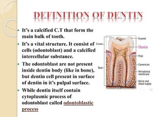
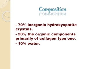
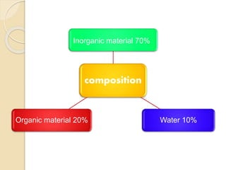
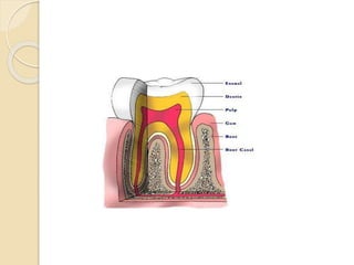
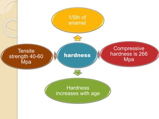
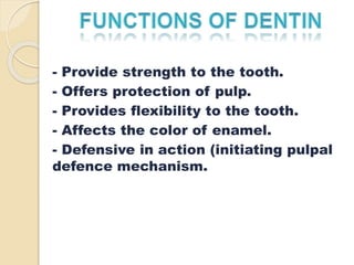
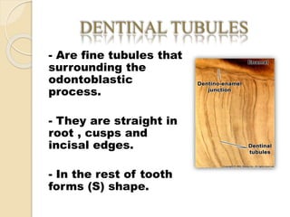
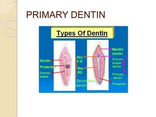
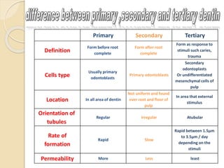
REPUBLIC OF YEMEN 
UNIVERSITY OF ADEN 
FACULTY OF DENTISTRY 
CONSERVATIVE DEPARTMENT 
DONE BY: 
Nehal Fathi Hanif (A-30) 
Nasima Ahmed Habtoor (A-23) 
Marwa Abdullah Bin Shehna (A-12) 
Islam Abubaker Aidros (A-26) 
Maryam Aidros Abdullah (A-25) 
Ahmed Mohammed Abdul Rahman (A-31)
 It’s a calcified C.T that form the 
main bulk of tooth. 
 It’s a vital structure. It consist of 
cells (odontoblast) and a calcified 
intercellular substance. 
 The odontoblast are not present 
inside dentin body (like in bone), 
but dentin cell present in surface 
of dentin in it’s pulpal surface. 
 While dentin itself contain 
cytoplasmic process of 
odontoblast called odontoblastic 
process
- 70% inorganic hydroxyapatite 
crystals. 
- 20% the organic components 
primarily of collagen type one. 
- 10% water.
Inorganic material 70% 
composition 
Organic material 20% Water 10%
- Light yellowish in young 
individuals while it becomes 
darker with age.
THICKNESS 
- Is around 3 - 3.5 mm on the 
coronal surface. 
- The thickness of secondary 
and tertiary dentin increases.
- Is one-fifth that of enamel. 
- DEJ is 3 times more than that near 
the pulp. 
- Hardness of dentin also increases 
with advancing age due to 
mineralization. 
- The flexibility of dentin provides 
support or cushion to the brittle 
enamel.
1/5th of 
enamel 
hardness 
Compressive 
hardness is 266 
Mpa 
Hardness 
increases with age 
Tensite 
strength 40-60 
Mpa
- Provide strength to the tooth. 
- Offers protection of pulp. 
- Provides flexibility to the tooth. 
- Affects the color of enamel. 
- Defensive in action (initiating pulpal 
defence mechanism.
DENTINAL TUBULES 
- Are fine tubules that 
surrounding the 
odontoblastic 
process. 
- They are straight in 
root , cusps and 
incisal edges. 
- In the rest of tooth 
forms (S) shape.
- PULP : is 2 - 3 μm 
- The numbers : 45.000 – 65.000 /mm² 
- DEJ : is 0.5 - 0.9 μm 
- The numbers : 15.000 - 20.000 /mm²
- It’s a layer of unmineralized dentin matrix 
of variable (10-30 μm). 
- It’s consist of Collagen, Glycoprotein and 
proteoglycan. 
- It’s similar to a bone layer and stainless 
than mineralized dentine. 
-This layer has protection function.
- It forms the wall of D.T and highly 
mineralized than rest of dentin by 40%
- It’s form the main body of dentin. 
- Determine the elasticity of dental 
matrix. 
- Consist of large collagen fibers.
PRIMARY DENTIN
III. Tertiary dentin: 
 Other names are reparative , reactive or irregular secondary 
dentin 
 Localized area of dentin formed opposite to reaction to 
stimulus as, trauma, caries, attrition, abrasion and restorative 
procedures. 
 Formed by odontoblast cell directly affected by stimulus but 
if the odontoblast are severed or dead it will replaced by 
underlying U.M.C 
 Histologically, D.T are few in number, irregular arrangement 
(twisted) 
 Dentin formation can be rapid as odontoblast are included in 
reparative dentin (osteodentin) or not contain D.T. as in 
atrubular Dentin. 
 It separated from other dentin types by deeply stain line.
Caused :by a mild stimulus as slowly progressing 
caries, mild attrition or abrasion or as normal age 
change.
Mechanism: mild stimulus will lead to mild injury 
which lead to fatty degeneration and calcification 
of entire D.T in affected area or in old aged dentin 
due to continued deposition of calcium ions which 
lead to complete closure of D.T this will prevent 
stimulus from extend toward pulp make pulp less 
sensitive.
Histologically: in G.S appear as transparent in 
transmitted light and black under reflected light. 
Both dead tract and sclerotic dentin are age 
change occur in primary dentin.
- Caused by sever stimuli as rapid progressing 
caries. Where is no time to form reparative 
dentin. Or in area of narrow pulp horn due to 
crowding of odontoblast.
- Odontoblastic process and 
cell undergo total destruction & D.T 
appear empty filled with air, so 
appear black under transmitted light 
and light in reflected light.
- As odontoblast cell die UMC form odontoblast 
like cell and neibhouring cell deposited 
reparative dentin at pulpal end to seal and 
close the opened D.T
 Care should be taken during tooth preparation, and 
should be done under constant air water spry to avoid 
the heat formation that will damage dental pulp. 
 The dentin should be protected by liner or bases or 
bonding agents. There is a layer on enamel of dentin 
for bonding of restorative materials to tooth structure 
called smear layer, it’s made up of small particles of 
mineralized collagen matrix , this layer should be 
remove and that done by etching, that cause removal 
of smear layer and etching of intertubular and 
peritubular dentin for micromechanical bonding. 
 The restorative should be well adapted to prepared 
walls to prevent microleakage and damage underlying 
dentin/pulp.
Primary Secondary Tertiary 
Definition 
Form before root 
complete 
Form after root 
complete 
Form as response to 
stimuli such caries, 
trauma 
Cells type 
Usually primary 
odontoblasts 
Primary odontoblasts 
Secondary 
odontoplasts 
Or undifferentiated 
mesenchymal cells of 
pulp 
Location In all area of dentin 
Not uniform and found 
over root and floor of 
pulp 
In area that external 
stimulus 
Orientation of 
tubules 
Regular Irregular Atubular 
Rate of 
formation 
Rapid Slow 
Rapid between 1.5μm 
to 3.5μm / day 
depending on the 
stimuli 
Permeability More Less least
Dentin^_^

Dentin^_^

  • 1.
    REPUBLIC OF YEMEN UNIVERSITY OF ADEN FACULTY OF DENTISTRY CONSERVATIVE DEPARTMENT DONE BY: Nehal Fathi Hanif (A-30) Nasima Ahmed Habtoor (A-23) Marwa Abdullah Bin Shehna (A-12) Islam Abubaker Aidros (A-26) Maryam Aidros Abdullah (A-25) Ahmed Mohammed Abdul Rahman (A-31)
  • 2.
     It’s acalcified C.T that form the main bulk of tooth.  It’s a vital structure. It consist of cells (odontoblast) and a calcified intercellular substance.  The odontoblast are not present inside dentin body (like in bone), but dentin cell present in surface of dentin in it’s pulpal surface.  While dentin itself contain cytoplasmic process of odontoblast called odontoblastic process
  • 3.
    - 70% inorganichydroxyapatite crystals. - 20% the organic components primarily of collagen type one. - 10% water.
  • 4.
    Inorganic material 70% composition Organic material 20% Water 10%
  • 5.
    - Light yellowishin young individuals while it becomes darker with age.
  • 6.
    THICKNESS - Isaround 3 - 3.5 mm on the coronal surface. - The thickness of secondary and tertiary dentin increases.
  • 8.
    - Is one-fifththat of enamel. - DEJ is 3 times more than that near the pulp. - Hardness of dentin also increases with advancing age due to mineralization. - The flexibility of dentin provides support or cushion to the brittle enamel.
  • 9.
    1/5th of enamel hardness Compressive hardness is 266 Mpa Hardness increases with age Tensite strength 40-60 Mpa
  • 10.
    - Provide strengthto the tooth. - Offers protection of pulp. - Provides flexibility to the tooth. - Affects the color of enamel. - Defensive in action (initiating pulpal defence mechanism.
  • 11.
    DENTINAL TUBULES -Are fine tubules that surrounding the odontoblastic process. - They are straight in root , cusps and incisal edges. - In the rest of tooth forms (S) shape.
  • 13.
    - PULP :is 2 - 3 μm - The numbers : 45.000 – 65.000 /mm² - DEJ : is 0.5 - 0.9 μm - The numbers : 15.000 - 20.000 /mm²
  • 14.
    - It’s alayer of unmineralized dentin matrix of variable (10-30 μm). - It’s consist of Collagen, Glycoprotein and proteoglycan. - It’s similar to a bone layer and stainless than mineralized dentine. -This layer has protection function.
  • 15.
    - It formsthe wall of D.T and highly mineralized than rest of dentin by 40%
  • 16.
    - It’s formthe main body of dentin. - Determine the elasticity of dental matrix. - Consist of large collagen fibers.
  • 18.
  • 23.
    III. Tertiary dentin:  Other names are reparative , reactive or irregular secondary dentin  Localized area of dentin formed opposite to reaction to stimulus as, trauma, caries, attrition, abrasion and restorative procedures.  Formed by odontoblast cell directly affected by stimulus but if the odontoblast are severed or dead it will replaced by underlying U.M.C  Histologically, D.T are few in number, irregular arrangement (twisted)  Dentin formation can be rapid as odontoblast are included in reparative dentin (osteodentin) or not contain D.T. as in atrubular Dentin.  It separated from other dentin types by deeply stain line.
  • 25.
    Caused :by amild stimulus as slowly progressing caries, mild attrition or abrasion or as normal age change.
  • 26.
    Mechanism: mild stimuluswill lead to mild injury which lead to fatty degeneration and calcification of entire D.T in affected area or in old aged dentin due to continued deposition of calcium ions which lead to complete closure of D.T this will prevent stimulus from extend toward pulp make pulp less sensitive.
  • 27.
    Histologically: in G.Sappear as transparent in transmitted light and black under reflected light. Both dead tract and sclerotic dentin are age change occur in primary dentin.
  • 29.
    - Caused bysever stimuli as rapid progressing caries. Where is no time to form reparative dentin. Or in area of narrow pulp horn due to crowding of odontoblast.
  • 30.
    - Odontoblastic processand cell undergo total destruction & D.T appear empty filled with air, so appear black under transmitted light and light in reflected light.
  • 31.
    - As odontoblastcell die UMC form odontoblast like cell and neibhouring cell deposited reparative dentin at pulpal end to seal and close the opened D.T
  • 33.
     Care shouldbe taken during tooth preparation, and should be done under constant air water spry to avoid the heat formation that will damage dental pulp.  The dentin should be protected by liner or bases or bonding agents. There is a layer on enamel of dentin for bonding of restorative materials to tooth structure called smear layer, it’s made up of small particles of mineralized collagen matrix , this layer should be remove and that done by etching, that cause removal of smear layer and etching of intertubular and peritubular dentin for micromechanical bonding.  The restorative should be well adapted to prepared walls to prevent microleakage and damage underlying dentin/pulp.
  • 34.
    Primary Secondary Tertiary Definition Form before root complete Form after root complete Form as response to stimuli such caries, trauma Cells type Usually primary odontoblasts Primary odontoblasts Secondary odontoplasts Or undifferentiated mesenchymal cells of pulp Location In all area of dentin Not uniform and found over root and floor of pulp In area that external stimulus Orientation of tubules Regular Irregular Atubular Rate of formation Rapid Slow Rapid between 1.5μm to 3.5μm / day depending on the stimuli Permeability More Less least