Download as PDF, PPTX


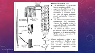

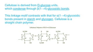
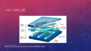


The document discusses the structure, functions, and composition of plant cell walls, which serve as protective and supportive barriers for plant cells. It details the various layers of the cell wall, including the middle lamella, primary wall, and secondary wall, and their roles in cell integrity and growth. Additionally, the document outlines the chemical makeup of cell walls and the significance of plasmodesmata in cell communication.


