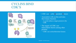
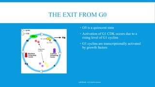
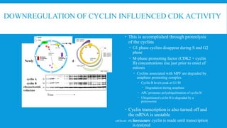
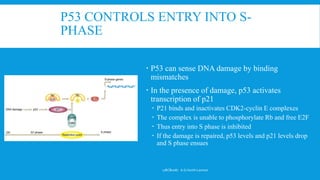
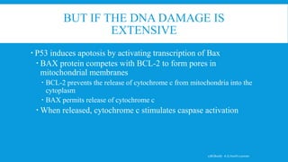
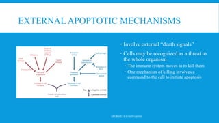
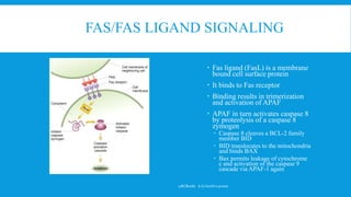
CELL CYCLE AND
REGULATION
17BCB0087 A.G.Harith Laxman
CELL CYCLE AND APOPTOSIS
 Cells undergo 3 controlled processes
 The first two are part of the cell cycle, the last an exit from the cell cycle
 Division (the cell cycle)
 Quiescence
 This is where most of the work of being a cell lies
 During division the energy of the cell is devoted to making a new cell
 Death
 This can be a normal process creating a final functional form or an induced suicide
 Epithelium and reticuloendothelial cells undergo active transitions towards terminally
differentiated states in which the final forms are unable to divide
 The stratum corneum consists of cells that have become bags of crosslinked keratin protein with
no internal metabolism
 Suicide can be induced because the organism senses a threat to the entire organism
 Infection, cancer, avoidance of autoimmunity
17BCB0087 A.G.Harith Laxman
CONTROL OF ENTRY INTO CELL CYCLE
AND APOPTOSIS
 Cell cycle is initiated by
phosphorylation of transcription
factors
 These activate transcription of a set of
proteins known as cyclins
 The appearance of cyclins is
progressive and determines the types
of proteins that will be phosphorylated
at a particular point during the cell
cycle
17BCB0087 A.G.Harith Laxman
CYCLINS AND CDK’S  CDK levels don’t change while
cyclins are destroyed at the end
of each phase
 There are 3 general groups of
each
 G1 cyclins
 Cyclin D
 S-phase cyclins
 Cyclin A
 G2 cyclins
 Cyclin B (maturation promoting
factor MPF)
 Cyclin E is shared between G1
and M phase
 Cyclin A is shared between M
phase and G2
17BCB0087 A.G.Harith Laxman
CYCLINS BIND
CDK’S
 CDK’s are Cyclin Dependent Kinases
 Association with cyclins activates
their kinase function
 A cyclin tethers a target protein to the
CDK
 The targets of CDK’s are
transcription factors among other
proteins
 CDK’s are serine/threonine kinases
17BCB0087 A.G.Harith Laxman
THE EXIT FROM G0
 G0 is a quiescent state
 Activation of G1 CDK occurs due to a
rising level of G1 cyclins
 G1 cyclins are transcriptionally activated
by growth factors
17BCB0087 A.G.Harith Laxman
EVENTS DURING G1
 A rising level of G1 cyclins increases
the activity of G1 CDK’s
 CDK’s in turn activate proteins and in
turn genes that prepare the cell to
begin DNA replication
 At the G1 S boundary, the cell
encounters a checkpoint
17BCB0087 A.G.Harith Laxman
G1/S
CHECKPOINT
 This is controlled by the activity of the
transcription factor E2F
 E2F is a family of related proteins (E2F 1 to E2F5)
 E2F is found complexed throughout the cell cycle
to another family of proteins: Rb
 At the G1/S checkpoint, Rb is phosphorylated by
CDK2/cyclinA
 E2F is freed from sequestration and activates
transcription at genes containing an E2F consensus
sequence
17BCB0087 A.G.Harith Laxman
AND THOSE GENES ARE
 Three groups
 Cell cycle regulators
 Cyclin A
 E2F, Rb, myc, myb
 Note that these are not all positive
regulators of cell cycle
 Enzymatic machinery for DNA
synthesis
 DNA polymerase
 PCNA
 Enzymes involved in nucleotide
metabolism
 DNA synthesis regulators
 Enzymes that recognize the origins
of replication for example17BCB0087 A.G.Harith Laxman
OTHER CHECKPOINTS
 These monitor the completion of
DNA synthesis
 The presence of Okazaki fragments
prevents entry into G2
 DNA damage
 This occurs before, during and after
completion of S phase
 Spindle attachment
 Failure to attach spindle to
centromere results in blockage of
mitosis at metaphase
 Failure to align the spindle during
cytokinesis results in blockage at
anaphase17BCB0087 A.G.Harith Laxman
DOWNREGULATION OF CYCLIN INFLUENCED CDK ACTIVITY
cyclin B
cyclin A
ribonucleotide
reductase
Mitosis MitosisInterphase Interphase
Time
Newly synthesized proteins labeled with 35S-methionine:
 This is accomplished through proteolysis
of the cyclins
 G1 phase cyclins disappear during S and G2
phase
 M-phase promoting factor (CDK2 + cyclin
B) concentrations rise just prior to onset of
mitosis
 Cyclins associated with MPF are degraded by
anaphase promoting complex
 Cyclin B levels peak at G1/M
 Degradation during anaphase
 APC promotes polyubiquitination of cyclin B
 Ubiquitinated cyclin B is degraded by a
proteosome
 Cyclin transcription is also turned off and
the mRNA is unstable
 So no new cyclin is made until transcription
is restored
17BCB0087 A.G.Harith Laxman
IN THE OVERALL
 Stimulated entry into G1 results in
appearance of an initial level of
cyclins that promote the progressive
activation of genes enabling the cell
to synthesize DNA
 A series of progressive steps result in
 Activation of genes further into the
cycle
 Degradation of cyclins that promoted
earlier steps
 Passage through checkpoints that
insure mechanistic fidelity of each step
17BCB0087 A.G.Harith Laxman
APOPTOSIS
 This is programmed cell death
 Distinguish it from necrosis
 Necrosis results from traumatic forces
outside the cell
 Necrotic tissue provokes
inflammation as the immune system
moves in to clear out damaged and
dead cells
 Apoptosis is an ordered stepwise
self-destruction that permits
surrounding cells to utilize the
breakdown products of the dead
cell
 There is no inflammation involved17BCB0087 A.G.Harith Laxman
THE APOPTOTIC CELL
 Mitochondria break open
 DNA fragments in a regular way
 The cell loses a regular shape
 Undergoes blebbing
 This is an irregular bubbling appearance of
the plasma membrane
17BCB0087 A.G.Harith Laxman
THE MECHANISMS OF APOTOSIS
 Can be classified as externally or
internally signaled
 One internal route involves p53
 p53 is a transcription factor that is
involved in cell cycle control and
sensing the presence of DNA damage
 The central role p53 plays is at the
G1/S checkpoint
17BCB0087 A.G.Harith Laxman
P53 CONTROLS ENTRY INTO S-
PHASE
 P53 can sense DNA damage by binding
mismatches
 In the presence of damage, p53 activates
transcription of p21
 P21 binds and inactivates CDK2-cyclin E complexes
 The complex is unable to phosphorylate Rb and free E2F
 Thus entry into S phase is inhibited
 If the damage is repaired, p53 levels and p21 levels drop
and S phase ensues
17BCB0087 A.G.Harith Laxman
BUT IF THE DNA DAMAGE IS
EXTENSIVE
 P53 induces apotosis by activating transcription of Bax
 BAX protein competes with BCL-2 to form pores in
mitochondrial membranes
 BCL-2 prevents the release of cytochrome c from mitochondria into the
cytoplasm
 BAX permits release of cytochrome c
 When released, cytochrome c stimulates caspase activation
17BCB0087 A.G.Harith Laxman
THE CASPASES
 These are proteolytic enzymes that
are held in check by external or
internal inhibitors
 Activation results in an explosive
proteolytic cascade
 Caspase 9 cleaves and activates
other caspases
 The caspases also activate
quiescent nucleases
17BCB0087 A.G.Harith Laxman
EXTERNAL APOPTOTIC MECHANISMS
 Involve external “death signals”
 Cells may be recognized as a threat to
the whole organism
 The immune system moves in to kill them
 One mechanism of killing involves a
command to the cell to initiate apoptosis
17BCB0087 A.G.Harith Laxman
FAS/FAS LIGAND SIGNALING
 Fas ligand (FasL) is a membrane
bound cell surface protein
 It binds to Fas receptor
 Binding results in trimerization
and activation of APAF
 APAF in turn activates caspase 8
by proteolysis of a caspase 8
zymogen
 Caspase 8 cleaves a BCL-2 family
member BID
 BID translocates to the mitochondria
and binds BAX
 Bax permits leakage of cytochrome
c and activation of the caspase 9
cascade via APAF-1 again
17BCB0087 A.G.Harith Laxman

Cell Cycle Regulations

  • 1.
  • 2.
    CELL CYCLE ANDAPOPTOSIS  Cells undergo 3 controlled processes  The first two are part of the cell cycle, the last an exit from the cell cycle  Division (the cell cycle)  Quiescence  This is where most of the work of being a cell lies  During division the energy of the cell is devoted to making a new cell  Death  This can be a normal process creating a final functional form or an induced suicide  Epithelium and reticuloendothelial cells undergo active transitions towards terminally differentiated states in which the final forms are unable to divide  The stratum corneum consists of cells that have become bags of crosslinked keratin protein with no internal metabolism  Suicide can be induced because the organism senses a threat to the entire organism  Infection, cancer, avoidance of autoimmunity 17BCB0087 A.G.Harith Laxman
  • 3.
    CONTROL OF ENTRYINTO CELL CYCLE AND APOPTOSIS  Cell cycle is initiated by phosphorylation of transcription factors  These activate transcription of a set of proteins known as cyclins  The appearance of cyclins is progressive and determines the types of proteins that will be phosphorylated at a particular point during the cell cycle 17BCB0087 A.G.Harith Laxman
  • 4.
    CYCLINS AND CDK’S CDK levels don’t change while cyclins are destroyed at the end of each phase  There are 3 general groups of each  G1 cyclins  Cyclin D  S-phase cyclins  Cyclin A  G2 cyclins  Cyclin B (maturation promoting factor MPF)  Cyclin E is shared between G1 and M phase  Cyclin A is shared between M phase and G2 17BCB0087 A.G.Harith Laxman
  • 5.
    CYCLINS BIND CDK’S  CDK’sare Cyclin Dependent Kinases  Association with cyclins activates their kinase function  A cyclin tethers a target protein to the CDK  The targets of CDK’s are transcription factors among other proteins  CDK’s are serine/threonine kinases 17BCB0087 A.G.Harith Laxman
  • 6.
    THE EXIT FROMG0  G0 is a quiescent state  Activation of G1 CDK occurs due to a rising level of G1 cyclins  G1 cyclins are transcriptionally activated by growth factors 17BCB0087 A.G.Harith Laxman
  • 7.
    EVENTS DURING G1 A rising level of G1 cyclins increases the activity of G1 CDK’s  CDK’s in turn activate proteins and in turn genes that prepare the cell to begin DNA replication  At the G1 S boundary, the cell encounters a checkpoint 17BCB0087 A.G.Harith Laxman
  • 8.
    G1/S CHECKPOINT  This iscontrolled by the activity of the transcription factor E2F  E2F is a family of related proteins (E2F 1 to E2F5)  E2F is found complexed throughout the cell cycle to another family of proteins: Rb  At the G1/S checkpoint, Rb is phosphorylated by CDK2/cyclinA  E2F is freed from sequestration and activates transcription at genes containing an E2F consensus sequence 17BCB0087 A.G.Harith Laxman
  • 9.
    AND THOSE GENESARE  Three groups  Cell cycle regulators  Cyclin A  E2F, Rb, myc, myb  Note that these are not all positive regulators of cell cycle  Enzymatic machinery for DNA synthesis  DNA polymerase  PCNA  Enzymes involved in nucleotide metabolism  DNA synthesis regulators  Enzymes that recognize the origins of replication for example17BCB0087 A.G.Harith Laxman
  • 10.
    OTHER CHECKPOINTS  Thesemonitor the completion of DNA synthesis  The presence of Okazaki fragments prevents entry into G2  DNA damage  This occurs before, during and after completion of S phase  Spindle attachment  Failure to attach spindle to centromere results in blockage of mitosis at metaphase  Failure to align the spindle during cytokinesis results in blockage at anaphase17BCB0087 A.G.Harith Laxman
  • 11.
    DOWNREGULATION OF CYCLININFLUENCED CDK ACTIVITY cyclin B cyclin A ribonucleotide reductase Mitosis MitosisInterphase Interphase Time Newly synthesized proteins labeled with 35S-methionine:  This is accomplished through proteolysis of the cyclins  G1 phase cyclins disappear during S and G2 phase  M-phase promoting factor (CDK2 + cyclin B) concentrations rise just prior to onset of mitosis  Cyclins associated with MPF are degraded by anaphase promoting complex  Cyclin B levels peak at G1/M  Degradation during anaphase  APC promotes polyubiquitination of cyclin B  Ubiquitinated cyclin B is degraded by a proteosome  Cyclin transcription is also turned off and the mRNA is unstable  So no new cyclin is made until transcription is restored 17BCB0087 A.G.Harith Laxman
  • 12.
  • 13.
    IN THE OVERALL Stimulated entry into G1 results in appearance of an initial level of cyclins that promote the progressive activation of genes enabling the cell to synthesize DNA  A series of progressive steps result in  Activation of genes further into the cycle  Degradation of cyclins that promoted earlier steps  Passage through checkpoints that insure mechanistic fidelity of each step 17BCB0087 A.G.Harith Laxman
  • 14.
    APOPTOSIS  This isprogrammed cell death  Distinguish it from necrosis  Necrosis results from traumatic forces outside the cell  Necrotic tissue provokes inflammation as the immune system moves in to clear out damaged and dead cells  Apoptosis is an ordered stepwise self-destruction that permits surrounding cells to utilize the breakdown products of the dead cell  There is no inflammation involved17BCB0087 A.G.Harith Laxman
  • 15.
    THE APOPTOTIC CELL Mitochondria break open  DNA fragments in a regular way  The cell loses a regular shape  Undergoes blebbing  This is an irregular bubbling appearance of the plasma membrane 17BCB0087 A.G.Harith Laxman
  • 16.
    THE MECHANISMS OFAPOTOSIS  Can be classified as externally or internally signaled  One internal route involves p53  p53 is a transcription factor that is involved in cell cycle control and sensing the presence of DNA damage  The central role p53 plays is at the G1/S checkpoint 17BCB0087 A.G.Harith Laxman
  • 17.
    P53 CONTROLS ENTRYINTO S- PHASE  P53 can sense DNA damage by binding mismatches  In the presence of damage, p53 activates transcription of p21  P21 binds and inactivates CDK2-cyclin E complexes  The complex is unable to phosphorylate Rb and free E2F  Thus entry into S phase is inhibited  If the damage is repaired, p53 levels and p21 levels drop and S phase ensues 17BCB0087 A.G.Harith Laxman
  • 18.
    BUT IF THEDNA DAMAGE IS EXTENSIVE  P53 induces apotosis by activating transcription of Bax  BAX protein competes with BCL-2 to form pores in mitochondrial membranes  BCL-2 prevents the release of cytochrome c from mitochondria into the cytoplasm  BAX permits release of cytochrome c  When released, cytochrome c stimulates caspase activation 17BCB0087 A.G.Harith Laxman
  • 19.
    THE CASPASES  Theseare proteolytic enzymes that are held in check by external or internal inhibitors  Activation results in an explosive proteolytic cascade  Caspase 9 cleaves and activates other caspases  The caspases also activate quiescent nucleases 17BCB0087 A.G.Harith Laxman
  • 20.
    EXTERNAL APOPTOTIC MECHANISMS Involve external “death signals”  Cells may be recognized as a threat to the whole organism  The immune system moves in to kill them  One mechanism of killing involves a command to the cell to initiate apoptosis 17BCB0087 A.G.Harith Laxman
  • 21.
    FAS/FAS LIGAND SIGNALING Fas ligand (FasL) is a membrane bound cell surface protein  It binds to Fas receptor  Binding results in trimerization and activation of APAF  APAF in turn activates caspase 8 by proteolysis of a caspase 8 zymogen  Caspase 8 cleaves a BCL-2 family member BID  BID translocates to the mitochondria and binds BAX  Bax permits leakage of cytochrome c and activation of the caspase 9 cascade via APAF-1 again 17BCB0087 A.G.Harith Laxman