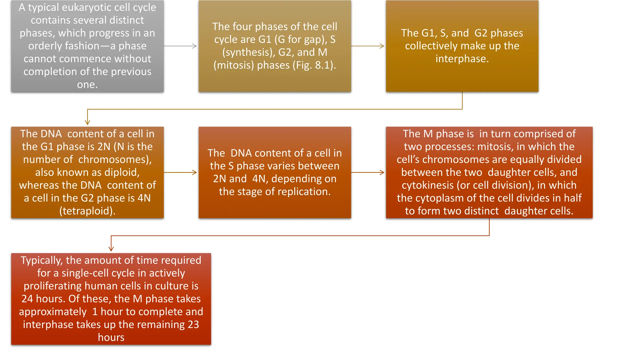
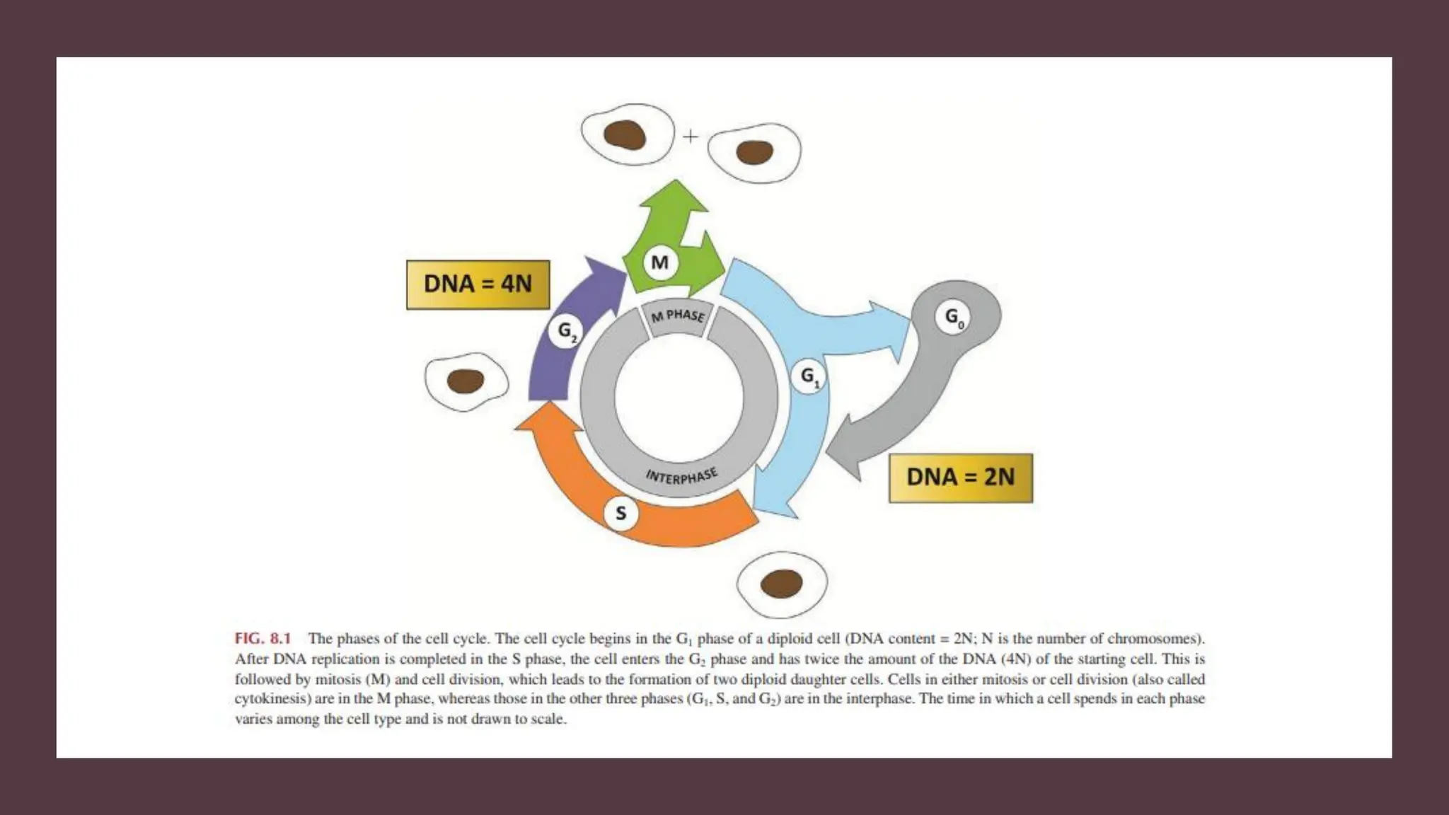
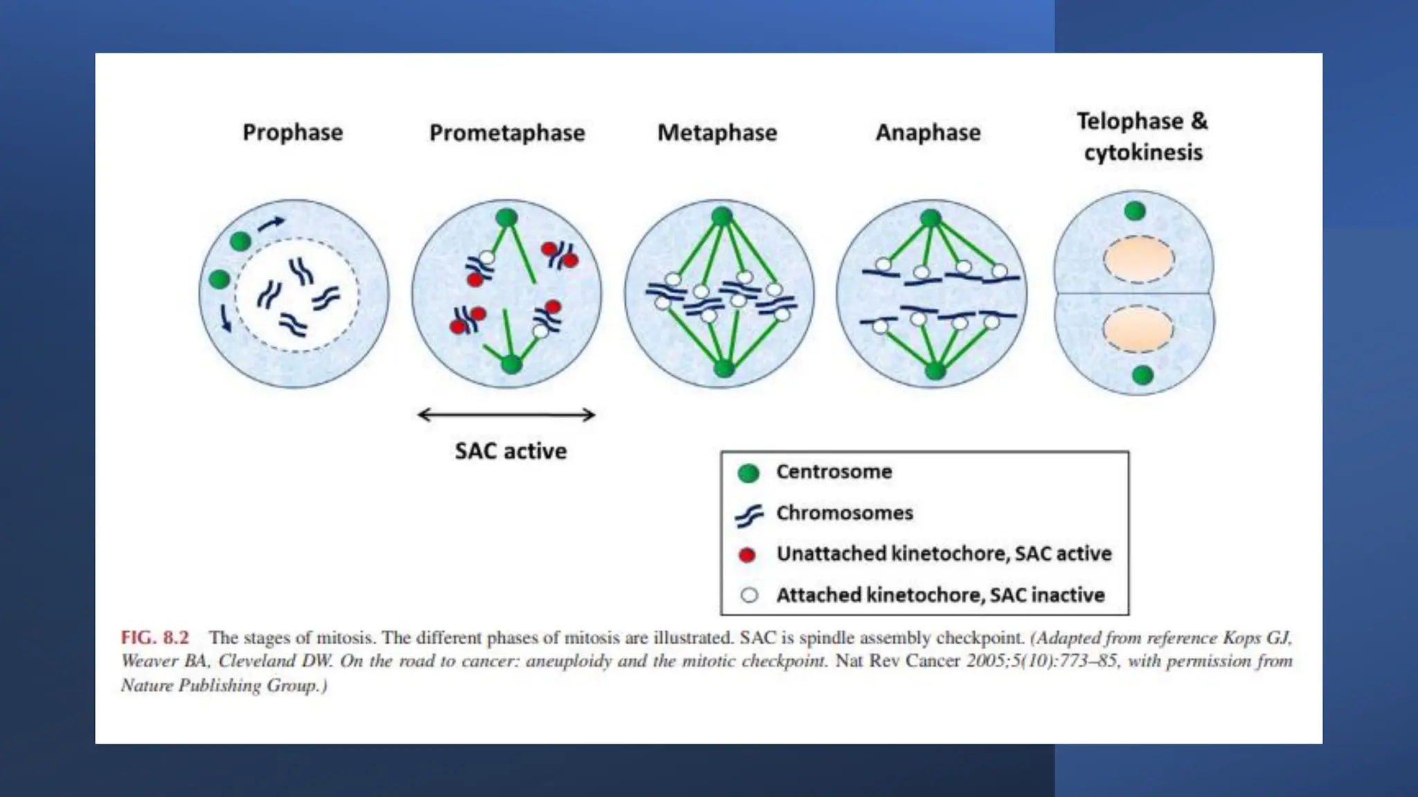
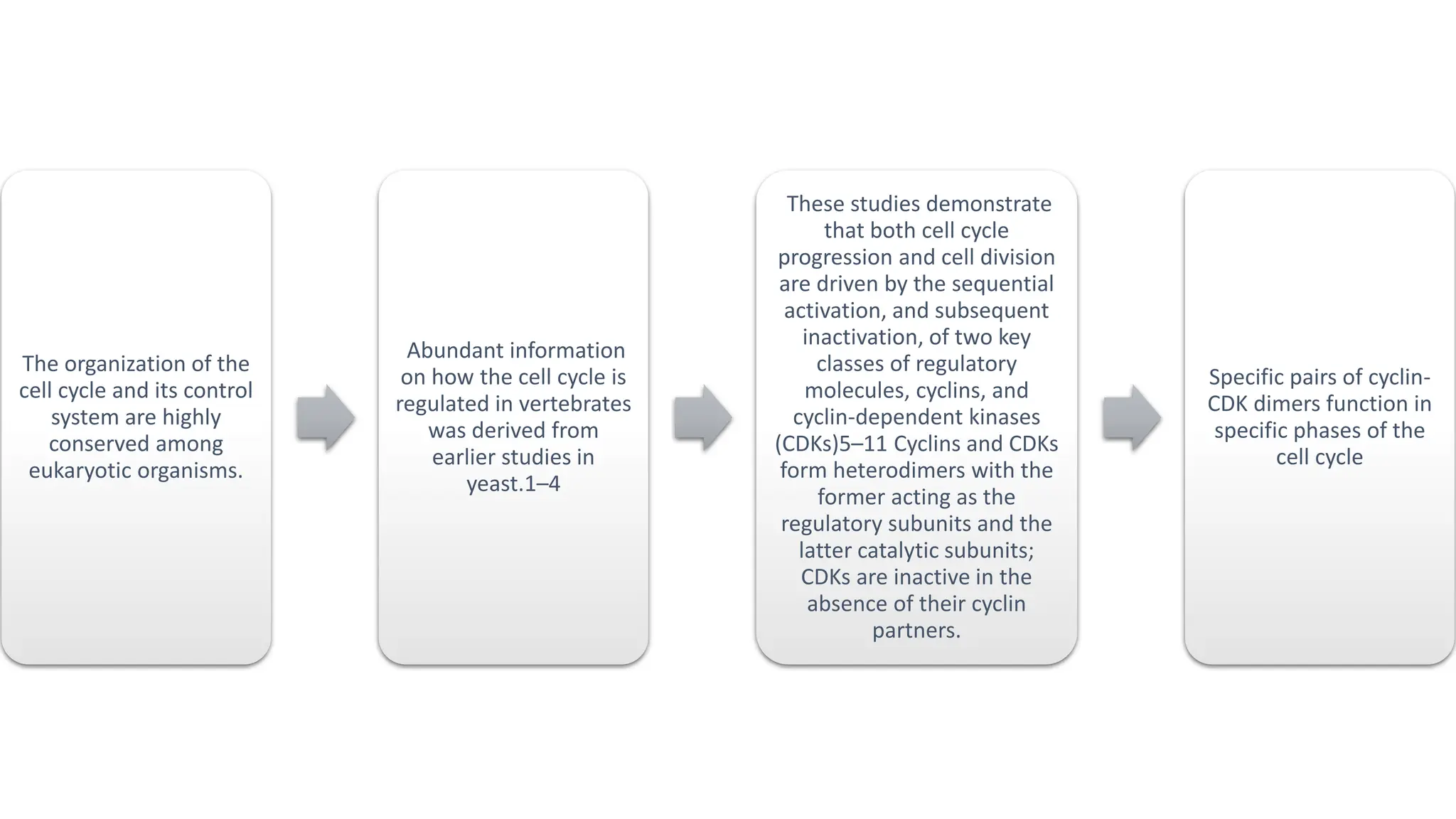
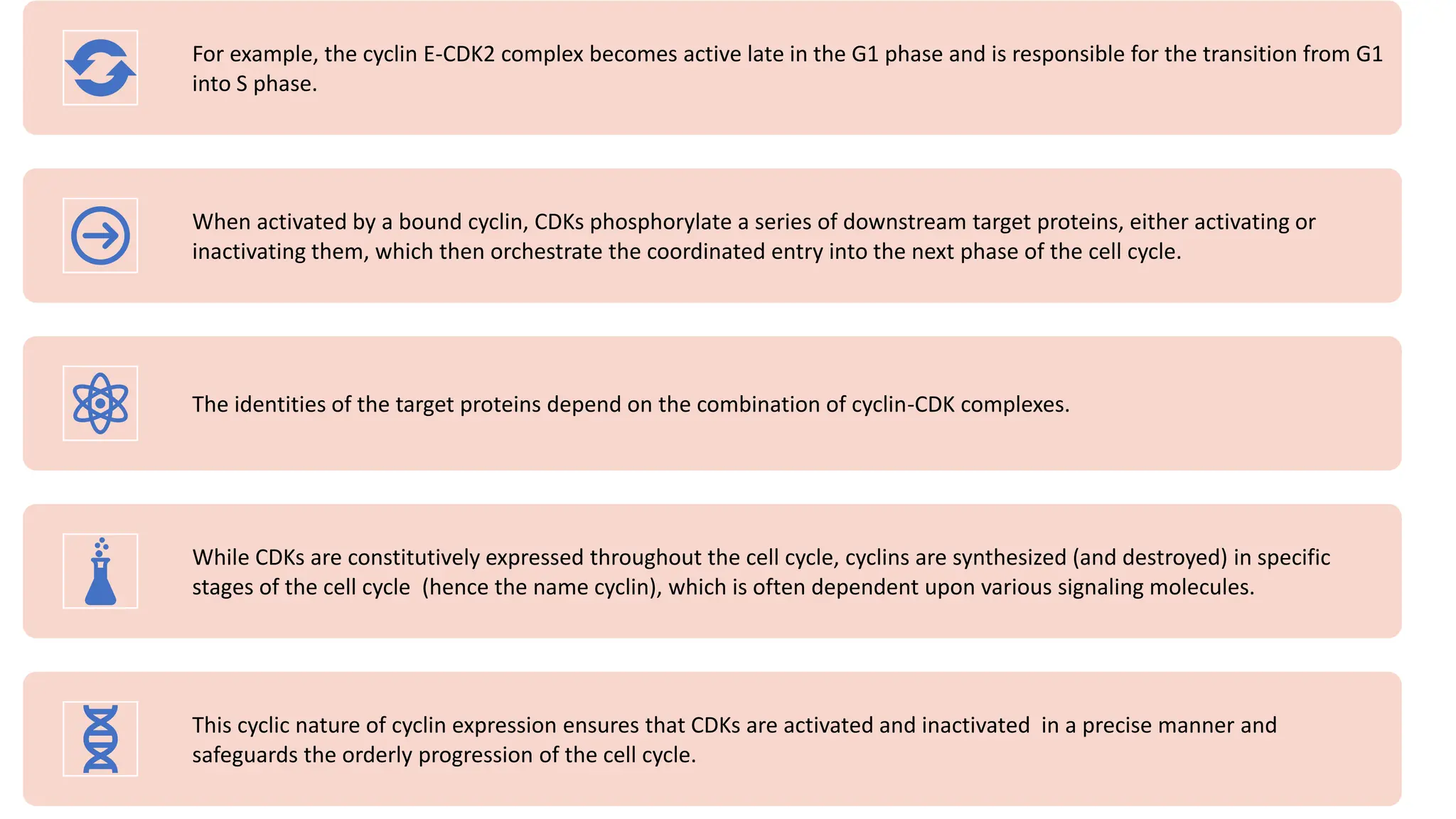
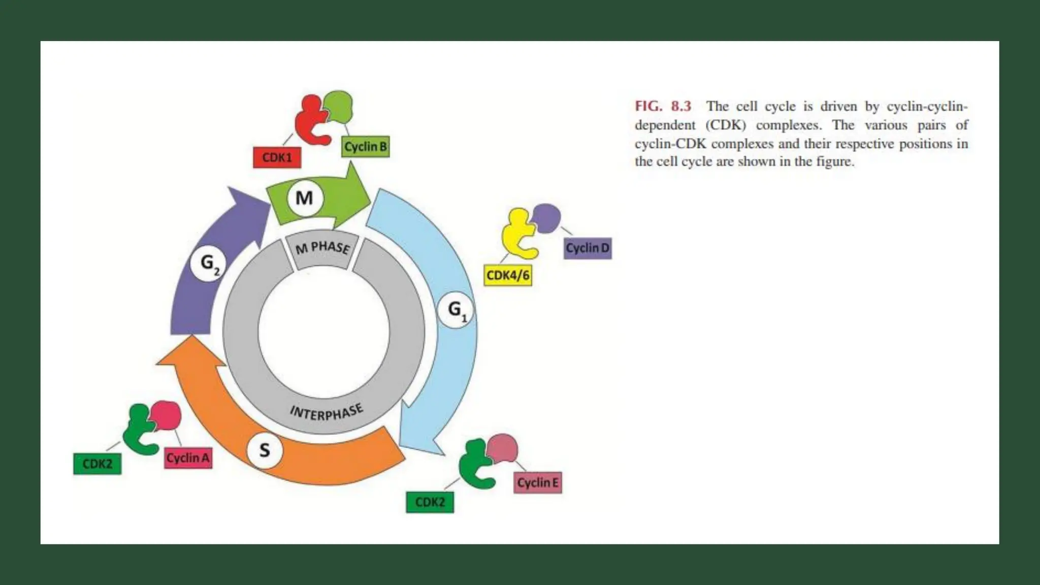
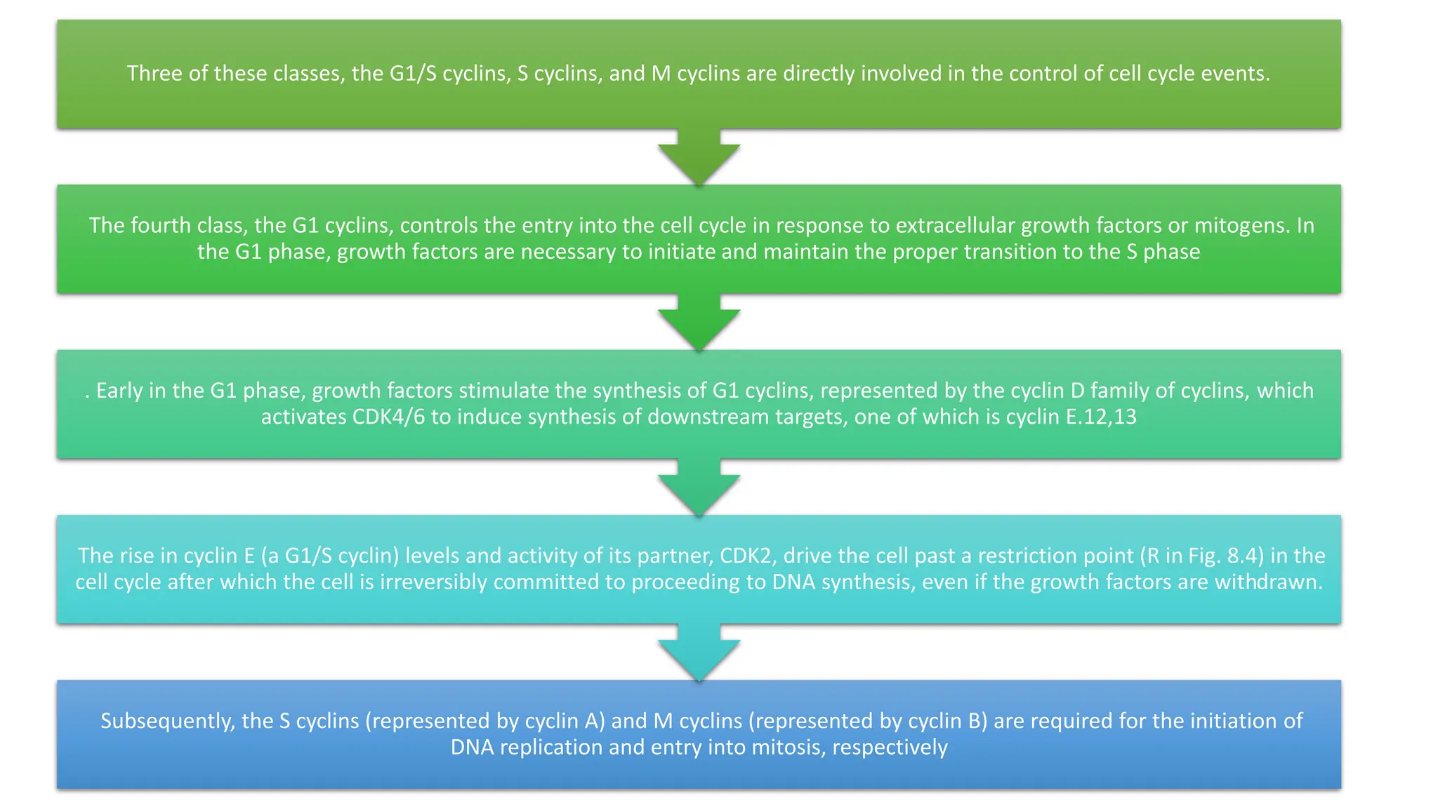
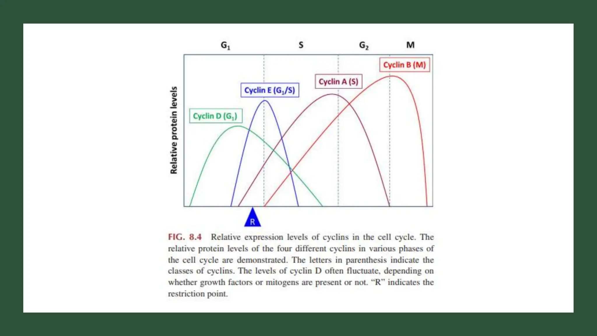
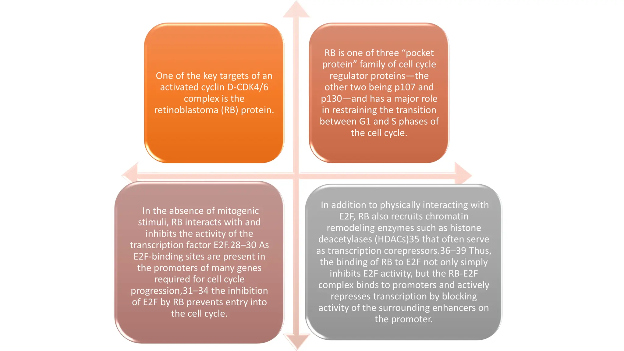
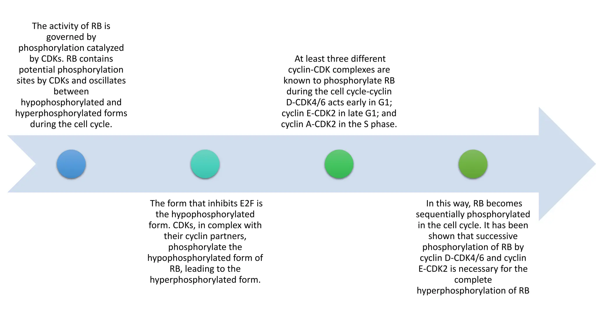
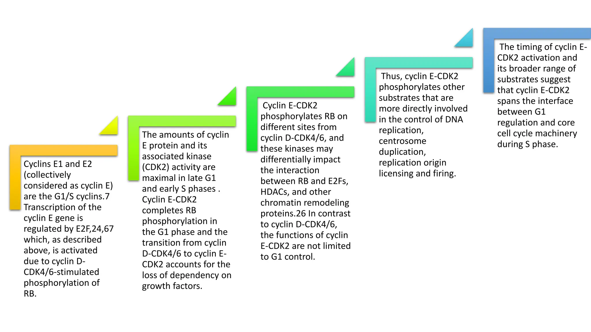
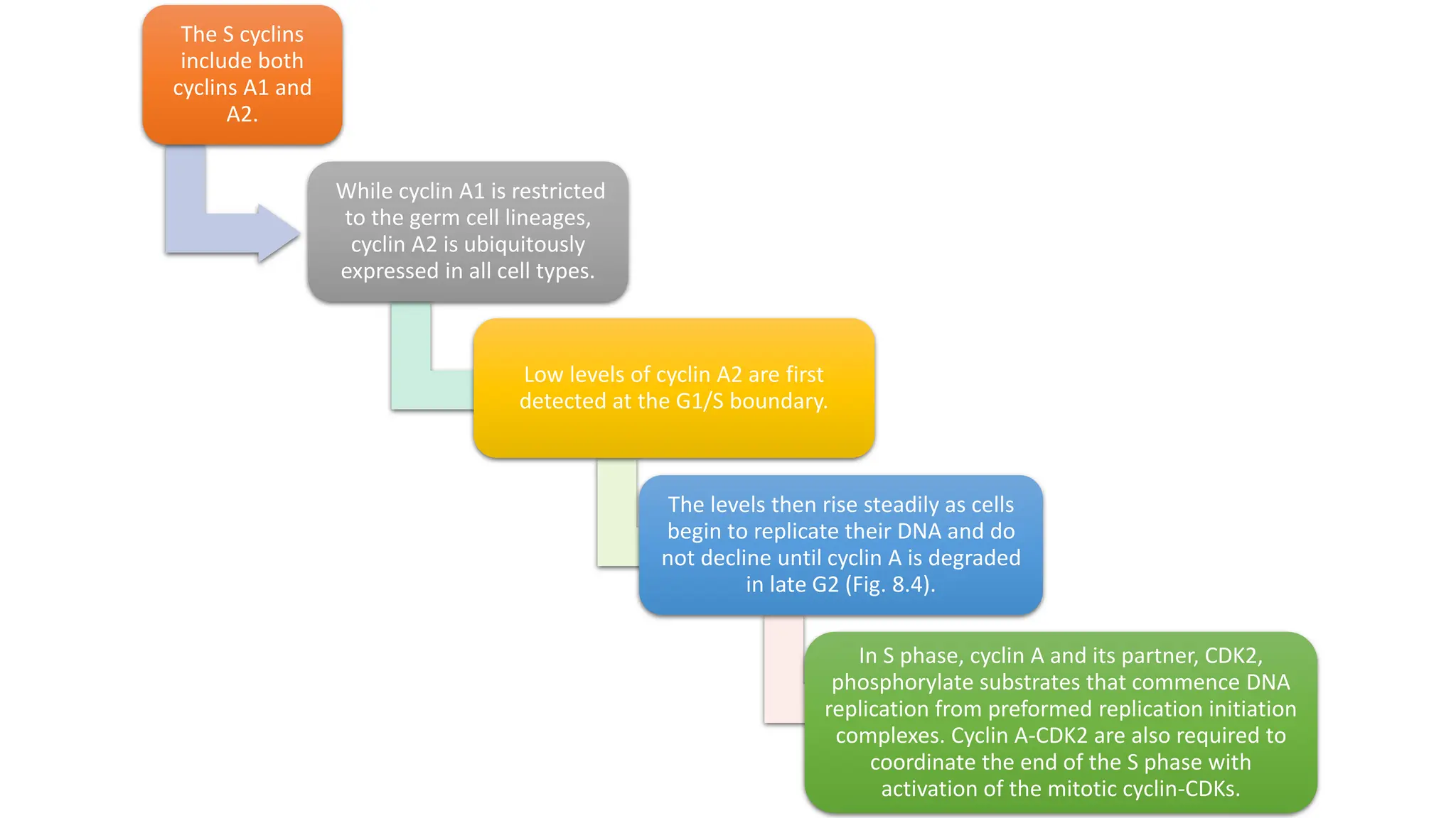
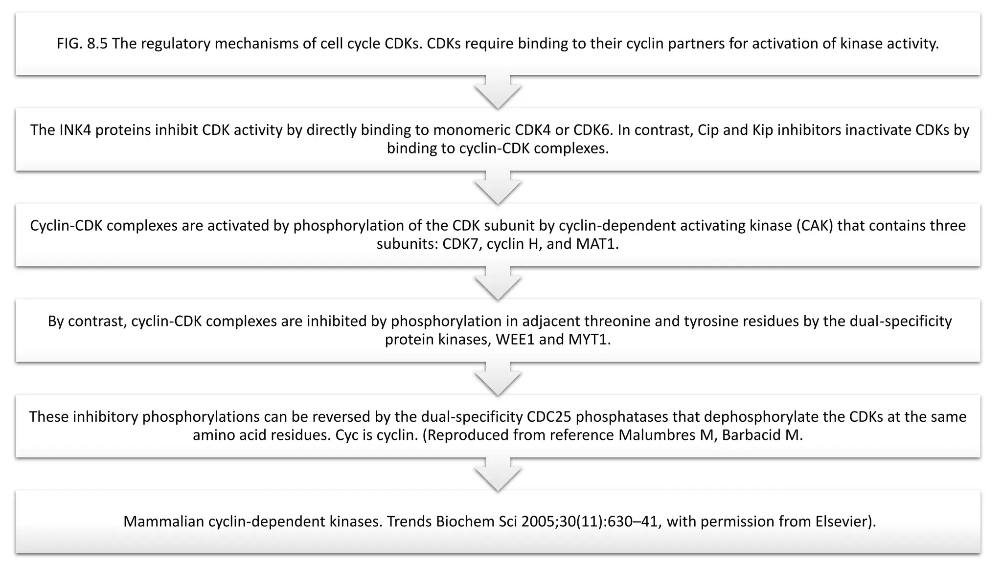
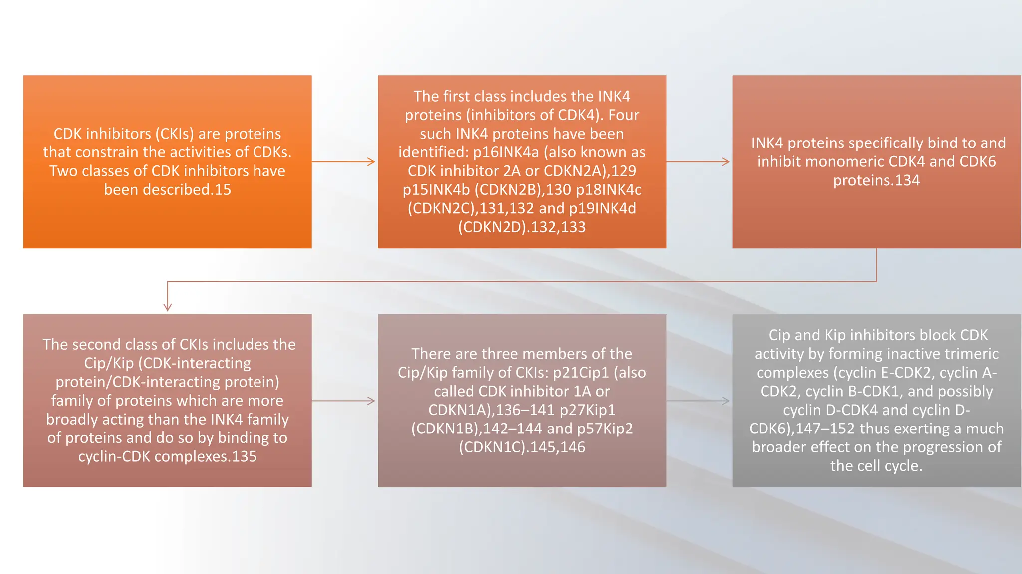
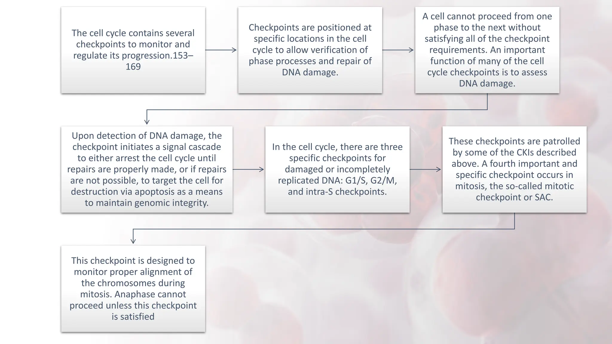
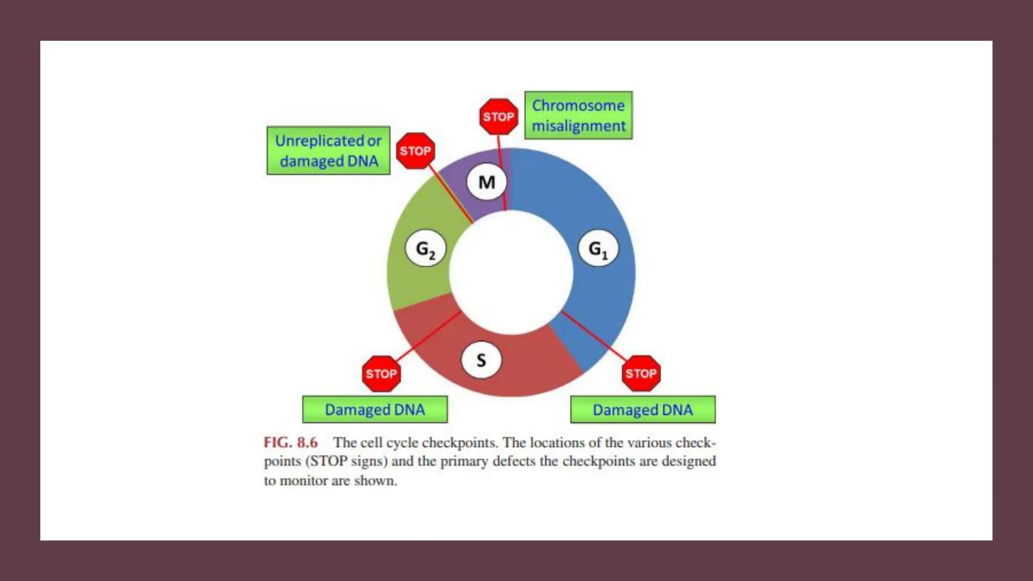
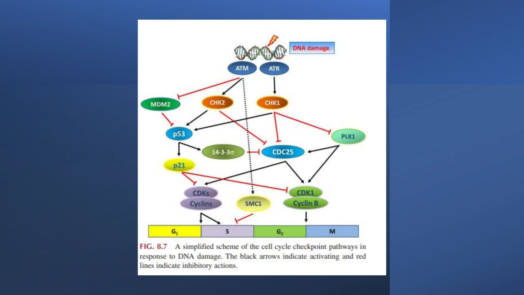
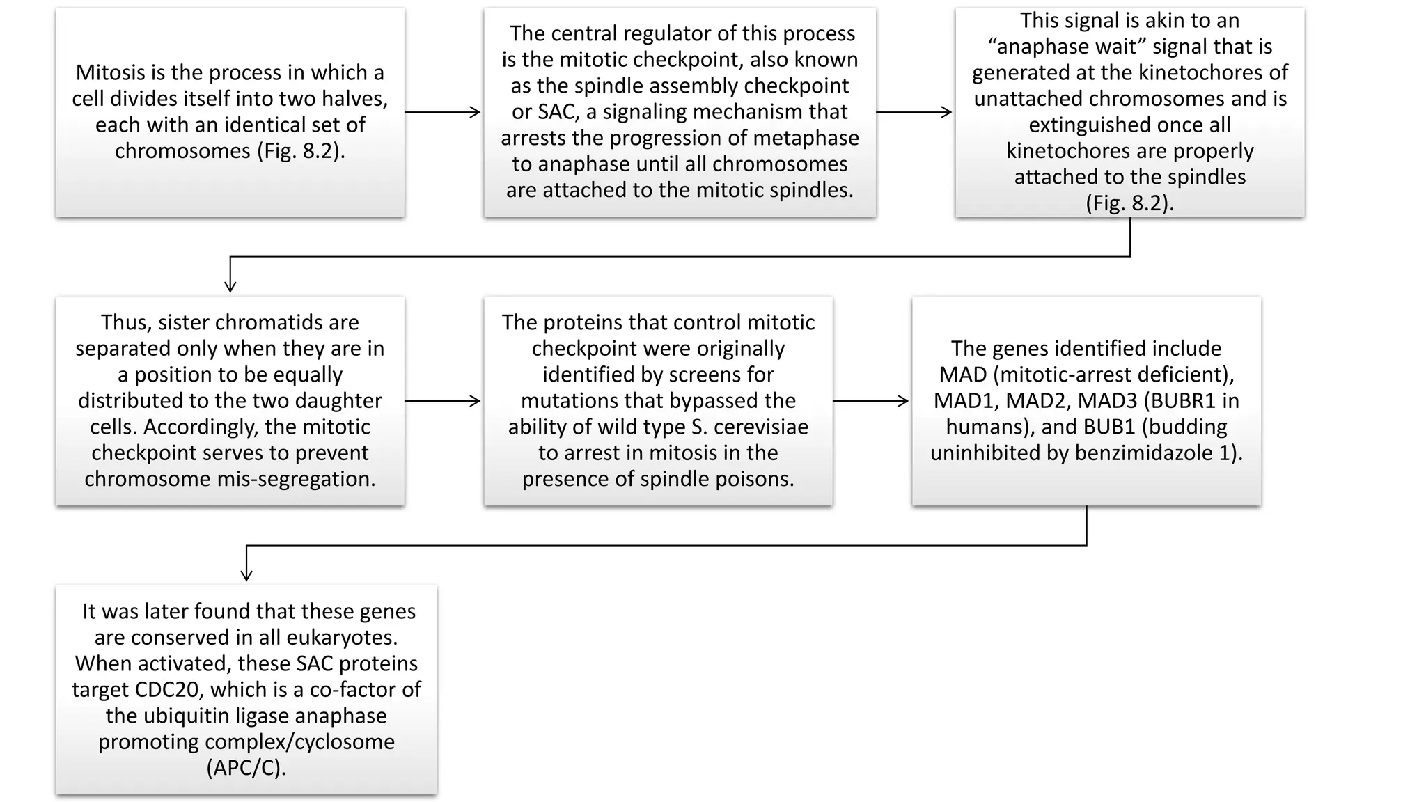
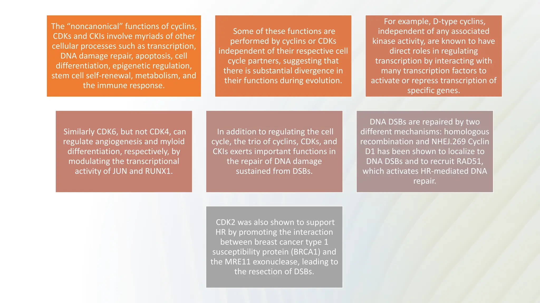
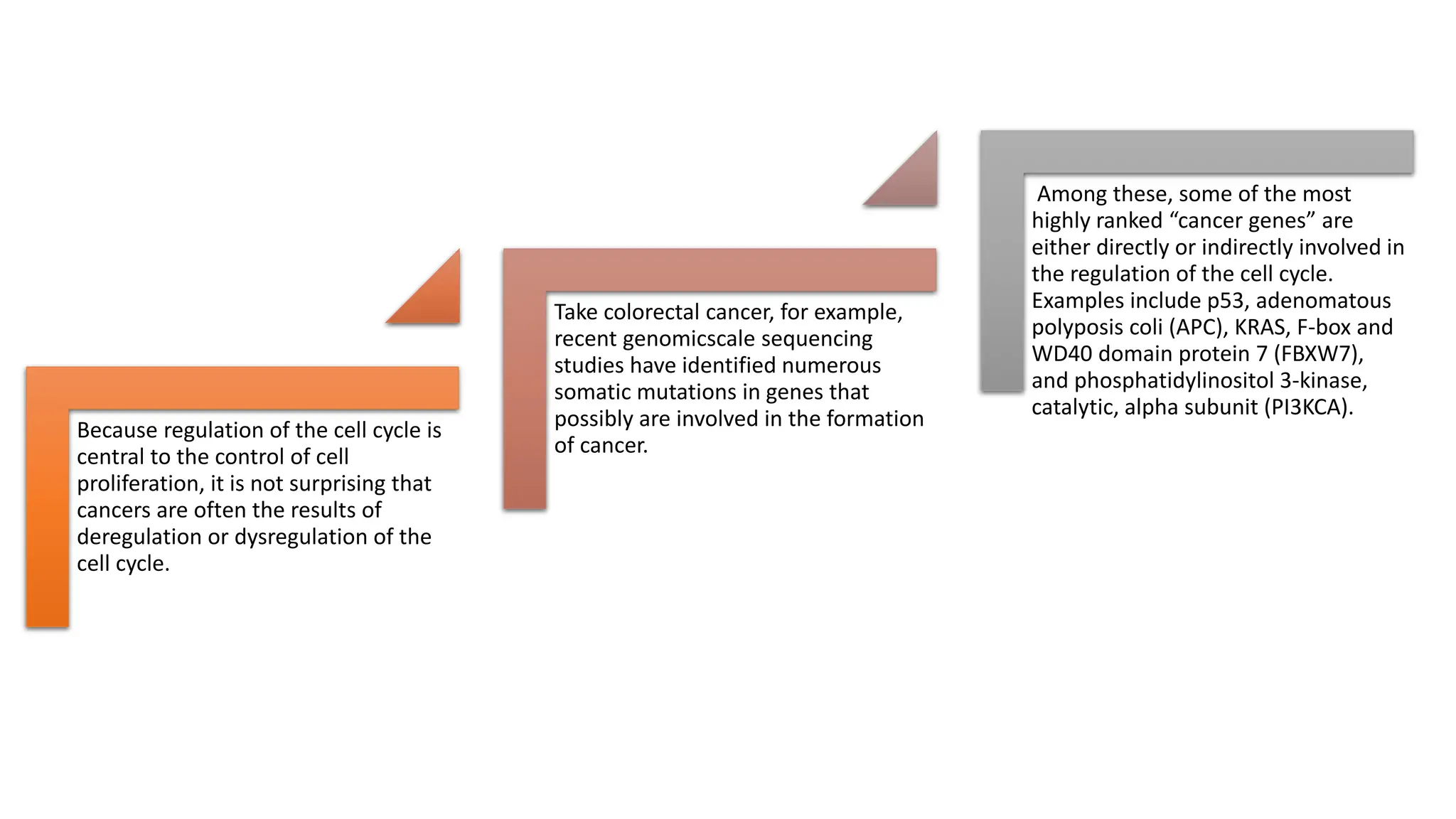
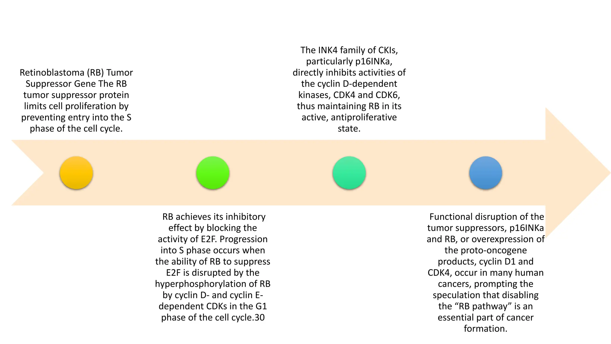
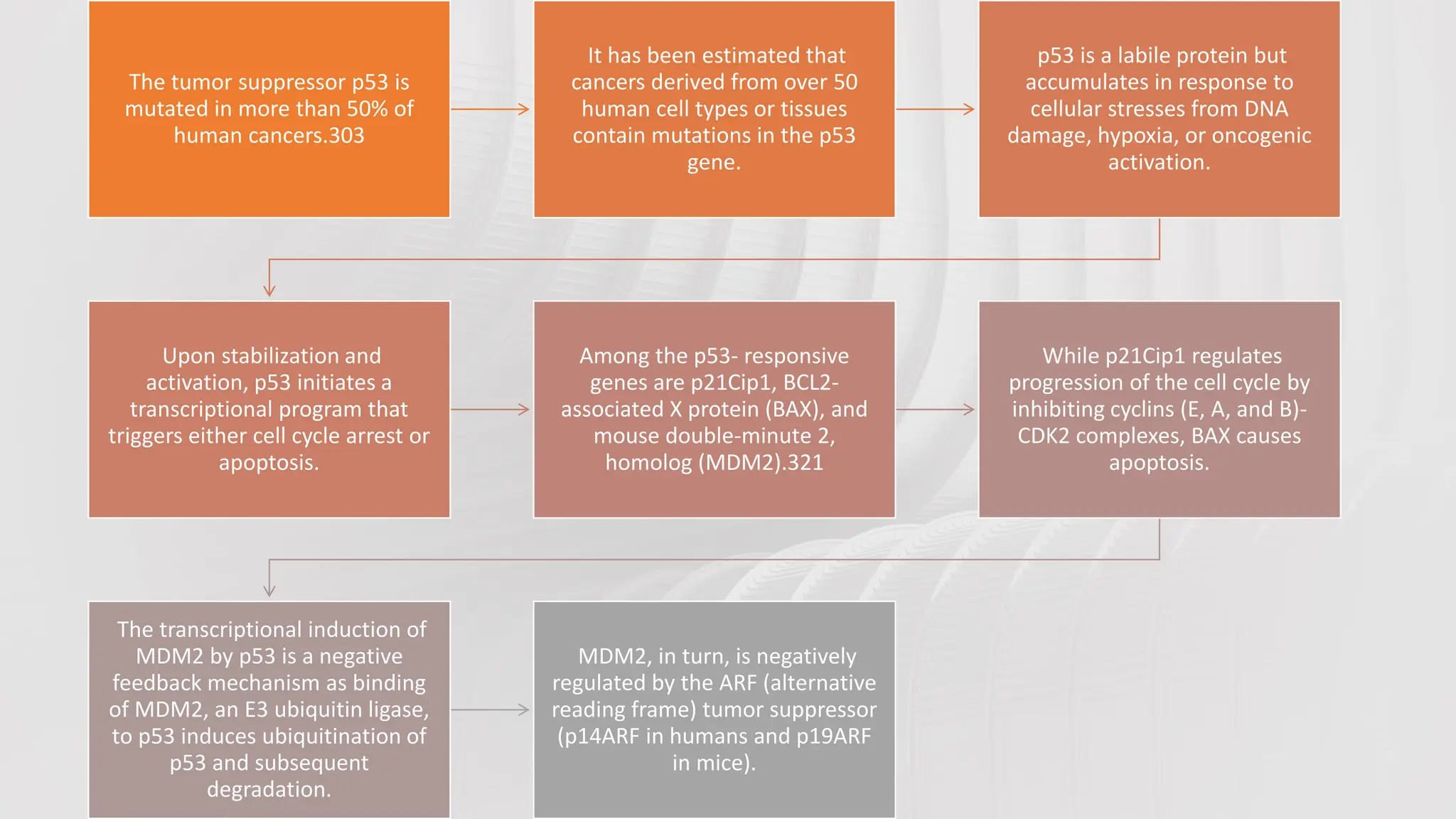
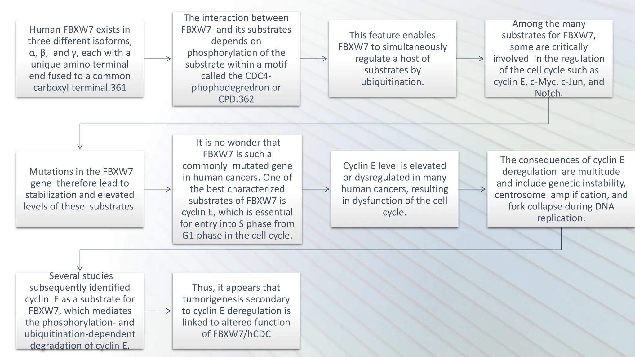
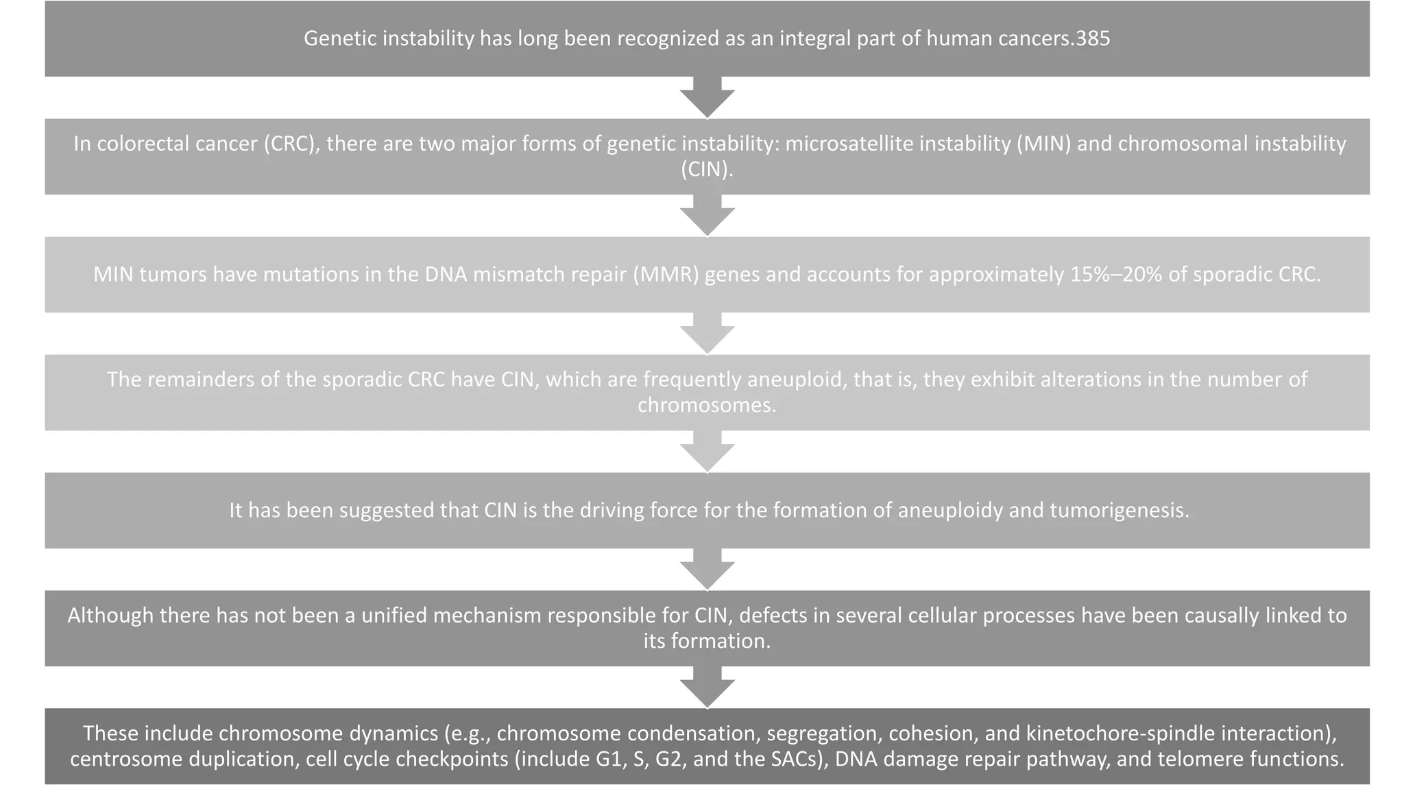
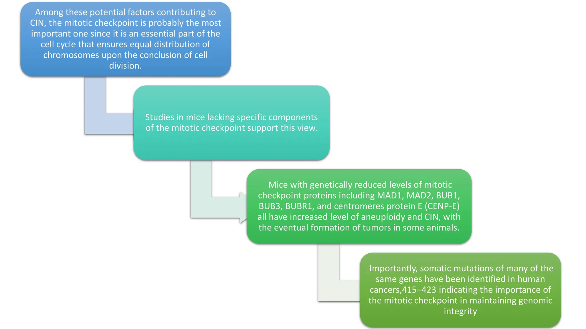
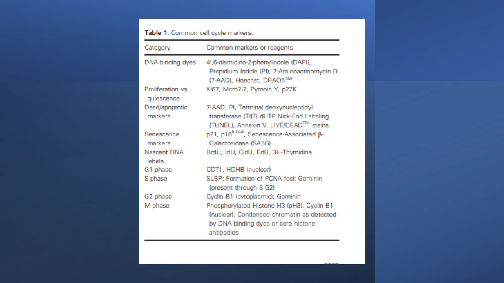
CELL CYCLE: Phases &control and
analysis
By
Romissaa Aly
Lecturer of Oral Medicine, Periodontology, Diagnosis
and Dental Radiology (Al-Azhar University)
• Contents:
• COMPONENTS OF THE CELL CYCLE
• CONTROL OF THE CELL CYCLE
• PATHOLOGICAL CONSEQUENCES OF CELL CYCLE
DEREGULATION OR DYSREGULATION
• Quantitative analysis of cell cycle phase durations
COMPONENTS OF THE CELL CYCLE
A typical eukaryotic cell cycle
contains several distinct
phases, which progress in an
orderly fashion—a phase
cannot commence without
completion of the previous
one.
The four phases of the cell
cycle are G1 (G for gap), S
(synthesis), G2, and M
(mitosis) phases (Fig. 8.1).
The G1, S, and G2 phases
collectively make up the
interphase.
The DNA content of a cell in
the G1 phase is 2N (N is the
number of chromosomes),
also known as diploid,
whereas the DNA content of
a cell in the G2 phase is 4N
(tetraploid).
The DNA content of a cell in
the S phase varies between
2N and 4N, depending on
the stage of replication.
The M phase is in turn comprised of
two processes: mitosis, in which the
cell’s chromosomes are equally divided
between the two daughter cells, and
cytokinesis (or cell division), in which
the cytoplasm of the cell divides in half
to form two distinct daughter cells.
Typically, the amount of time required
for a single-cell cycle in actively
proliferating human cells in culture is
24 hours. Of these, the M phase takes
approximately 1 hour to complete and
interphase takes up the remaining 23
hours
In addition to the four
phases of the cell cycle
listed above, one phase
that lies outside the cell
cycle is called the G0 (0
for zero) phase.
Cells in this phase are in
the resting phase, which
is often the result of their
leaving the G1 phase of
the cell cycle.
Typically, a cell enters G0
phase if the environment
is not conducive for the
progression of the cell
cycle, as in the event of
deprivation of essential
nutrients or growth
factors, or if a cell has
reached a fully
differentiated state such
as a hepatocytes or
neuron.
In these conditions, the
cell is sometimes referred
to as in a quiescent state.
Additionally, a cell can
enter the G0 phase and
become senescent due
to DNA damage or
telomere attrition.
This is often an
alternative to self-
destruction of the
damaged cell by
apoptosis.
In the M phase,
mitosis commences
when DNA
condenses into
visible
chromosomes,
followed by the
separation of the
chromosomes into
two identical sets.
Cytokinesis is the
last phase of mitosis
when the two
daughter cells
separate, each with
a nucleus and
cytoplasmic
organelles.
Mitosis begins with
nuclear membrane
breakdown followed
by condensation of
the chromosomes
and separation of
the centrosomes
(prophase).
This is accompanied
by the formation of
mitotic spindles,
which are attached
on one end, the
centrosomes, and
the other end,
kinetochore, a
protein structure
located at or near
the centromeres of
mitotic
chromosomes
(prometaphase).
At this point,
kinetochores that
are unattached to
the mitotic spindles
generate a “wait”
signal that delays
the onset of
anaphase until all
chromosomes are
properly attached
and aligned.
This signal is also
called mitotic
checkpoint or
spindle assembly
checkpoint (SAC),
which is satisfied
once all
chromosomes are
congregated at the
equatorial plate
(metaphase).
This is followed by separation of the chromosomes to the opposite poles (anaphase) and
formation of new nuclear membranes around the daughter nuclei and uncoiling of the
chromosomes (telophase), eventually forming a cleavage furrow that leads to the formation of
two daughter cells (cytokinesis). Fig. 8.2 illustrates the various stages of mitosis
CONTROL OF THE CELL CYCLE
The organization of the
cell cycle and its control
system are highly
conserved among
eukaryotic organisms.
Abundant information
on how the cell cycle is
regulated in vertebrates
was derived from
earlier studies in
yeast.1–4
These studies demonstrate
that both cell cycle
progression and cell division
are driven by the sequential
activation, and subsequent
inactivation, of two key
classes of regulatory
molecules, cyclins, and
cyclin-dependent kinases
(CDKs)5–11 Cyclins and CDKs
form heterodimers with the
former acting as the
regulatory subunits and the
latter catalytic subunits;
CDKs are inactive in the
absence of their cyclin
partners.
Specific pairs of cyclin-
CDK dimers function in
specific phases of the
cell cycle
For example, the cyclin E-CDK2 complex becomes active late in the G1 phase and is responsible for the transition from G1
into S phase.
When activated by a bound cyclin, CDKs phosphorylate a series of downstream target proteins, either activating or
inactivating them, which then orchestrate the coordinated entry into the next phase of the cell cycle.
The identities of the target proteins depend on the combination of cyclin-CDK complexes.
While CDKs are constitutively expressed throughout the cell cycle, cyclins are synthesized (and destroyed) in specific
stages of the cell cycle (hence the name cyclin), which is often dependent upon various signaling molecules.
This cyclic nature of cyclin expression ensures that CDKs are activated and inactivated in a precise manner and
safeguards the orderly progression of the cell cycle.
Subsequently, the S cyclins (represented by cyclin A) and M cyclins (represented by cyclin B) are required for the initiation of
DNA replication and entry into mitosis, respectively
The rise in cyclin E (a G1/S cyclin) levels and activity of its partner, CDK2, drive the cell past a restriction point (R in Fig. 8.4) in the
cell cycle after which the cell is irreversibly committed to proceeding to DNA synthesis, even if the growth factors are withdrawn.
. Early in the G1 phase, growth factors stimulate the synthesis of G1 cyclins, represented by the cyclin D family of cyclins, which
activates CDK4/6 to induce synthesis of downstream targets, one of which is cyclin E.12,13
The fourth class, the G1 cyclins, controls the entry into the cell cycle in response to extracellular growth factors or mitogens. In
the G1 phase, growth factors are necessary to initiate and maintain the proper transition to the S phase
Three of these classes, the G1/S cyclins, S cyclins, and M cyclins are directly involved in the control of cell cycle events.
The G1 cyclins are composed of the
D-type cyclins that include cyclins
D1, D2, and D3.7 Along with their
partners, CDK4 and CDK6, G1
cyclins act early in the G1 phase of
the cell cycle.
The levels of G1 cyclin are low in
G0 phase and increase
progressively upon addition of
growth factors or mitogens to the
cells.
The mechanisms by which
mitogens or growth factors activate
cyclin D1 are complex and occur at
both transcriptional and
posttranscriptional levels.
At the transcriptional level,
induction of cyclin D1 by growth
factors is dependent on the
RAS/RAF/mitogen activated kinase
(MEK)/extracellular signal-
regulated kinase (ERK) signaling
pathway.
Once synthesized, cyclin D1 protein has a short half-
life20—its turnover being governed by
ubiquitination and proteasomal degradation, which
in turn are dependent on phosphorylation of cyclin
D1 by glycogen synthase kinase-3β (GSK-3β).21,22
Growth factors prevent cyclin D1 degradation by
inhibiting GSK3β-dependent phosphorylation of
cyclin D1 through the Ras/phosphatidylinositol-3-OH
kinase (PI3K)/AKT pathway
One of the key targets of an
activated cyclin D-CDK4/6
complex is the
retinoblastoma (RB) protein.
RB is one of three “pocket
protein” family of cell cycle
regulator proteins—the
other two being p107 and
p130—and has a major role
in restraining the transition
between G1 and S phases of
the cell cycle.
In the absence of mitogenic
stimuli, RB interacts with and
inhibits the activity of the
transcription factor E2F.28–30 As
E2F-binding sites are present in
the promoters of many genes
required for cell cycle
progression,31–34 the inhibition
of E2F by RB prevents entry into
the cell cycle.
In addition to physically interacting with
E2F, RB also recruits chromatin
remodeling enzymes such as histone
deacetylases (HDACs)35 that often serve
as transcription corepressors.36–39 Thus,
the binding of RB to E2F not only simply
inhibits E2F activity, but the RB-E2F
complex binds to promoters and actively
represses transcription by blocking
activity of the surrounding enhancers on
the promoter.
The activity of RB is
governed by
phosphorylation catalyzed
by CDKs. RB contains
potential phosphorylation
sites by CDKs and oscillates
between
hypophosphorylated and
hyperphosphorylated forms
during the cell cycle.
The form that inhibits E2F is
the hypophosphorylated
form. CDKs, in complex with
their cyclin partners,
phosphorylate the
hypophosphorylated form of
RB, leading to the
hyperphosphorylated form.
At least three different
cyclin-CDK complexes are
known to phosphorylate RB
during the cell cycle-cyclin
D-CDK4/6 acts early in G1;
cyclin E-CDK2 in late G1; and
cyclin A-CDK2 in the S phase.
In this way, RB becomes
sequentially phosphorylated
in the cell cycle. It has been
shown that successive
phosphorylation of RB by
cyclin D-CDK4/6 and cyclin
E-CDK2 is necessary for the
complete
hyperphosphorylation of RB
Cyclins E1 and E2
(collectively
considered as cyclin E)
are the G1/S cyclins.7
Transcription of the
cyclin E gene is
regulated by E2F,24,67
which, as described
above, is activated
due to cyclin D-
CDK4/6-stimulated
phosphorylation of
RB.
The amounts of cyclin
E protein and its
associated kinase
(CDK2) activity are
maximal in late G1
and early S phases .
Cyclin E-CDK2
completes RB
phosphorylation in
the G1 phase and the
transition from cyclin
D-CDK4/6 to cyclin E-
CDK2 accounts for the
loss of dependency on
growth factors.
Cyclin E-CDK2
phosphorylates RB on
different sites from
cyclin D-CDK4/6, and
these kinases may
differentially impact
the interaction
between RB and E2Fs,
HDACs, and other
chromatin remodeling
proteins.26 In contrast
to cyclin D-CDK4/6,
the functions of cyclin
E-CDK2 are not limited
to G1 control.
Thus, cyclin E-CDK2
phosphorylates other
substrates that are
more directly involved
in the control of DNA
replication,
centrosome
duplication,
replication origin
licensing and firing.
The timing of cyclin E-
CDK2 activation and
its broader range of
substrates suggest
that cyclin E-CDK2
spans the interface
between G1
regulation and core
cell cycle machinery
during S phase.
The S cyclins
include both
cyclins A1 and
A2.
While cyclin A1 is restricted
to the germ cell lineages,
cyclin A2 is ubiquitously
expressed in all cell types.
Low levels of cyclin A2 are first
detected at the G1/S boundary.
The levels then rise steadily as cells
begin to replicate their DNA and do
not decline until cyclin A is degraded
in late G2 (Fig. 8.4).
In S phase, cyclin A and its partner, CDK2,
phosphorylate substrates that commence DNA
replication from preformed replication initiation
complexes. Cyclin A-CDK2 are also required to
coordinate the end of the S phase with
activation of the mitotic cyclin-CDKs.
During G2, A-type cyclins (the S
cyclins) are degraded by
ubiquitin-mediated proteolysis
whereas B-type cyclins (the M
cyclins) are actively synthesized
(Fig. 8.4).
As a consequence, CDK1 (also
known as Cdc2) binds to B-type
cyclins—an association required
for the commencement of
mitosis.
CDK1 preferentially binds to two
main B-type cyclins, cyclins B1
and B2. In contrast, the third
isoforms, cyclin B3, may have a
function in the meiotic cell cycle.
Cyclin B-CDK1 regulate events
during both the G2/M transition
and progression through mitosis.
This is accomplished by the
phosphorylation of over 70
proteins by the cyclin B-CDK1
complexes 6 although the
number of substrates could be
much larger.
Phosphorylation of target
proteins leads to numerous
events that include separation of
centrosomes.
condensation of chromosomes,
breakdown of the nuclear lamina,
and disassembly of the Golgi
apparatus, among others.
Finally, inactivation of the cyclin B-
CDK1 complexes is required for
proper exit from mitosis and this
inactivation is achieved by the
degradation of B-type cyclins by
ubiquitin-mediated proteolysis that
is regulated by the anaphase-
promoting complex/cyclosome
(APC/C).
CDKs are the
catalytic
subunits of a
relatively large
family of
serine/threonin
e protein
kinases with a
primary role in
cell cycle
progression.
The mammalian
genome contains 11
genes encoding
CDKs and nine
others encoding
CDK-like proteins
with conserved
structure.
The prototype CDK,
CDK1, was first
identified in yeasts and
designated as Cdc2 in
Saccharomyces pombe
or Cdc28 in
Saccharomyces
cerevisiae.
The mammalian
homologue of yeast
Cdc2, CDK1, was
subsequently
identified due to its
ability to complement
the yeast mutants.
Other mammalian CDKs
were then identified by
a host of techniques
including
complementation,
homology probing,
differential display, and
PCR amplification with
degenerate primers.
In contrast, cyclin-CDK complexes can be
negatively regulated by phosphorylation in
adjacent threonine and tyrosine residues of
the CDK subunit by the dual-specificity
protein kinases WEE1 and MYT1.
Conversely, these inhibitory
phosphorylations can be reversed by the
ability of the dual-specificity CDC25
phosphatases (CDC25A, CDC25B, and
CDC25C) to dephosphorylate the same
threonine and tyrosine residues and thus
act as positive regulators of cyclin-CDK
activity.
If both activating and inactivating
phosphorylations exist in the same
molecule, they result in an inactive kinase.
Mammalian cyclin-dependent kinases. Trends Biochem Sci 2005;30(11):630–41, with permission from Elsevier).
These inhibitory phosphorylations can be reversed by the dual-specificity CDC25 phosphatases that dephosphorylate the CDKs at the same
amino acid residues. Cyc is cyclin. (Reproduced from reference Malumbres M, Barbacid M.
By contrast, cyclin-CDK complexes are inhibited by phosphorylation in adjacent threonine and tyrosine residues by the dual-specificity
protein kinases, WEE1 and MYT1.
Cyclin-CDK complexes are activated by phosphorylation of the CDK subunit by cyclin-dependent activating kinase (CAK) that contains three
subunits: CDK7, cyclin H, and MAT1.
The INK4 proteins inhibit CDK activity by directly binding to monomeric CDK4 or CDK6. In contrast, Cip and Kip inhibitors inactivate CDKs by
binding to cyclin-CDK complexes.
FIG. 8.5 The regulatory mechanisms of cell cycle CDKs. CDKs require binding to their cyclin partners for activation of kinase activity.
CDK inhibitors (CKIs) are proteins
that constrain the activities of CDKs.
Two classes of CDK inhibitors have
been described.15
The first class includes the INK4
proteins (inhibitors of CDK4). Four
such INK4 proteins have been
identified: p16INK4a (also known as
CDK inhibitor 2A or CDKN2A),129
p15INK4b (CDKN2B),130 p18INK4c
(CDKN2C),131,132 and p19INK4d
(CDKN2D).132,133
INK4 proteins specifically bind to and
inhibit monomeric CDK4 and CDK6
proteins.134
The second class of CKIs includes the
Cip/Kip (CDK-interacting
protein/CDK-interacting protein)
family of proteins which are more
broadly acting than the INK4 family
of proteins and do so by binding to
cyclin-CDK complexes.135
There are three members of the
Cip/Kip family of CKIs: p21Cip1 (also
called CDK inhibitor 1A or
CDKN1A),136–141 p27Kip1
(CDKN1B),142–144 and p57Kip2
(CDKN1C).145,146
Cip and Kip inhibitors block CDK
activity by forming inactive trimeric
complexes (cyclin E-CDK2, cyclin A-
CDK2, cyclin B-CDK1, and possibly
cyclin D-CDK4 and cyclin D-
CDK6),147–152 thus exerting a much
broader effect on the progression of
the cell cycle.
The cell cycle contains several
checkpoints to monitor and
regulate its progression.153–
169
Checkpoints are positioned at
specific locations in the cell
cycle to allow verification of
phase processes and repair of
DNA damage.
A cell cannot proceed from one
phase to the next without
satisfying all of the checkpoint
requirements. An important
function of many of the cell
cycle checkpoints is to assess
DNA damage.
Upon detection of DNA damage, the
checkpoint initiates a signal cascade
to either arrest the cell cycle until
repairs are properly made, or if repairs
are not possible, to target the cell for
destruction via apoptosis as a means
to maintain genomic integrity.
In the cell cycle, there are three
specific checkpoints for
damaged or incompletely
replicated DNA: G1/S, G2/M,
and intra-S checkpoints.
These checkpoints are patrolled
by some of the CKIs described
above. A fourth important and
specific checkpoint occurs in
mitosis, the so-called mitotic
checkpoint or SAC.
This checkpoint is designed to
monitor proper alignment of
the chromosomes during
mitosis. Anaphase cannot
proceed unless this checkpoint
is satisfied
The G1/S checkpoint (also called the G1 checkpoint) is located near the end of the G1 phase; just before the entry into S
phase (Fig. 8.6).
In mammalian cells, the G1 checkpoint is the restriction or R point (Fig. 8.4).
This is a point where cells typically arrest the cell cycle if environmental conditions are unfavorable for cell division, such as
the presence of DNA damage or lack of growth factors.
The G1 checkpoint is controlled by both the INK4 and Cip/Kip families of CKIs. INK4 proteins specifically bind to CDK4 and
CDK6 and inhibit their activity.134
Enforced expression of INK4 proteins arrest cells in the G1 phase in an RB-dependent manner.170,171
Here CDK4 is redistributed from cyclin D-CDK4 complexes to INK4-CDK4 complexes, and unbound D-type cyclin is rapidly
degraded by ubiquitination mediated proteasomal pathway.21
Also, in early G1 phase, the cyclin E-CDK2 and cyclin A-CDK2 complexes are inhibited by bound p21Cip1 and p27Kip1. 152 In
addition, cyclin D-CDK4/6 complexes bind p21Cip1 and p27Kip1. 172–174
Loss of D-type cyclins therefore prevents the titration of p21Cip1 and p27Kip1 by cyclin D-CDK4/6 complexes away from the
cyclin E-CDK2 and cyclin A-CDK2 complexes.
The G1/S checkpoint is activated
upon detection of DNA damage.
The mammalian DNA-damage response
is a complex network, involving a
multitude of proteins that include
“sensor” proteins that sense the damage
and transmit signals to “transducer”
proteins, which, in turn, convey the
signals to numerous “effector” proteins
implicated in specific cellular pathways,
including DNA repair mechanisms, cell
cycle checkpoints, cellular senescence,
and apoptosis.176–181
In response to DNA damage, signals
initiated by the sensors rapidly
transduce to the ATM (ataxia
telangiectasia, mutated) and ATR
(ataxia telangiectasia and Rad3-
related) kinases, which
phosphorylate a great number of
substrates.182–188
Among the substrates
phosphorylated by activated ATM
and ATR are the checkpoint serine/
threonine kinases, CHK1 (checkpoint
kinase 1) and CHK2 (checkpoint
kinase 2).
To prevent entry into S phase, CHK1
and CHK2 phosphorylate the cell
cycle regulatory phosphatase
CDC25A, leading to its ubiquitin-
mediated proteolysis.189–191
Inactivation of CDC25A leads to
sustained inhibitory phosphorylation
of cyclin E-CDK2 complexes, thus
preventing G1/S transition189
(Fig. 8.5).
The G2/M checkpoint
(also known as G2
checkpoint) prevents
cells from initiating
mitosis when they
experience DNA damage
while in G2, when they
progress into G2 with
either unrepaired DNA
sustained during the
previous S or G1 phase,
or when they possess
incompletely replicated
DNA from S
phase.207,208
The critical target of the
G2 checkpoint is the
mitosis-promoting
activity of the cyclin B-
CDK1 complexes, whose
activation after
genotoxic stresses is
inhibited by ATM/ATR,
CHK1/CHK2-mediated
degradation of CDC25
family of phosphatases,
which normally activate
CDK1 at the G2/M
boundary.189,191,209
In addition, other
regulators of CDC25 and
cyclin B-CDK1, such as
the Pololike kinases
(PLKs) are targeted by
DNA damage-induced
mechanisms.208
Finally, the maintenance
of the G2 checkpoint is
dependent on the
transcriptional programs
regulated by p53, leading
to an induction of cell
cycle inhibitors such as
p21Cip1, growth arrest
and DNA damage-
inducible 45 (GADD45),
and 14-3-3σ
proteins.208,210
These proteins
cooperatively inhibit
cyclin B-CDK1 activity by
directly binding to cyclin
B-CDK1 (p21Cip1),
dissociating CDK1 from
cyclin B (GADD45), and
sequestering CDK1 in the
cytoplasm (14-3-3σ),
resulting in G2 arrest.
Mitosis is the process in which a
cell divides itself into two halves,
each with an identical set of
chromosomes (Fig. 8.2).
The central regulator of this process
is the mitotic checkpoint, also known
as the spindle assembly checkpoint
or SAC, a signaling mechanism that
arrests the progression of metaphase
to anaphase until all chromosomes
are attached to the mitotic spindles.
This signal is akin to an
“anaphase wait” signal that is
generated at the kinetochores of
unattached chromosomes and is
extinguished once all
kinetochores are properly
attached to the spindles
(Fig. 8.2).
Thus, sister chromatids are
separated only when they are in
a position to be equally
distributed to the two daughter
cells. Accordingly, the mitotic
checkpoint serves to prevent
chromosome mis-segregation.
The proteins that control mitotic
checkpoint were originally
identified by screens for
mutations that bypassed the
ability of wild type S. cerevisiae
to arrest in mitosis in the
presence of spindle poisons.
The genes identified include
MAD (mitotic-arrest deficient),
MAD1, MAD2, MAD3 (BUBR1 in
humans), and BUB1 (budding
uninhibited by benzimidazole 1).
It was later found that these genes
are conserved in all eukaryotes.
When activated, these SAC proteins
target CDC20, which is a co-factor of
the ubiquitin ligase anaphase
promoting complex/cyclosome
(APC/C).
The “noncanonical” functions of cyclins,
CDKs and CKIs involve myriads of other
cellular processes such as transcription,
DNA damage repair, apoptosis, cell
differentiation, epigenetic regulation,
stem cell self-renewal, metabolism, and
the immune response.
Some of these functions are
performed by cyclins or CDKs
independent of their respective cell
cycle partners, suggesting that
there is substantial divergence in
their functions during evolution.
For example, D-type cyclins,
independent of any associated
kinase activity, are known to have
direct roles in regulating
transcription by interacting with
many transcription factors to
activate or repress transcription of
specific genes.
Similarly CDK6, but not CDK4, can
regulate angiogenesis and myloid
differentiation, respectively, by
modulating the transcriptional
activity of JUN and RUNX1.
In addition to regulating the cell
cycle, the trio of cyclins, CDKs, and
CKIs exerts important functions in
the repair of DNA damage
sustained from DSBs.
DNA DSBs are repaired by two
different mechanisms: homologous
recombination and NHEJ.269 Cyclin
D1 has been shown to localize to
DNA DSBs and to recruit RAD51,
which activates HR-mediated DNA
repair.
CDK2 was also shown to support
HR by promoting the interaction
between breast cancer type 1
susceptibility protein (BRCA1) and
the MRE11 exonuclease, leading to
the resection of DSBs.
PATHOLOGICAL
CONSEQUENCES OF CELL
CYCLE DEREGULATION
OR DYSREGULATION
Because regulation of the cell cycle is
central to the control of cell
proliferation, it is not surprising that
cancers are often the results of
deregulation or dysregulation of the
cell cycle.
Take colorectal cancer, for example,
recent genomicscale sequencing
studies have identified numerous
somatic mutations in genes that
possibly are involved in the formation
of cancer.
Among these, some of the most
highly ranked “cancer genes” are
either directly or indirectly involved in
the regulation of the cell cycle.
Examples include p53, adenomatous
polyposis coli (APC), KRAS, F-box and
WD40 domain protein 7 (FBXW7),
and phosphatidylinositol 3-kinase,
catalytic, alpha subunit (PI3KCA).
Retinoblastoma (RB) Tumor
Suppressor Gene The RB
tumor suppressor protein
limits cell proliferation by
preventing entry into the S
phase of the cell cycle.
RB achieves its inhibitory
effect by blocking the
activity of E2F. Progression
into S phase occurs when
the ability of RB to suppress
E2F is disrupted by the
hyperphosphorylation of RB
by cyclin D- and cyclin E-
dependent CDKs in the G1
phase of the cell cycle.30
The INK4 family of CKIs,
particularly p16INKa,
directly inhibits activities of
the cyclin D-dependent
kinases, CDK4 and CDK6,
thus maintaining RB in its
active, antiproliferative
state.
Functional disruption of the
tumor suppressors, p16INKa
and RB, or overexpression of
the proto-oncogene
products, cyclin D1 and
CDK4, occur in many human
cancers, prompting the
speculation that disabling
the “RB pathway” is an
essential part of cancer
formation.
The tumor suppressor p53 is
mutated in more than 50% of
human cancers.303
It has been estimated that
cancers derived from over 50
human cell types or tissues
contain mutations in the p53
gene.
p53 is a labile protein but
accumulates in response to
cellular stresses from DNA
damage, hypoxia, or oncogenic
activation.
Upon stabilization and
activation, p53 initiates a
transcriptional program that
triggers either cell cycle arrest or
apoptosis.
Among the p53- responsive
genes are p21Cip1, BCL2-
associated X protein (BAX), and
mouse double-minute 2,
homolog (MDM2).321
While p21Cip1 regulates
progression of the cell cycle by
inhibiting cyclins (E, A, and B)-
CDK2 complexes, BAX causes
apoptosis.
The transcriptional induction of
MDM2 by p53 is a negative
feedback mechanism as binding
of MDM2, an E3 ubiquitin ligase,
to p53 induces ubiquitination of
p53 and subsequent
degradation.
MDM2, in turn, is negatively
regulated by the ARF (alternative
reading frame) tumor suppressor
(p14ARF in humans and p19ARF
in mice).
Human FBXW7 exists in
three different isoforms,
α, β, and γ, each with a
unique amino terminal
end fused to a common
carboxyl terminal.361
The interaction between
FBXW7 and its substrates
depends on
phosphorylation of the
substrate within a motif
called the CDC4-
phophodegredron or
CPD.362
This feature enables
FBXW7 to simultaneously
regulate a host of
substrates by
ubiquitination.
Among the many
substrates for FBXW7,
some are critically
involved in the regulation
of the cell cycle such as
cyclin E, c-Myc, c-Jun, and
Notch.
Mutations in the FBXW7
gene therefore lead to
stabilization and elevated
levels of these substrates.
It is no wonder that
FBXW7 is such a
commonly mutated gene
in human cancers. One of
the best characterized
substrates of FBXW7 is
cyclin E, which is essential
for entry into S phase from
G1 phase in the cell cycle.
Cyclin E level is elevated
or dysregulated in many
human cancers, resulting
in dysfunction of the cell
cycle.
The consequences of cyclin E
deregulation are multitude
and include genetic instability,
centrosome amplification, and
fork collapse during DNA
replication.
Several studies
subsequently identified
cyclin E as a substrate for
FBXW7, which mediates
the phosphorylation- and
ubiquitination-dependent
degradation of cyclin E.
Thus, it appears that
tumorigenesis secondary
to cyclin E deregulation is
linked to altered function
of FBXW7/hCDC
These include chromosome dynamics (e.g., chromosome condensation, segregation, cohesion, and kinetochore-spindle interaction),
centrosome duplication, cell cycle checkpoints (include G1, S, G2, and the SACs), DNA damage repair pathway, and telomere functions.
Although there has not been a unified mechanism responsible for CIN, defects in several cellular processes have been causally linked to
its formation.
It has been suggested that CIN is the driving force for the formation of aneuploidy and tumorigenesis.
The remainders of the sporadic CRC have CIN, which are frequently aneuploid, that is, they exhibit alterations in the number of
chromosomes.
MIN tumors have mutations in the DNA mismatch repair (MMR) genes and accounts for approximately 15%–20% of sporadic CRC.
In colorectal cancer (CRC), there are two major forms of genetic instability: microsatellite instability (MIN) and chromosomal instability
(CIN).
Genetic instability has long been recognized as an integral part of human cancers.385
Among these potential factors contributing to
CIN, the mitotic checkpoint is probably the most
important one since it is an essential part of the
cell cycle that ensures equal distribution of
chromosomes upon the conclusion of cell
division.
Studies in mice lacking specific components
of the mitotic checkpoint support this view.
Mice with genetically reduced levels of mitotic
checkpoint proteins including MAD1, MAD2, BUB1,
BUB3, BUBR1, and centromeres protein E (CENP-E)
all have increased level of aneuploidy and CIN, with
the eventual formation of tumors in some animals.
Importantly, somatic mutations of many of the
same genes have been identified in human
cancers,415–423 indicating the importance of
the mitotic checkpoint in maintaining genomic
integrity
Wnt signaling is
normally absent
in a quiescent,
noncycling cell.
This is
accompanied by
the sequestration
of β-catenin in a
“destruction
complex” in the
cytoplasm that
includes APC,
Axin, casein
kinase 1 (CK1),
and glycogen
synthase kinase 3
(GSK3)
(Fig. 8.8A).451
This complex
leads to the
phosphorylation
of β-catenin,
which is then
degraded by
ubiquitin-
mediated
proteasomal
degradation.452–
459
When Wnt, a
secreted
glycoprotein, is
present, it binds
to the cell
surface receptors
Frizzled (Fz) and
lipoprotein
receptor-related
protein (LRP).
This leads to
activation of the
protein
disheveled (Dsh)
and subsequent
release of β-
catenin from the
destruction
complex,
resulting in
accumulation of
free β-catenin in
the cytoplasm
(Fig. 8.8B).
Some of this free
β-catenin is
shuttled into the
nucleus where it
becomes
associated with
the transcription
factor, T-cell
factor (TCF) to
activate target
gene expression
(Fig. 8.8B).
Among the genes
stimulated by the
β-catenin/TCF
complexes are
those encoding
cyclin D1 and c-
Myc,
both of which are
critical for the
progression of
the cell cycle
Cell Cycle Regulators as Targets for
Cancer Treatment
• Because deregulation or dysregulation of the cell cycle is
frequently found in cancer, the cell cycle regulatory proteins
are logical targets for development of novel theories for
cancer.
• Recent studies have demonstrated that orally available
small-molecule inhibitors of the cyclin D-dependent CDK4
and CDK6, when combined with established therapies, have
potential in the treatment of certain cancers.
• A recently completed phase 3 clinical trial is an example of
a success story that the combination of palbociclib, a CDK4-
CDK6 inhibitor, and letrozole, an aromatase inhibitor, is highly
effective in the treatment of advanced breast cancer.
Quantitative analysis of cell cycle phase
durations
Figure 1. Phase-dependent
biosensor localization and
combinatorial usage. (A) The G1
phase biosensor construct
consists of the HDHB C-terminus
and tdimer2 (a dimeric red
fluorescent protein).
In G1 phase (post nuclear
envelope formation in the
daughter cells) the biosensor is
nuclear, as shown in the
schematic and transfected HeLa
cell (left).
As the cell progresses through
the cell cycle, the G1 phase
biosensor translocates to the
cytoplasm (G1/S), becoming
nuclear excluded in S and G2
phases.
Breakdown of the nuclear envelope in M
phase allows for fluorescence to spread
throughout the rounded cell, with
exclusion only at the condensed
chromatin (right). Time is in
hours:minutes. (B) The nuclear localized S
phase biosensor consists of an NLS (SV40
nuclear location signal), EYFP, a linker (18
hydrophilic amino acids) and PCNA.
Epifluorescence timelapse
images of a HeLa cell (top) and a
schematic (bottom) show S
phase biosensor localization
through one cell cycle.
The nuclei of the schematic S phase cell have been
enlarged to better illustrate puncta formation and
their change in morphology, which indicates
replicative progression. Time is in hours:minutes.
(C) Confocal images of a HeLa cell with a punctate S
phase biosensor (green) and cytoplasmic G1 phase
biosensor (red) denotes S phase DNA replication.
(D) Single frame analysis of coexpressed G1 and S
phase biosensors allows for the identification of the
four phases.
When the G1 phase biosensor is nuclear and the S phase
biosensor is nuclear but not punctuate, the cell is in G1
phase (arrows). A cytoplasmically localized G1 phase
biosensor and a punctate S phase biosensor identify S
phase cells (*). G2 cells have cytoplasmic G1 phase
biosensor fluorescence and non-punctate S phase
biosensor fluorescence (circle).
Coalignment of the G1 and S phase
fluorescence in the rounded mitotic
cell body occurs post nuclear
envelope breakdown (square). S
phase puncta are not always easy to
identify, especially in early and mid S
phase (
cell cycle: Phases, control and analysis.pdf

cell cycle: Phases, control and analysis.pdf

  • 2.
    CELL CYCLE: Phases&control and analysis By Romissaa Aly Lecturer of Oral Medicine, Periodontology, Diagnosis and Dental Radiology (Al-Azhar University)
  • 3.
    • Contents: • COMPONENTSOF THE CELL CYCLE • CONTROL OF THE CELL CYCLE • PATHOLOGICAL CONSEQUENCES OF CELL CYCLE DEREGULATION OR DYSREGULATION • Quantitative analysis of cell cycle phase durations
  • 4.
  • 5.
    A typical eukaryoticcell cycle contains several distinct phases, which progress in an orderly fashion—a phase cannot commence without completion of the previous one. The four phases of the cell cycle are G1 (G for gap), S (synthesis), G2, and M (mitosis) phases (Fig. 8.1). The G1, S, and G2 phases collectively make up the interphase. The DNA content of a cell in the G1 phase is 2N (N is the number of chromosomes), also known as diploid, whereas the DNA content of a cell in the G2 phase is 4N (tetraploid). The DNA content of a cell in the S phase varies between 2N and 4N, depending on the stage of replication. The M phase is in turn comprised of two processes: mitosis, in which the cell’s chromosomes are equally divided between the two daughter cells, and cytokinesis (or cell division), in which the cytoplasm of the cell divides in half to form two distinct daughter cells. Typically, the amount of time required for a single-cell cycle in actively proliferating human cells in culture is 24 hours. Of these, the M phase takes approximately 1 hour to complete and interphase takes up the remaining 23 hours
  • 6.
    In addition tothe four phases of the cell cycle listed above, one phase that lies outside the cell cycle is called the G0 (0 for zero) phase. Cells in this phase are in the resting phase, which is often the result of their leaving the G1 phase of the cell cycle. Typically, a cell enters G0 phase if the environment is not conducive for the progression of the cell cycle, as in the event of deprivation of essential nutrients or growth factors, or if a cell has reached a fully differentiated state such as a hepatocytes or neuron. In these conditions, the cell is sometimes referred to as in a quiescent state. Additionally, a cell can enter the G0 phase and become senescent due to DNA damage or telomere attrition. This is often an alternative to self- destruction of the damaged cell by apoptosis.
  • 7.
    In the Mphase, mitosis commences when DNA condenses into visible chromosomes, followed by the separation of the chromosomes into two identical sets. Cytokinesis is the last phase of mitosis when the two daughter cells separate, each with a nucleus and cytoplasmic organelles. Mitosis begins with nuclear membrane breakdown followed by condensation of the chromosomes and separation of the centrosomes (prophase). This is accompanied by the formation of mitotic spindles, which are attached on one end, the centrosomes, and the other end, kinetochore, a protein structure located at or near the centromeres of mitotic chromosomes (prometaphase). At this point, kinetochores that are unattached to the mitotic spindles generate a “wait” signal that delays the onset of anaphase until all chromosomes are properly attached and aligned. This signal is also called mitotic checkpoint or spindle assembly checkpoint (SAC), which is satisfied once all chromosomes are congregated at the equatorial plate (metaphase).
  • 8.
    This is followedby separation of the chromosomes to the opposite poles (anaphase) and formation of new nuclear membranes around the daughter nuclei and uncoiling of the chromosomes (telophase), eventually forming a cleavage furrow that leads to the formation of two daughter cells (cytokinesis). Fig. 8.2 illustrates the various stages of mitosis
  • 11.
    CONTROL OF THECELL CYCLE
  • 12.
    The organization ofthe cell cycle and its control system are highly conserved among eukaryotic organisms. Abundant information on how the cell cycle is regulated in vertebrates was derived from earlier studies in yeast.1–4 These studies demonstrate that both cell cycle progression and cell division are driven by the sequential activation, and subsequent inactivation, of two key classes of regulatory molecules, cyclins, and cyclin-dependent kinases (CDKs)5–11 Cyclins and CDKs form heterodimers with the former acting as the regulatory subunits and the latter catalytic subunits; CDKs are inactive in the absence of their cyclin partners. Specific pairs of cyclin- CDK dimers function in specific phases of the cell cycle
  • 13.
    For example, thecyclin E-CDK2 complex becomes active late in the G1 phase and is responsible for the transition from G1 into S phase. When activated by a bound cyclin, CDKs phosphorylate a series of downstream target proteins, either activating or inactivating them, which then orchestrate the coordinated entry into the next phase of the cell cycle. The identities of the target proteins depend on the combination of cyclin-CDK complexes. While CDKs are constitutively expressed throughout the cell cycle, cyclins are synthesized (and destroyed) in specific stages of the cell cycle (hence the name cyclin), which is often dependent upon various signaling molecules. This cyclic nature of cyclin expression ensures that CDKs are activated and inactivated in a precise manner and safeguards the orderly progression of the cell cycle.
  • 15.
    Subsequently, the Scyclins (represented by cyclin A) and M cyclins (represented by cyclin B) are required for the initiation of DNA replication and entry into mitosis, respectively The rise in cyclin E (a G1/S cyclin) levels and activity of its partner, CDK2, drive the cell past a restriction point (R in Fig. 8.4) in the cell cycle after which the cell is irreversibly committed to proceeding to DNA synthesis, even if the growth factors are withdrawn. . Early in the G1 phase, growth factors stimulate the synthesis of G1 cyclins, represented by the cyclin D family of cyclins, which activates CDK4/6 to induce synthesis of downstream targets, one of which is cyclin E.12,13 The fourth class, the G1 cyclins, controls the entry into the cell cycle in response to extracellular growth factors or mitogens. In the G1 phase, growth factors are necessary to initiate and maintain the proper transition to the S phase Three of these classes, the G1/S cyclins, S cyclins, and M cyclins are directly involved in the control of cell cycle events.
  • 17.
    The G1 cyclinsare composed of the D-type cyclins that include cyclins D1, D2, and D3.7 Along with their partners, CDK4 and CDK6, G1 cyclins act early in the G1 phase of the cell cycle. The levels of G1 cyclin are low in G0 phase and increase progressively upon addition of growth factors or mitogens to the cells. The mechanisms by which mitogens or growth factors activate cyclin D1 are complex and occur at both transcriptional and posttranscriptional levels. At the transcriptional level, induction of cyclin D1 by growth factors is dependent on the RAS/RAF/mitogen activated kinase (MEK)/extracellular signal- regulated kinase (ERK) signaling pathway. Once synthesized, cyclin D1 protein has a short half- life20—its turnover being governed by ubiquitination and proteasomal degradation, which in turn are dependent on phosphorylation of cyclin D1 by glycogen synthase kinase-3β (GSK-3β).21,22 Growth factors prevent cyclin D1 degradation by inhibiting GSK3β-dependent phosphorylation of cyclin D1 through the Ras/phosphatidylinositol-3-OH kinase (PI3K)/AKT pathway
  • 18.
    One of thekey targets of an activated cyclin D-CDK4/6 complex is the retinoblastoma (RB) protein. RB is one of three “pocket protein” family of cell cycle regulator proteins—the other two being p107 and p130—and has a major role in restraining the transition between G1 and S phases of the cell cycle. In the absence of mitogenic stimuli, RB interacts with and inhibits the activity of the transcription factor E2F.28–30 As E2F-binding sites are present in the promoters of many genes required for cell cycle progression,31–34 the inhibition of E2F by RB prevents entry into the cell cycle. In addition to physically interacting with E2F, RB also recruits chromatin remodeling enzymes such as histone deacetylases (HDACs)35 that often serve as transcription corepressors.36–39 Thus, the binding of RB to E2F not only simply inhibits E2F activity, but the RB-E2F complex binds to promoters and actively represses transcription by blocking activity of the surrounding enhancers on the promoter.
  • 19.
    The activity ofRB is governed by phosphorylation catalyzed by CDKs. RB contains potential phosphorylation sites by CDKs and oscillates between hypophosphorylated and hyperphosphorylated forms during the cell cycle. The form that inhibits E2F is the hypophosphorylated form. CDKs, in complex with their cyclin partners, phosphorylate the hypophosphorylated form of RB, leading to the hyperphosphorylated form. At least three different cyclin-CDK complexes are known to phosphorylate RB during the cell cycle-cyclin D-CDK4/6 acts early in G1; cyclin E-CDK2 in late G1; and cyclin A-CDK2 in the S phase. In this way, RB becomes sequentially phosphorylated in the cell cycle. It has been shown that successive phosphorylation of RB by cyclin D-CDK4/6 and cyclin E-CDK2 is necessary for the complete hyperphosphorylation of RB
  • 20.
    Cyclins E1 andE2 (collectively considered as cyclin E) are the G1/S cyclins.7 Transcription of the cyclin E gene is regulated by E2F,24,67 which, as described above, is activated due to cyclin D- CDK4/6-stimulated phosphorylation of RB. The amounts of cyclin E protein and its associated kinase (CDK2) activity are maximal in late G1 and early S phases . Cyclin E-CDK2 completes RB phosphorylation in the G1 phase and the transition from cyclin D-CDK4/6 to cyclin E- CDK2 accounts for the loss of dependency on growth factors. Cyclin E-CDK2 phosphorylates RB on different sites from cyclin D-CDK4/6, and these kinases may differentially impact the interaction between RB and E2Fs, HDACs, and other chromatin remodeling proteins.26 In contrast to cyclin D-CDK4/6, the functions of cyclin E-CDK2 are not limited to G1 control. Thus, cyclin E-CDK2 phosphorylates other substrates that are more directly involved in the control of DNA replication, centrosome duplication, replication origin licensing and firing. The timing of cyclin E- CDK2 activation and its broader range of substrates suggest that cyclin E-CDK2 spans the interface between G1 regulation and core cell cycle machinery during S phase.
  • 21.
    The S cyclins includeboth cyclins A1 and A2. While cyclin A1 is restricted to the germ cell lineages, cyclin A2 is ubiquitously expressed in all cell types. Low levels of cyclin A2 are first detected at the G1/S boundary. The levels then rise steadily as cells begin to replicate their DNA and do not decline until cyclin A is degraded in late G2 (Fig. 8.4). In S phase, cyclin A and its partner, CDK2, phosphorylate substrates that commence DNA replication from preformed replication initiation complexes. Cyclin A-CDK2 are also required to coordinate the end of the S phase with activation of the mitotic cyclin-CDKs.
  • 22.
    During G2, A-typecyclins (the S cyclins) are degraded by ubiquitin-mediated proteolysis whereas B-type cyclins (the M cyclins) are actively synthesized (Fig. 8.4). As a consequence, CDK1 (also known as Cdc2) binds to B-type cyclins—an association required for the commencement of mitosis. CDK1 preferentially binds to two main B-type cyclins, cyclins B1 and B2. In contrast, the third isoforms, cyclin B3, may have a function in the meiotic cell cycle. Cyclin B-CDK1 regulate events during both the G2/M transition and progression through mitosis. This is accomplished by the phosphorylation of over 70 proteins by the cyclin B-CDK1 complexes 6 although the number of substrates could be much larger. Phosphorylation of target proteins leads to numerous events that include separation of centrosomes. condensation of chromosomes, breakdown of the nuclear lamina, and disassembly of the Golgi apparatus, among others. Finally, inactivation of the cyclin B- CDK1 complexes is required for proper exit from mitosis and this inactivation is achieved by the degradation of B-type cyclins by ubiquitin-mediated proteolysis that is regulated by the anaphase- promoting complex/cyclosome (APC/C).
  • 23.
    CDKs are the catalytic subunitsof a relatively large family of serine/threonin e protein kinases with a primary role in cell cycle progression. The mammalian genome contains 11 genes encoding CDKs and nine others encoding CDK-like proteins with conserved structure. The prototype CDK, CDK1, was first identified in yeasts and designated as Cdc2 in Saccharomyces pombe or Cdc28 in Saccharomyces cerevisiae. The mammalian homologue of yeast Cdc2, CDK1, was subsequently identified due to its ability to complement the yeast mutants. Other mammalian CDKs were then identified by a host of techniques including complementation, homology probing, differential display, and PCR amplification with degenerate primers.
  • 24.
    In contrast, cyclin-CDKcomplexes can be negatively regulated by phosphorylation in adjacent threonine and tyrosine residues of the CDK subunit by the dual-specificity protein kinases WEE1 and MYT1. Conversely, these inhibitory phosphorylations can be reversed by the ability of the dual-specificity CDC25 phosphatases (CDC25A, CDC25B, and CDC25C) to dephosphorylate the same threonine and tyrosine residues and thus act as positive regulators of cyclin-CDK activity. If both activating and inactivating phosphorylations exist in the same molecule, they result in an inactive kinase.
  • 26.
    Mammalian cyclin-dependent kinases.Trends Biochem Sci 2005;30(11):630–41, with permission from Elsevier). These inhibitory phosphorylations can be reversed by the dual-specificity CDC25 phosphatases that dephosphorylate the CDKs at the same amino acid residues. Cyc is cyclin. (Reproduced from reference Malumbres M, Barbacid M. By contrast, cyclin-CDK complexes are inhibited by phosphorylation in adjacent threonine and tyrosine residues by the dual-specificity protein kinases, WEE1 and MYT1. Cyclin-CDK complexes are activated by phosphorylation of the CDK subunit by cyclin-dependent activating kinase (CAK) that contains three subunits: CDK7, cyclin H, and MAT1. The INK4 proteins inhibit CDK activity by directly binding to monomeric CDK4 or CDK6. In contrast, Cip and Kip inhibitors inactivate CDKs by binding to cyclin-CDK complexes. FIG. 8.5 The regulatory mechanisms of cell cycle CDKs. CDKs require binding to their cyclin partners for activation of kinase activity.
  • 27.
    CDK inhibitors (CKIs)are proteins that constrain the activities of CDKs. Two classes of CDK inhibitors have been described.15 The first class includes the INK4 proteins (inhibitors of CDK4). Four such INK4 proteins have been identified: p16INK4a (also known as CDK inhibitor 2A or CDKN2A),129 p15INK4b (CDKN2B),130 p18INK4c (CDKN2C),131,132 and p19INK4d (CDKN2D).132,133 INK4 proteins specifically bind to and inhibit monomeric CDK4 and CDK6 proteins.134 The second class of CKIs includes the Cip/Kip (CDK-interacting protein/CDK-interacting protein) family of proteins which are more broadly acting than the INK4 family of proteins and do so by binding to cyclin-CDK complexes.135 There are three members of the Cip/Kip family of CKIs: p21Cip1 (also called CDK inhibitor 1A or CDKN1A),136–141 p27Kip1 (CDKN1B),142–144 and p57Kip2 (CDKN1C).145,146 Cip and Kip inhibitors block CDK activity by forming inactive trimeric complexes (cyclin E-CDK2, cyclin A- CDK2, cyclin B-CDK1, and possibly cyclin D-CDK4 and cyclin D- CDK6),147–152 thus exerting a much broader effect on the progression of the cell cycle.
  • 28.
    The cell cyclecontains several checkpoints to monitor and regulate its progression.153– 169 Checkpoints are positioned at specific locations in the cell cycle to allow verification of phase processes and repair of DNA damage. A cell cannot proceed from one phase to the next without satisfying all of the checkpoint requirements. An important function of many of the cell cycle checkpoints is to assess DNA damage. Upon detection of DNA damage, the checkpoint initiates a signal cascade to either arrest the cell cycle until repairs are properly made, or if repairs are not possible, to target the cell for destruction via apoptosis as a means to maintain genomic integrity. In the cell cycle, there are three specific checkpoints for damaged or incompletely replicated DNA: G1/S, G2/M, and intra-S checkpoints. These checkpoints are patrolled by some of the CKIs described above. A fourth important and specific checkpoint occurs in mitosis, the so-called mitotic checkpoint or SAC. This checkpoint is designed to monitor proper alignment of the chromosomes during mitosis. Anaphase cannot proceed unless this checkpoint is satisfied
  • 31.
    The G1/S checkpoint(also called the G1 checkpoint) is located near the end of the G1 phase; just before the entry into S phase (Fig. 8.6). In mammalian cells, the G1 checkpoint is the restriction or R point (Fig. 8.4). This is a point where cells typically arrest the cell cycle if environmental conditions are unfavorable for cell division, such as the presence of DNA damage or lack of growth factors. The G1 checkpoint is controlled by both the INK4 and Cip/Kip families of CKIs. INK4 proteins specifically bind to CDK4 and CDK6 and inhibit their activity.134 Enforced expression of INK4 proteins arrest cells in the G1 phase in an RB-dependent manner.170,171 Here CDK4 is redistributed from cyclin D-CDK4 complexes to INK4-CDK4 complexes, and unbound D-type cyclin is rapidly degraded by ubiquitination mediated proteasomal pathway.21 Also, in early G1 phase, the cyclin E-CDK2 and cyclin A-CDK2 complexes are inhibited by bound p21Cip1 and p27Kip1. 152 In addition, cyclin D-CDK4/6 complexes bind p21Cip1 and p27Kip1. 172–174 Loss of D-type cyclins therefore prevents the titration of p21Cip1 and p27Kip1 by cyclin D-CDK4/6 complexes away from the cyclin E-CDK2 and cyclin A-CDK2 complexes.
  • 32.
    The G1/S checkpointis activated upon detection of DNA damage. The mammalian DNA-damage response is a complex network, involving a multitude of proteins that include “sensor” proteins that sense the damage and transmit signals to “transducer” proteins, which, in turn, convey the signals to numerous “effector” proteins implicated in specific cellular pathways, including DNA repair mechanisms, cell cycle checkpoints, cellular senescence, and apoptosis.176–181 In response to DNA damage, signals initiated by the sensors rapidly transduce to the ATM (ataxia telangiectasia, mutated) and ATR (ataxia telangiectasia and Rad3- related) kinases, which phosphorylate a great number of substrates.182–188 Among the substrates phosphorylated by activated ATM and ATR are the checkpoint serine/ threonine kinases, CHK1 (checkpoint kinase 1) and CHK2 (checkpoint kinase 2). To prevent entry into S phase, CHK1 and CHK2 phosphorylate the cell cycle regulatory phosphatase CDC25A, leading to its ubiquitin- mediated proteolysis.189–191 Inactivation of CDC25A leads to sustained inhibitory phosphorylation of cyclin E-CDK2 complexes, thus preventing G1/S transition189 (Fig. 8.5).
  • 33.
    The G2/M checkpoint (alsoknown as G2 checkpoint) prevents cells from initiating mitosis when they experience DNA damage while in G2, when they progress into G2 with either unrepaired DNA sustained during the previous S or G1 phase, or when they possess incompletely replicated DNA from S phase.207,208 The critical target of the G2 checkpoint is the mitosis-promoting activity of the cyclin B- CDK1 complexes, whose activation after genotoxic stresses is inhibited by ATM/ATR, CHK1/CHK2-mediated degradation of CDC25 family of phosphatases, which normally activate CDK1 at the G2/M boundary.189,191,209 In addition, other regulators of CDC25 and cyclin B-CDK1, such as the Pololike kinases (PLKs) are targeted by DNA damage-induced mechanisms.208 Finally, the maintenance of the G2 checkpoint is dependent on the transcriptional programs regulated by p53, leading to an induction of cell cycle inhibitors such as p21Cip1, growth arrest and DNA damage- inducible 45 (GADD45), and 14-3-3σ proteins.208,210 These proteins cooperatively inhibit cyclin B-CDK1 activity by directly binding to cyclin B-CDK1 (p21Cip1), dissociating CDK1 from cyclin B (GADD45), and sequestering CDK1 in the cytoplasm (14-3-3σ), resulting in G2 arrest.
  • 34.
    Mitosis is theprocess in which a cell divides itself into two halves, each with an identical set of chromosomes (Fig. 8.2). The central regulator of this process is the mitotic checkpoint, also known as the spindle assembly checkpoint or SAC, a signaling mechanism that arrests the progression of metaphase to anaphase until all chromosomes are attached to the mitotic spindles. This signal is akin to an “anaphase wait” signal that is generated at the kinetochores of unattached chromosomes and is extinguished once all kinetochores are properly attached to the spindles (Fig. 8.2). Thus, sister chromatids are separated only when they are in a position to be equally distributed to the two daughter cells. Accordingly, the mitotic checkpoint serves to prevent chromosome mis-segregation. The proteins that control mitotic checkpoint were originally identified by screens for mutations that bypassed the ability of wild type S. cerevisiae to arrest in mitosis in the presence of spindle poisons. The genes identified include MAD (mitotic-arrest deficient), MAD1, MAD2, MAD3 (BUBR1 in humans), and BUB1 (budding uninhibited by benzimidazole 1). It was later found that these genes are conserved in all eukaryotes. When activated, these SAC proteins target CDC20, which is a co-factor of the ubiquitin ligase anaphase promoting complex/cyclosome (APC/C).
  • 35.
    The “noncanonical” functionsof cyclins, CDKs and CKIs involve myriads of other cellular processes such as transcription, DNA damage repair, apoptosis, cell differentiation, epigenetic regulation, stem cell self-renewal, metabolism, and the immune response. Some of these functions are performed by cyclins or CDKs independent of their respective cell cycle partners, suggesting that there is substantial divergence in their functions during evolution. For example, D-type cyclins, independent of any associated kinase activity, are known to have direct roles in regulating transcription by interacting with many transcription factors to activate or repress transcription of specific genes. Similarly CDK6, but not CDK4, can regulate angiogenesis and myloid differentiation, respectively, by modulating the transcriptional activity of JUN and RUNX1. In addition to regulating the cell cycle, the trio of cyclins, CDKs, and CKIs exerts important functions in the repair of DNA damage sustained from DSBs. DNA DSBs are repaired by two different mechanisms: homologous recombination and NHEJ.269 Cyclin D1 has been shown to localize to DNA DSBs and to recruit RAD51, which activates HR-mediated DNA repair. CDK2 was also shown to support HR by promoting the interaction between breast cancer type 1 susceptibility protein (BRCA1) and the MRE11 exonuclease, leading to the resection of DSBs.
  • 36.
    PATHOLOGICAL CONSEQUENCES OF CELL CYCLEDEREGULATION OR DYSREGULATION
  • 37.
    Because regulation ofthe cell cycle is central to the control of cell proliferation, it is not surprising that cancers are often the results of deregulation or dysregulation of the cell cycle. Take colorectal cancer, for example, recent genomicscale sequencing studies have identified numerous somatic mutations in genes that possibly are involved in the formation of cancer. Among these, some of the most highly ranked “cancer genes” are either directly or indirectly involved in the regulation of the cell cycle. Examples include p53, adenomatous polyposis coli (APC), KRAS, F-box and WD40 domain protein 7 (FBXW7), and phosphatidylinositol 3-kinase, catalytic, alpha subunit (PI3KCA).
  • 38.
    Retinoblastoma (RB) Tumor SuppressorGene The RB tumor suppressor protein limits cell proliferation by preventing entry into the S phase of the cell cycle. RB achieves its inhibitory effect by blocking the activity of E2F. Progression into S phase occurs when the ability of RB to suppress E2F is disrupted by the hyperphosphorylation of RB by cyclin D- and cyclin E- dependent CDKs in the G1 phase of the cell cycle.30 The INK4 family of CKIs, particularly p16INKa, directly inhibits activities of the cyclin D-dependent kinases, CDK4 and CDK6, thus maintaining RB in its active, antiproliferative state. Functional disruption of the tumor suppressors, p16INKa and RB, or overexpression of the proto-oncogene products, cyclin D1 and CDK4, occur in many human cancers, prompting the speculation that disabling the “RB pathway” is an essential part of cancer formation.
  • 39.
    The tumor suppressorp53 is mutated in more than 50% of human cancers.303 It has been estimated that cancers derived from over 50 human cell types or tissues contain mutations in the p53 gene. p53 is a labile protein but accumulates in response to cellular stresses from DNA damage, hypoxia, or oncogenic activation. Upon stabilization and activation, p53 initiates a transcriptional program that triggers either cell cycle arrest or apoptosis. Among the p53- responsive genes are p21Cip1, BCL2- associated X protein (BAX), and mouse double-minute 2, homolog (MDM2).321 While p21Cip1 regulates progression of the cell cycle by inhibiting cyclins (E, A, and B)- CDK2 complexes, BAX causes apoptosis. The transcriptional induction of MDM2 by p53 is a negative feedback mechanism as binding of MDM2, an E3 ubiquitin ligase, to p53 induces ubiquitination of p53 and subsequent degradation. MDM2, in turn, is negatively regulated by the ARF (alternative reading frame) tumor suppressor (p14ARF in humans and p19ARF in mice).
  • 40.
    Human FBXW7 existsin three different isoforms, α, β, and γ, each with a unique amino terminal end fused to a common carboxyl terminal.361 The interaction between FBXW7 and its substrates depends on phosphorylation of the substrate within a motif called the CDC4- phophodegredron or CPD.362 This feature enables FBXW7 to simultaneously regulate a host of substrates by ubiquitination. Among the many substrates for FBXW7, some are critically involved in the regulation of the cell cycle such as cyclin E, c-Myc, c-Jun, and Notch. Mutations in the FBXW7 gene therefore lead to stabilization and elevated levels of these substrates. It is no wonder that FBXW7 is such a commonly mutated gene in human cancers. One of the best characterized substrates of FBXW7 is cyclin E, which is essential for entry into S phase from G1 phase in the cell cycle. Cyclin E level is elevated or dysregulated in many human cancers, resulting in dysfunction of the cell cycle. The consequences of cyclin E deregulation are multitude and include genetic instability, centrosome amplification, and fork collapse during DNA replication. Several studies subsequently identified cyclin E as a substrate for FBXW7, which mediates the phosphorylation- and ubiquitination-dependent degradation of cyclin E. Thus, it appears that tumorigenesis secondary to cyclin E deregulation is linked to altered function of FBXW7/hCDC
  • 41.
    These include chromosomedynamics (e.g., chromosome condensation, segregation, cohesion, and kinetochore-spindle interaction), centrosome duplication, cell cycle checkpoints (include G1, S, G2, and the SACs), DNA damage repair pathway, and telomere functions. Although there has not been a unified mechanism responsible for CIN, defects in several cellular processes have been causally linked to its formation. It has been suggested that CIN is the driving force for the formation of aneuploidy and tumorigenesis. The remainders of the sporadic CRC have CIN, which are frequently aneuploid, that is, they exhibit alterations in the number of chromosomes. MIN tumors have mutations in the DNA mismatch repair (MMR) genes and accounts for approximately 15%–20% of sporadic CRC. In colorectal cancer (CRC), there are two major forms of genetic instability: microsatellite instability (MIN) and chromosomal instability (CIN). Genetic instability has long been recognized as an integral part of human cancers.385
  • 42.
    Among these potentialfactors contributing to CIN, the mitotic checkpoint is probably the most important one since it is an essential part of the cell cycle that ensures equal distribution of chromosomes upon the conclusion of cell division. Studies in mice lacking specific components of the mitotic checkpoint support this view. Mice with genetically reduced levels of mitotic checkpoint proteins including MAD1, MAD2, BUB1, BUB3, BUBR1, and centromeres protein E (CENP-E) all have increased level of aneuploidy and CIN, with the eventual formation of tumors in some animals. Importantly, somatic mutations of many of the same genes have been identified in human cancers,415–423 indicating the importance of the mitotic checkpoint in maintaining genomic integrity
  • 43.
    Wnt signaling is normallyabsent in a quiescent, noncycling cell. This is accompanied by the sequestration of β-catenin in a “destruction complex” in the cytoplasm that includes APC, Axin, casein kinase 1 (CK1), and glycogen synthase kinase 3 (GSK3) (Fig. 8.8A).451 This complex leads to the phosphorylation of β-catenin, which is then degraded by ubiquitin- mediated proteasomal degradation.452– 459 When Wnt, a secreted glycoprotein, is present, it binds to the cell surface receptors Frizzled (Fz) and lipoprotein receptor-related protein (LRP). This leads to activation of the protein disheveled (Dsh) and subsequent release of β- catenin from the destruction complex, resulting in accumulation of free β-catenin in the cytoplasm (Fig. 8.8B). Some of this free β-catenin is shuttled into the nucleus where it becomes associated with the transcription factor, T-cell factor (TCF) to activate target gene expression (Fig. 8.8B). Among the genes stimulated by the β-catenin/TCF complexes are those encoding cyclin D1 and c- Myc, both of which are critical for the progression of the cell cycle
  • 45.
    Cell Cycle Regulatorsas Targets for Cancer Treatment
  • 46.
    • Because deregulationor dysregulation of the cell cycle is frequently found in cancer, the cell cycle regulatory proteins are logical targets for development of novel theories for cancer. • Recent studies have demonstrated that orally available small-molecule inhibitors of the cyclin D-dependent CDK4 and CDK6, when combined with established therapies, have potential in the treatment of certain cancers. • A recently completed phase 3 clinical trial is an example of a success story that the combination of palbociclib, a CDK4- CDK6 inhibitor, and letrozole, an aromatase inhibitor, is highly effective in the treatment of advanced breast cancer.
  • 47.
    Quantitative analysis ofcell cycle phase durations
  • 49.
    Figure 1. Phase-dependent biosensorlocalization and combinatorial usage. (A) The G1 phase biosensor construct consists of the HDHB C-terminus and tdimer2 (a dimeric red fluorescent protein). In G1 phase (post nuclear envelope formation in the daughter cells) the biosensor is nuclear, as shown in the schematic and transfected HeLa cell (left). As the cell progresses through the cell cycle, the G1 phase biosensor translocates to the cytoplasm (G1/S), becoming nuclear excluded in S and G2 phases. Breakdown of the nuclear envelope in M phase allows for fluorescence to spread throughout the rounded cell, with exclusion only at the condensed chromatin (right). Time is in hours:minutes. (B) The nuclear localized S phase biosensor consists of an NLS (SV40 nuclear location signal), EYFP, a linker (18 hydrophilic amino acids) and PCNA. Epifluorescence timelapse images of a HeLa cell (top) and a schematic (bottom) show S phase biosensor localization through one cell cycle. The nuclei of the schematic S phase cell have been enlarged to better illustrate puncta formation and their change in morphology, which indicates replicative progression. Time is in hours:minutes. (C) Confocal images of a HeLa cell with a punctate S phase biosensor (green) and cytoplasmic G1 phase biosensor (red) denotes S phase DNA replication. (D) Single frame analysis of coexpressed G1 and S phase biosensors allows for the identification of the four phases. When the G1 phase biosensor is nuclear and the S phase biosensor is nuclear but not punctuate, the cell is in G1 phase (arrows). A cytoplasmically localized G1 phase biosensor and a punctate S phase biosensor identify S phase cells (*). G2 cells have cytoplasmic G1 phase biosensor fluorescence and non-punctate S phase biosensor fluorescence (circle). Coalignment of the G1 and S phase fluorescence in the rounded mitotic cell body occurs post nuclear envelope breakdown (square). S phase puncta are not always easy to identify, especially in early and mid S phase (