Download to read offline
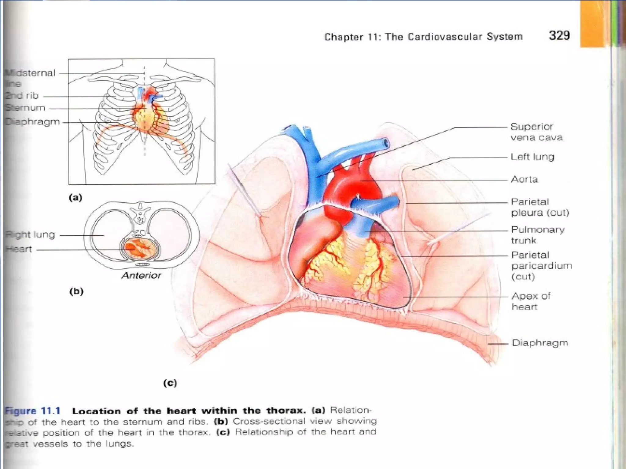
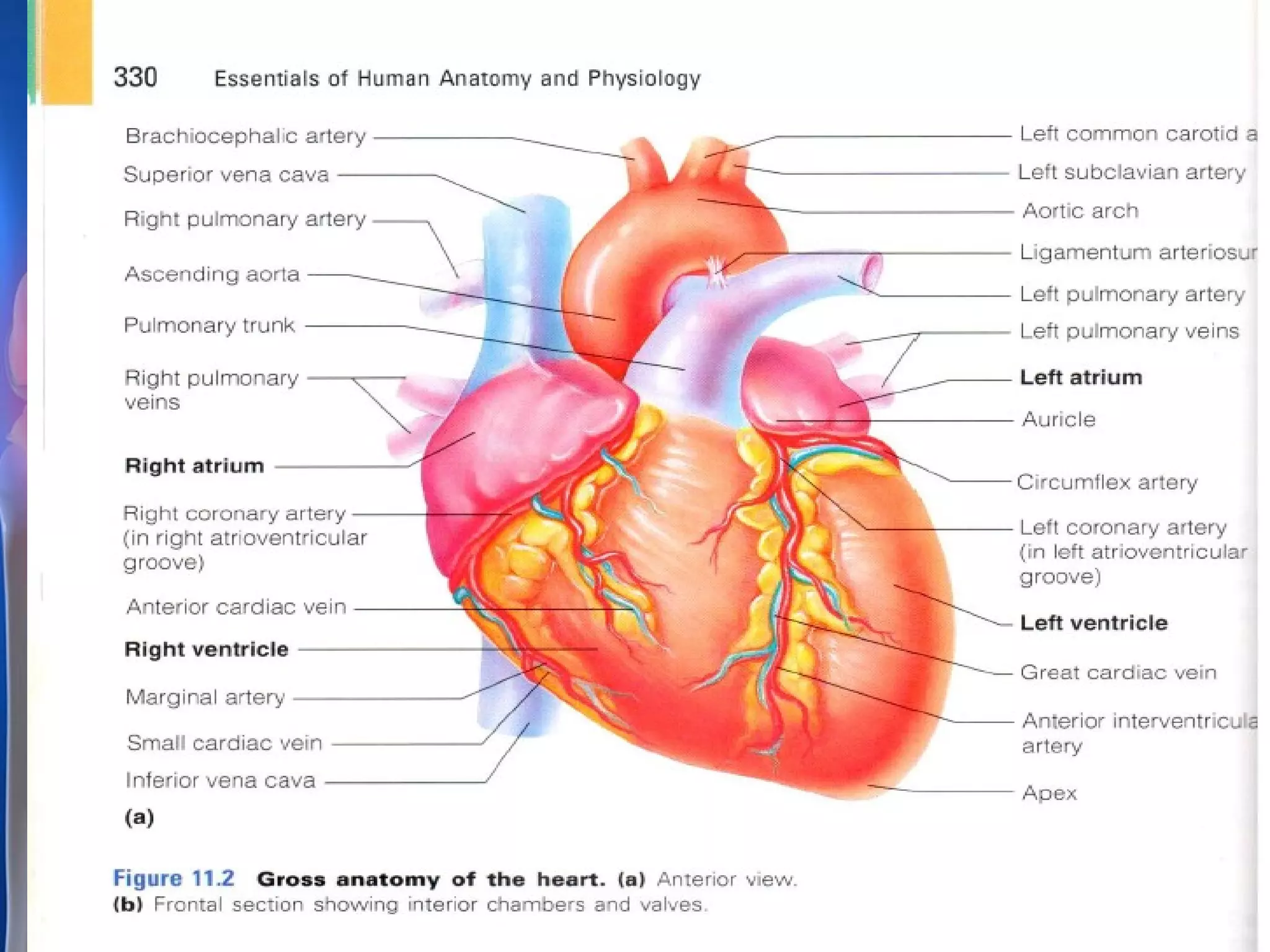
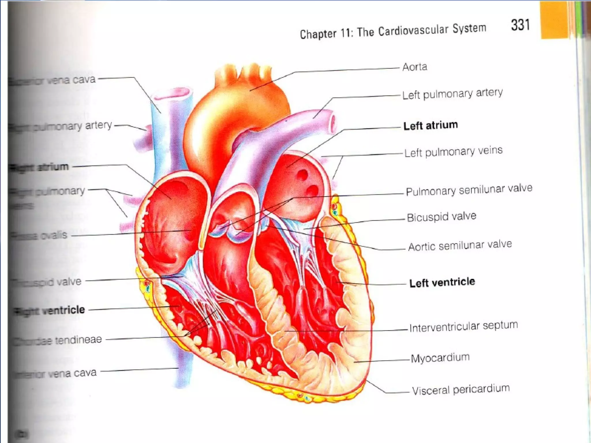
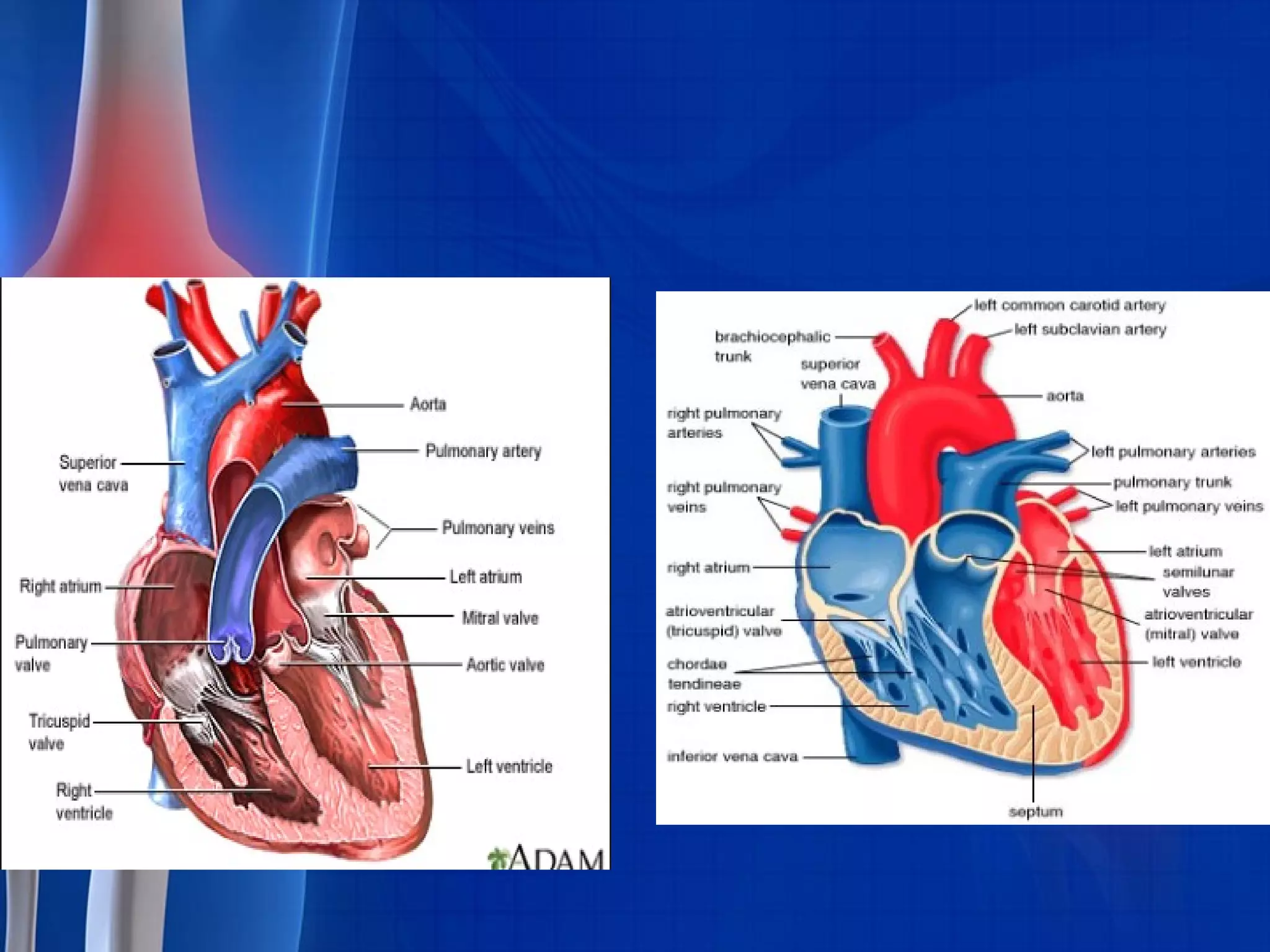
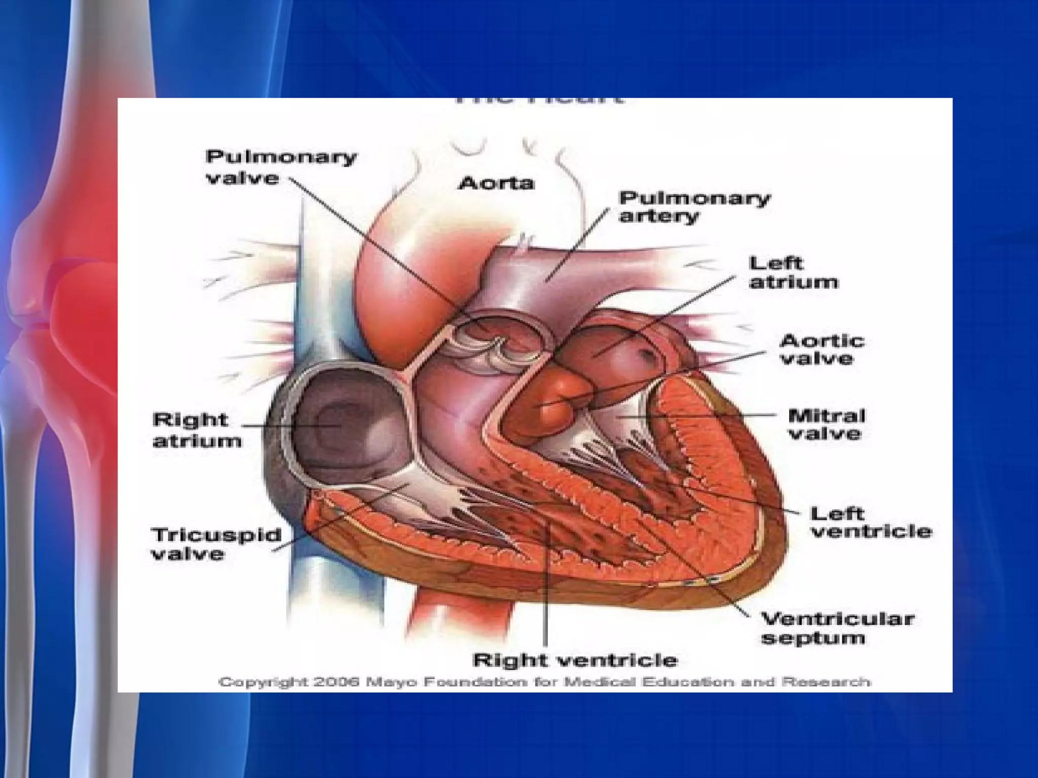
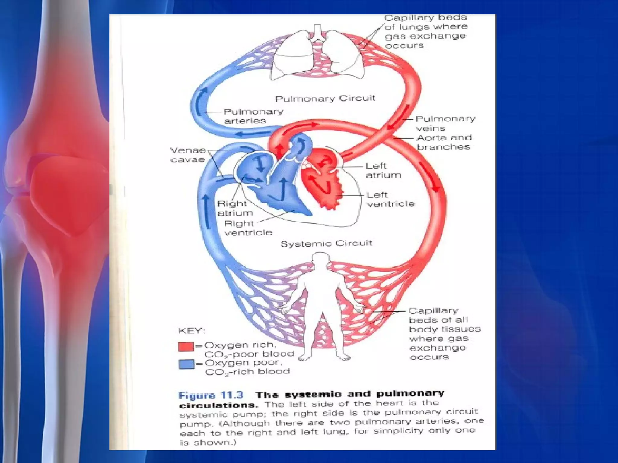
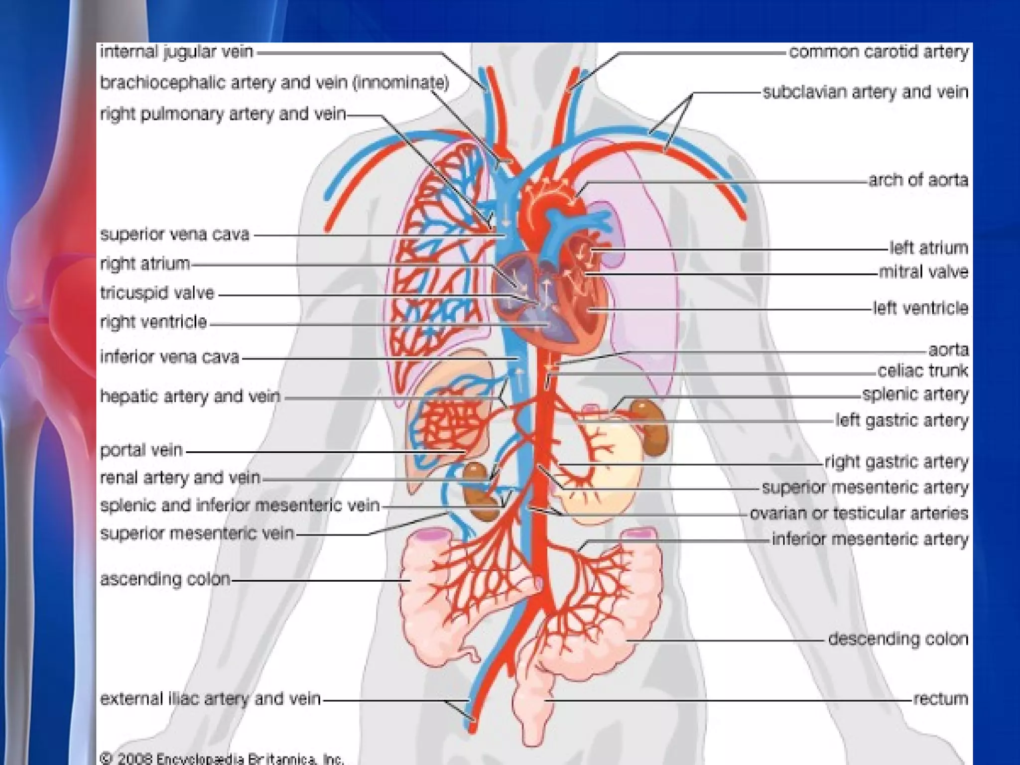


The cardiovascular system summary is as follows: 1. The heart is located in the chest behind the sternum and is about the size of a fist. It has four chambers - two atria for receiving blood and two ventricles for pumping blood out. 2. The pericardium is a sac that surrounds and protects the heart. It has two layers - an outer fibrous layer and an inner serous layer that secretes fluid. 3. Blood enters the right atrium from the body and is pumped into the pulmonary artery to the lungs. Oxygenated blood returns to the left atrium and is pumped by the left ventricle through the aorta to the body.


