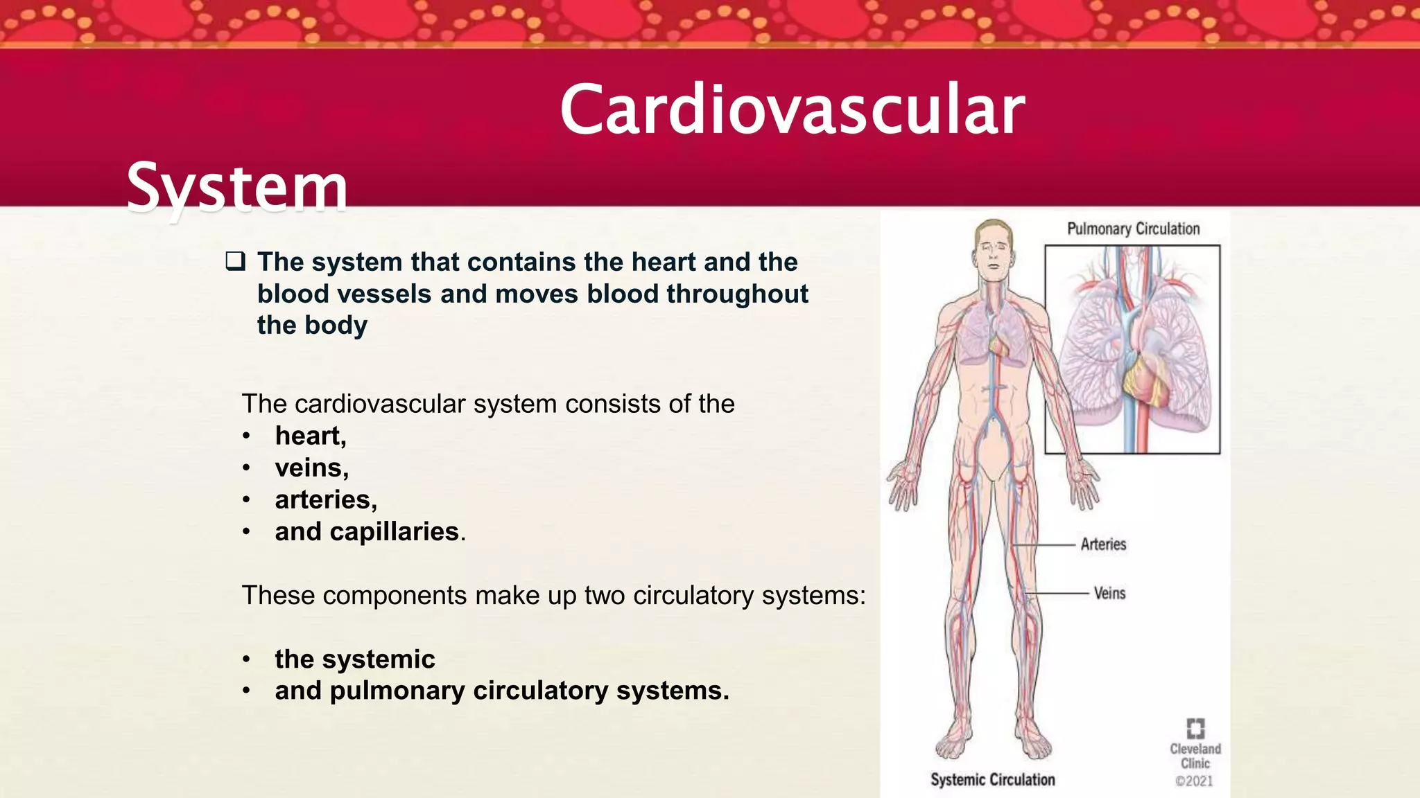
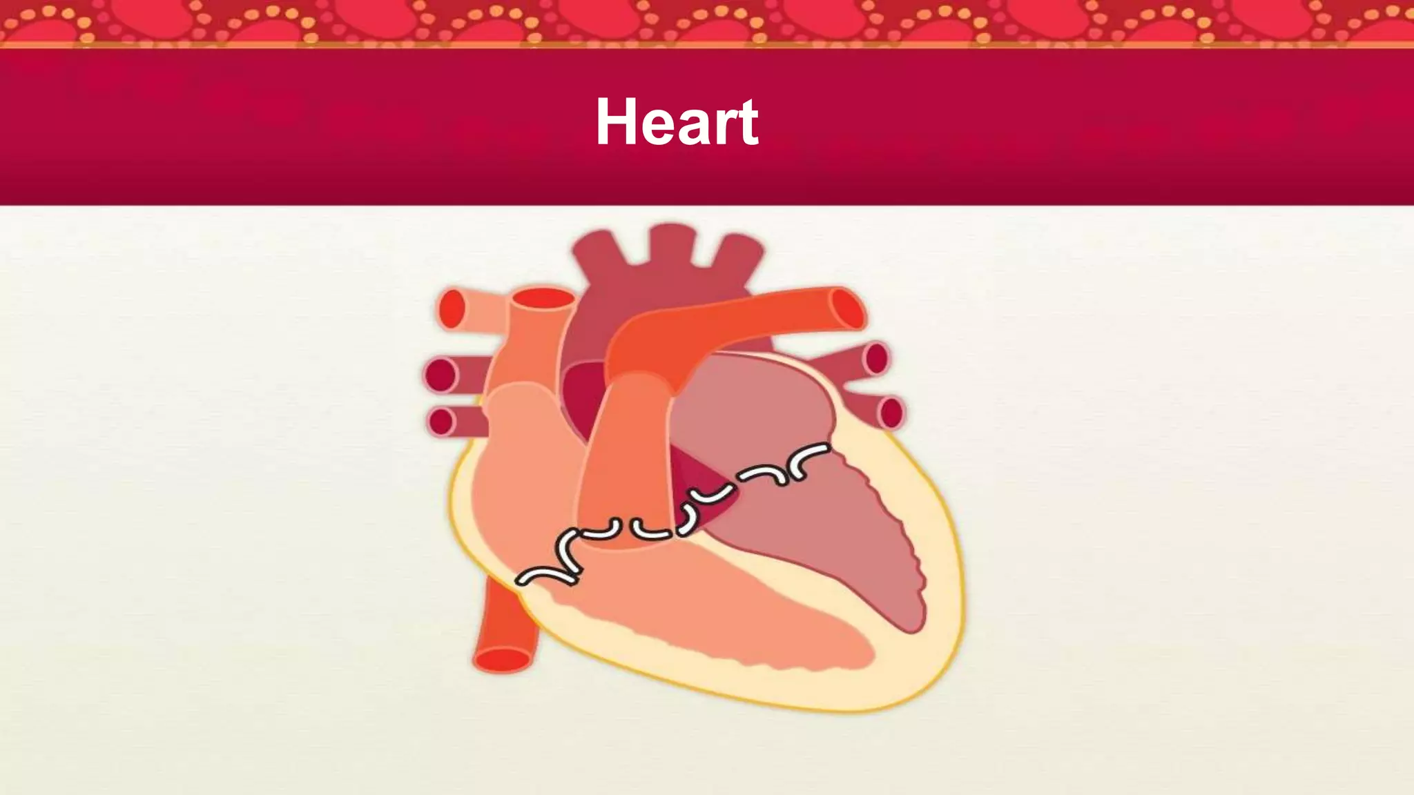
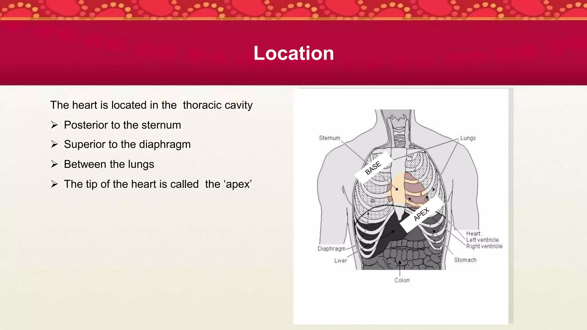
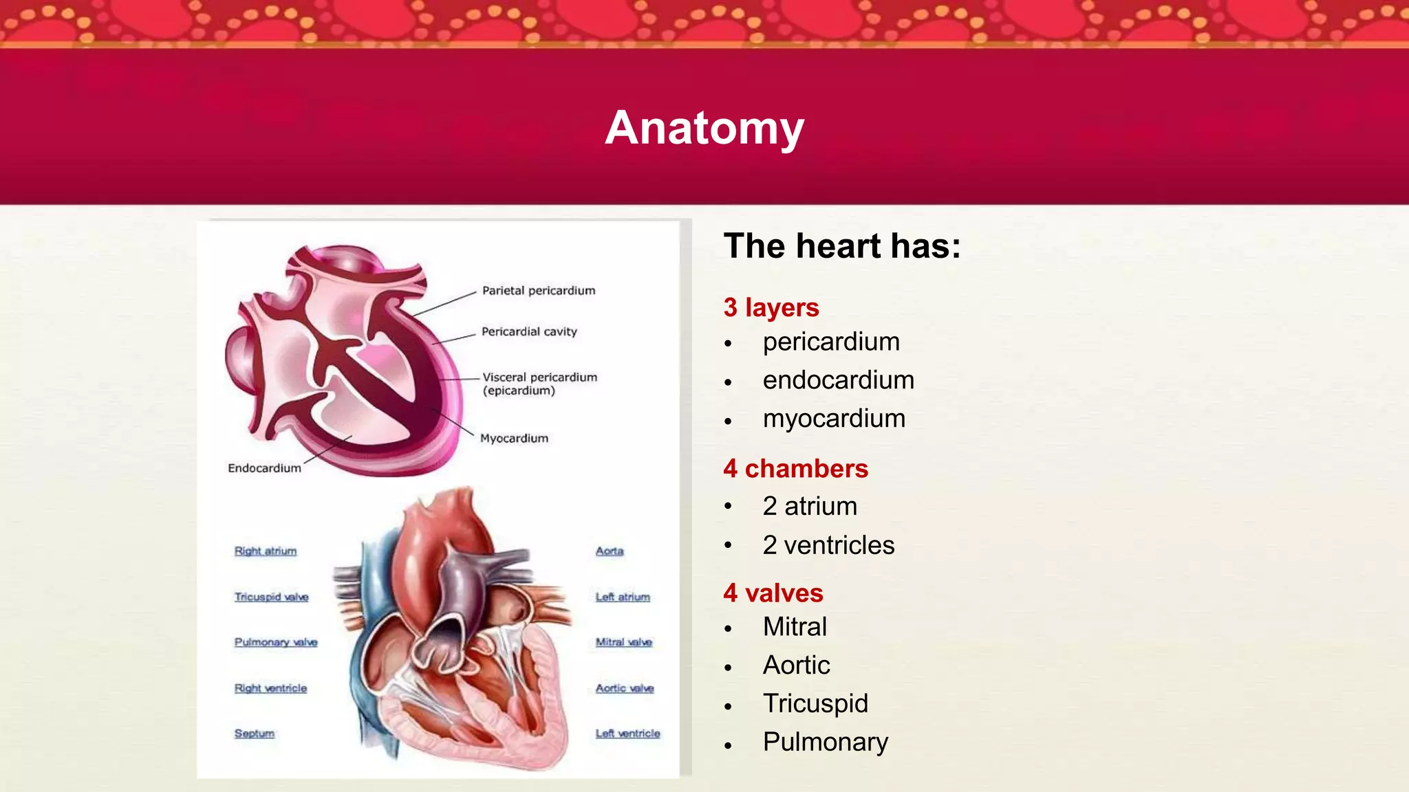
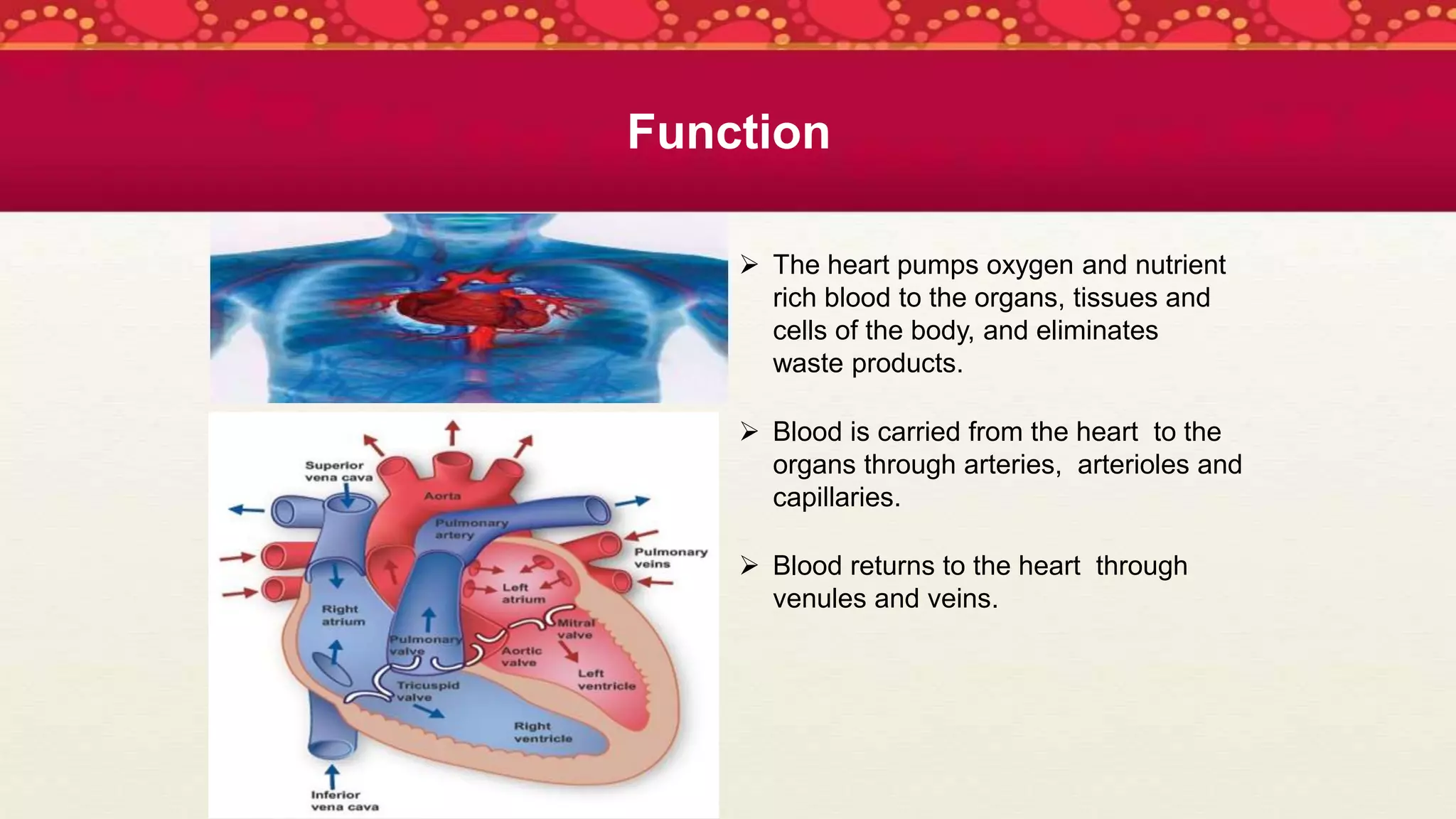
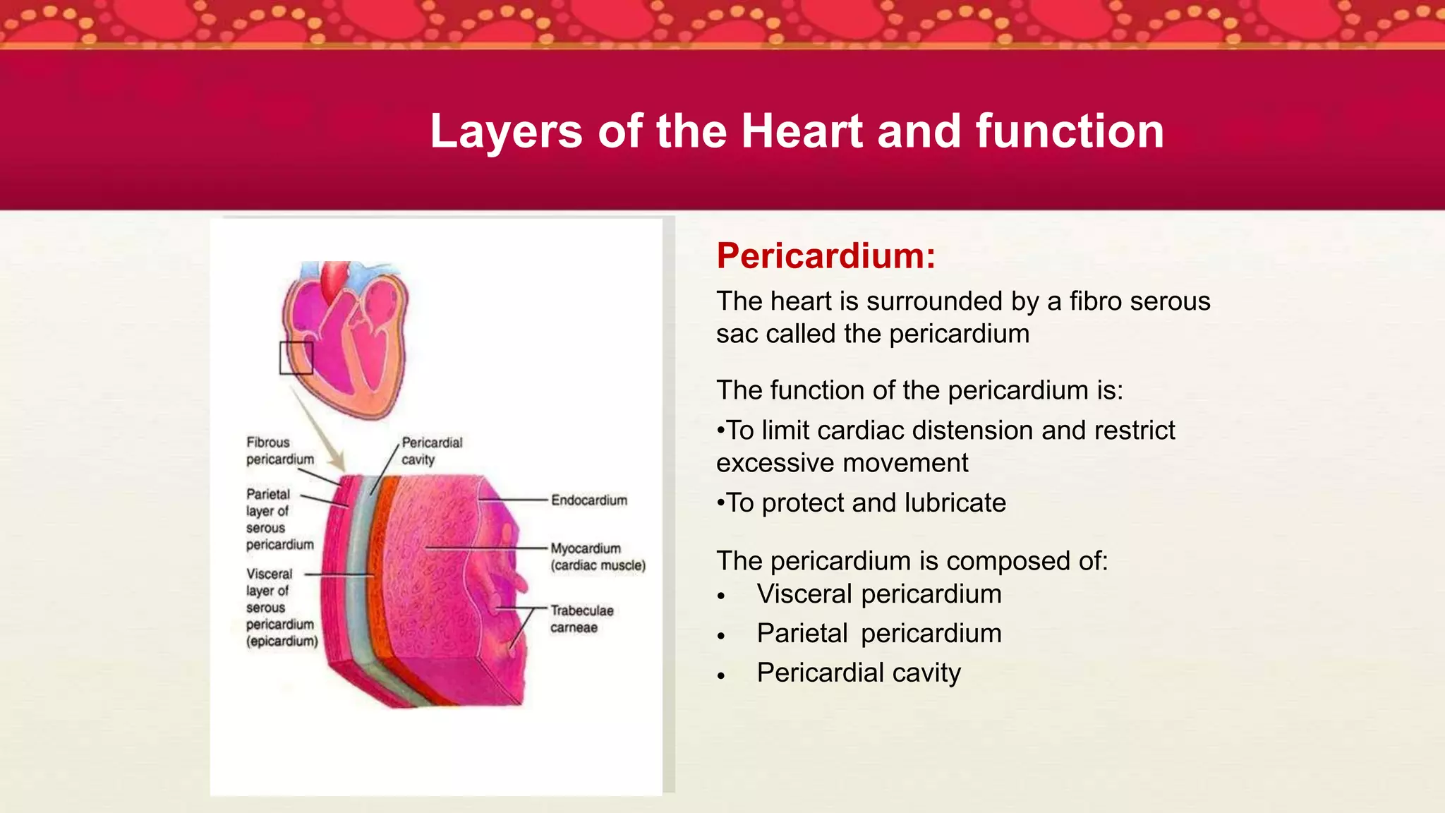
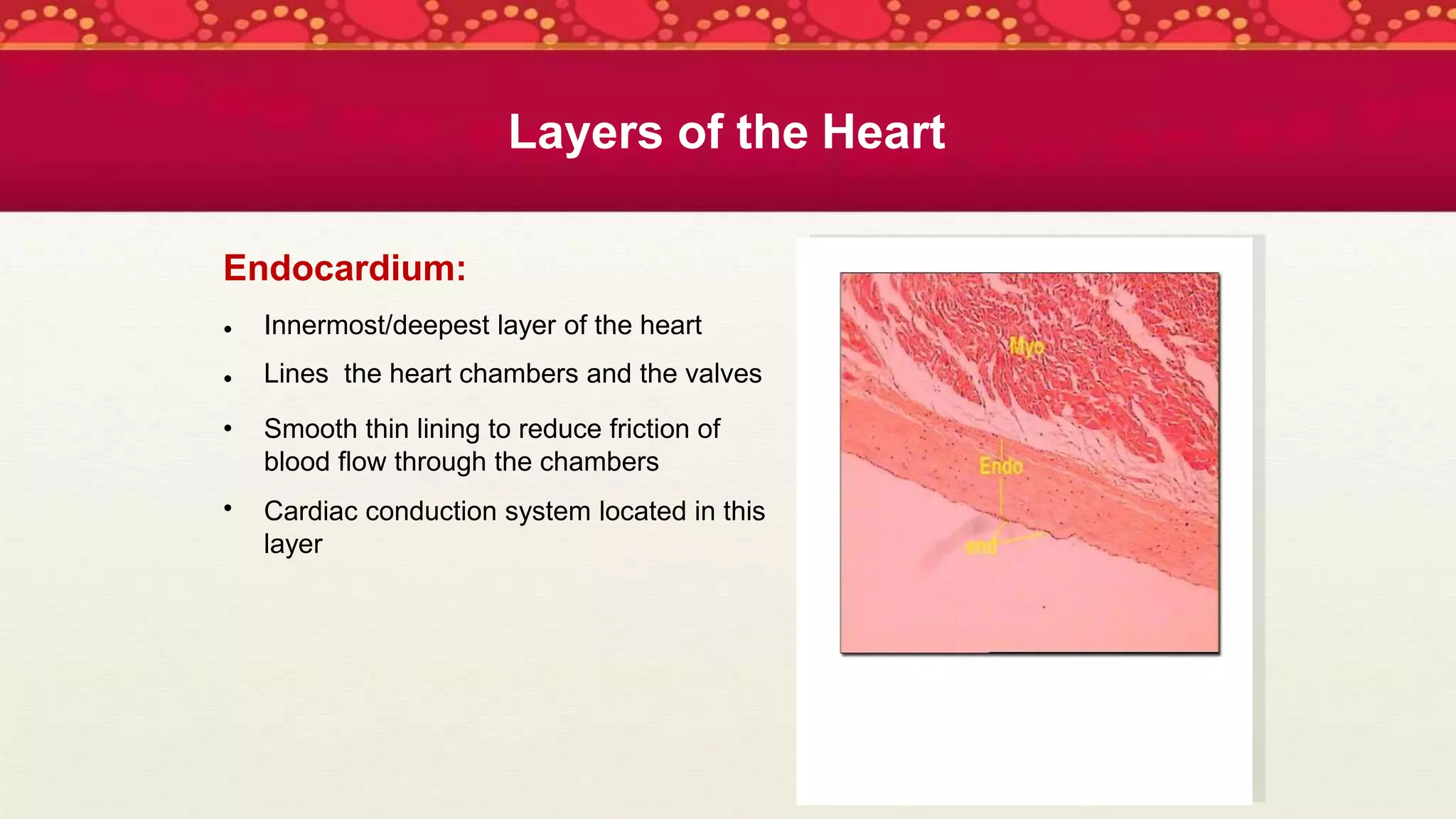
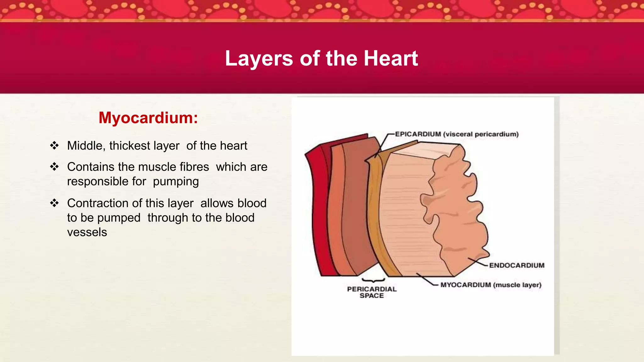
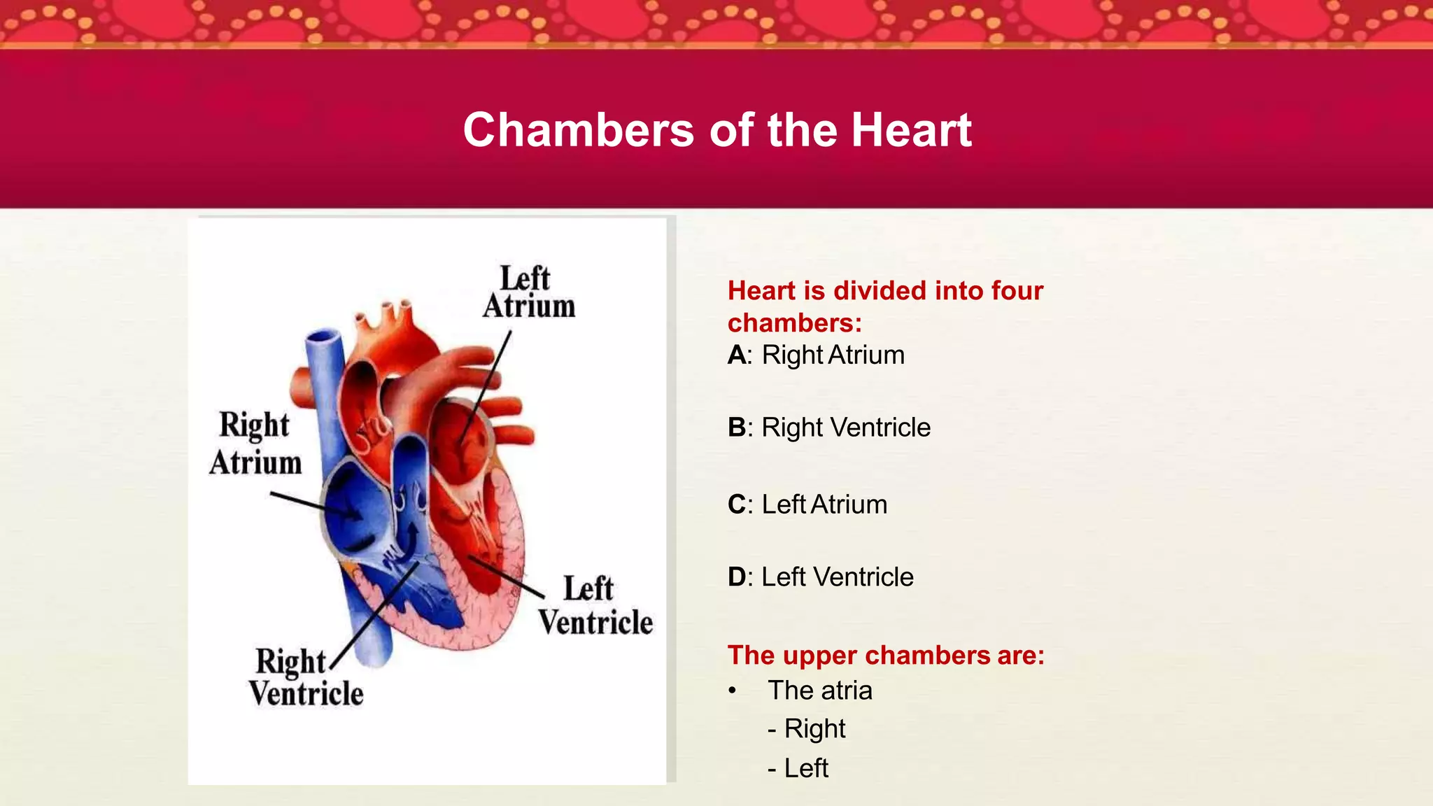
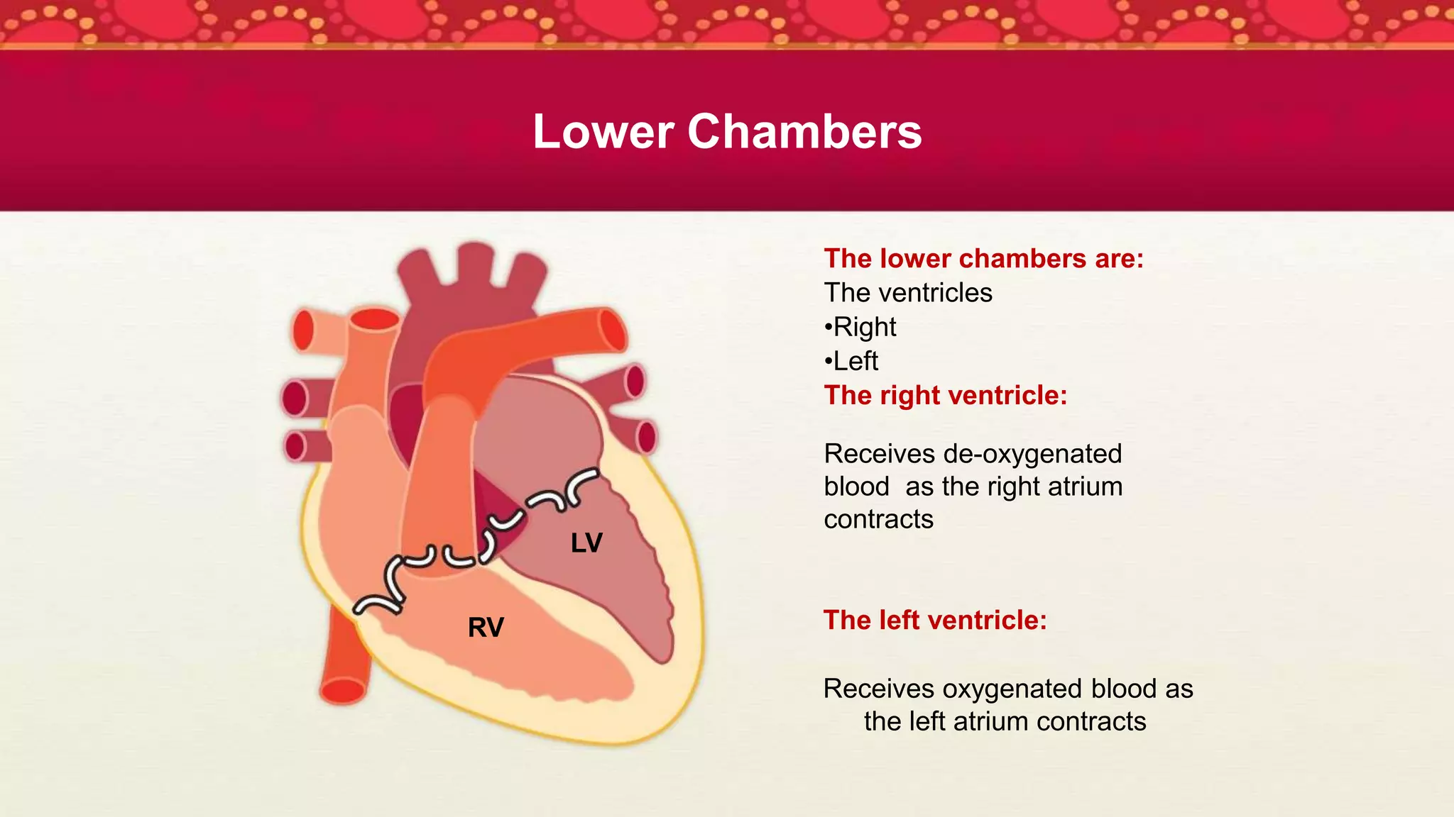
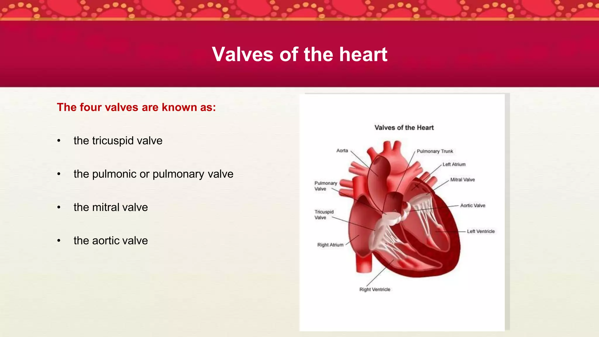
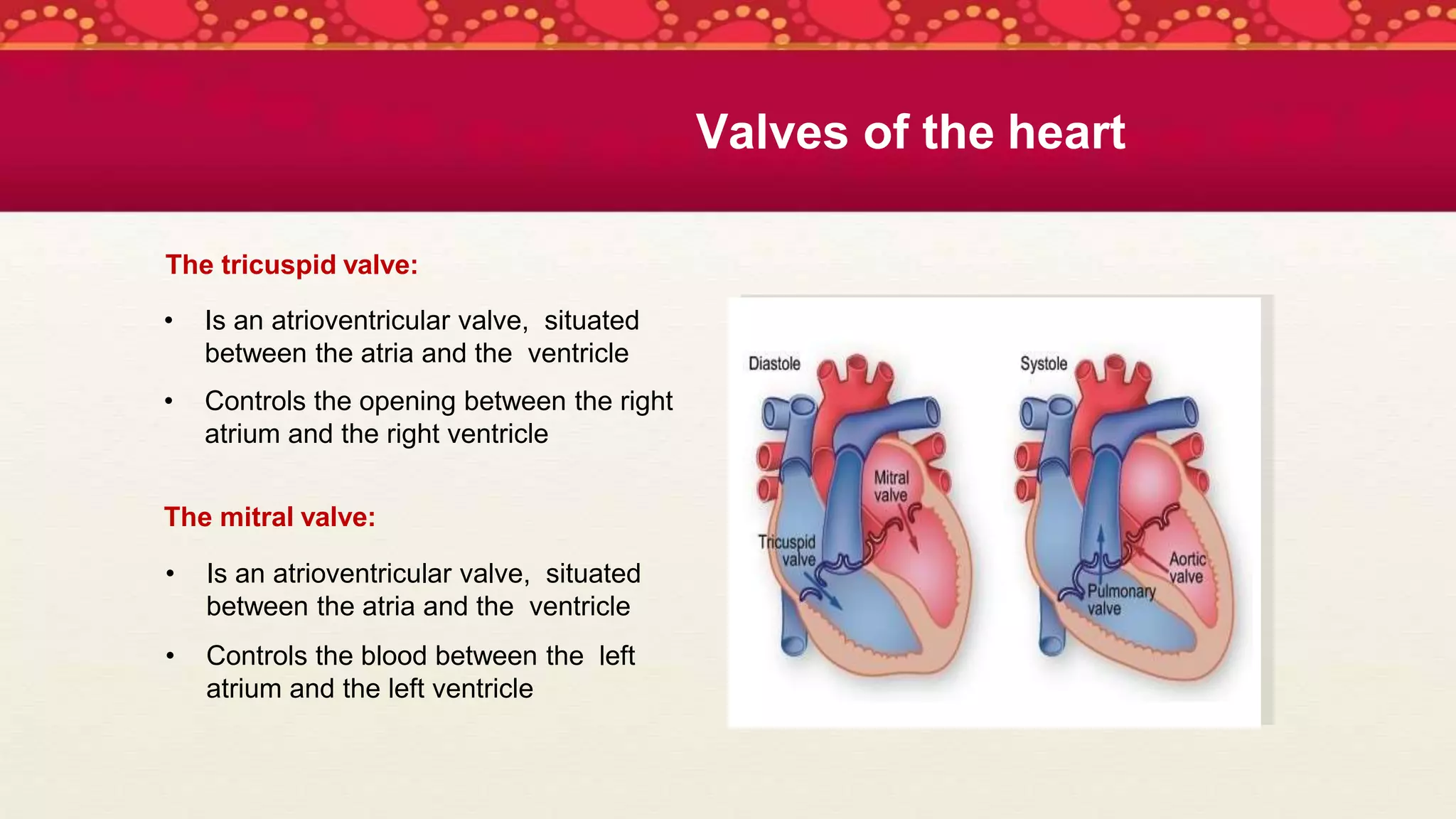
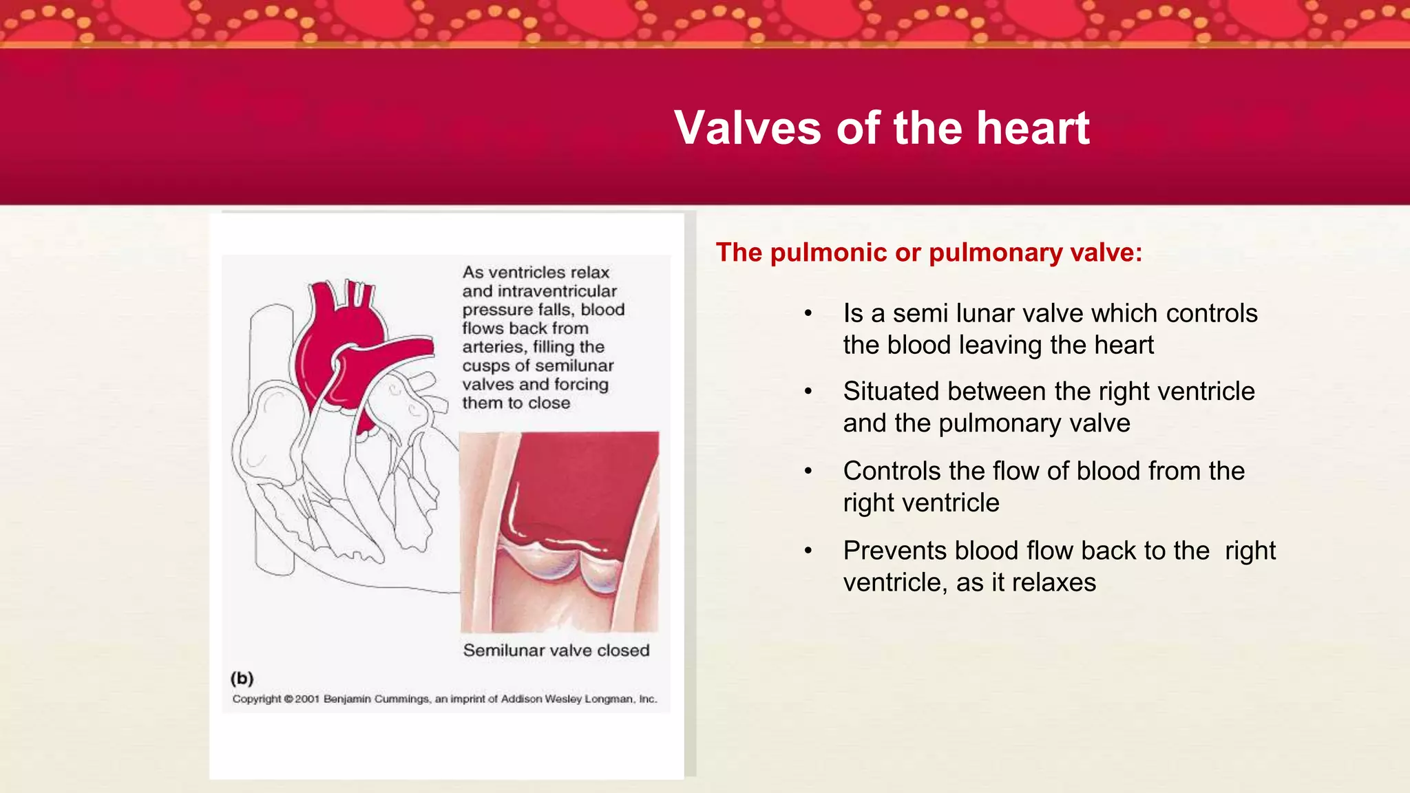
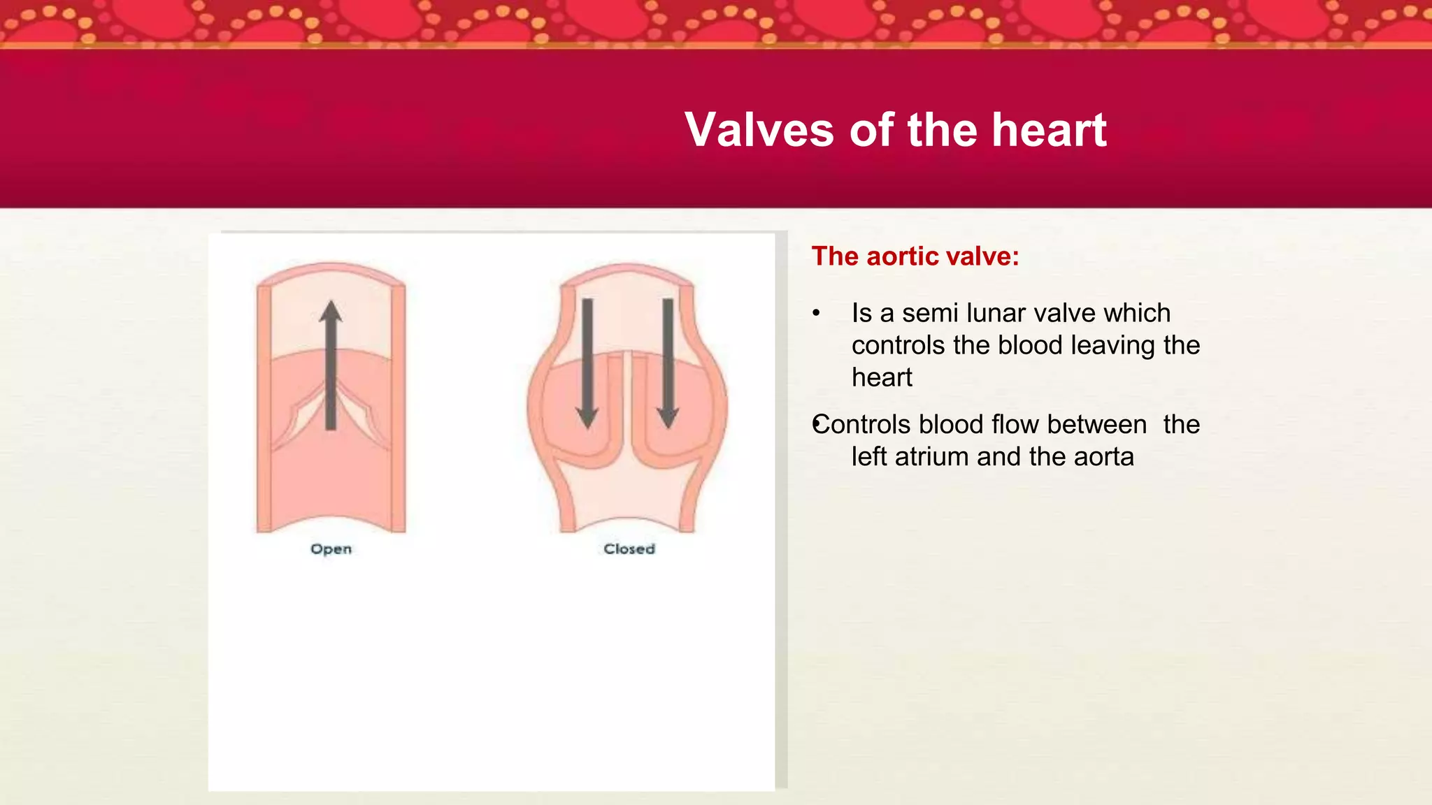
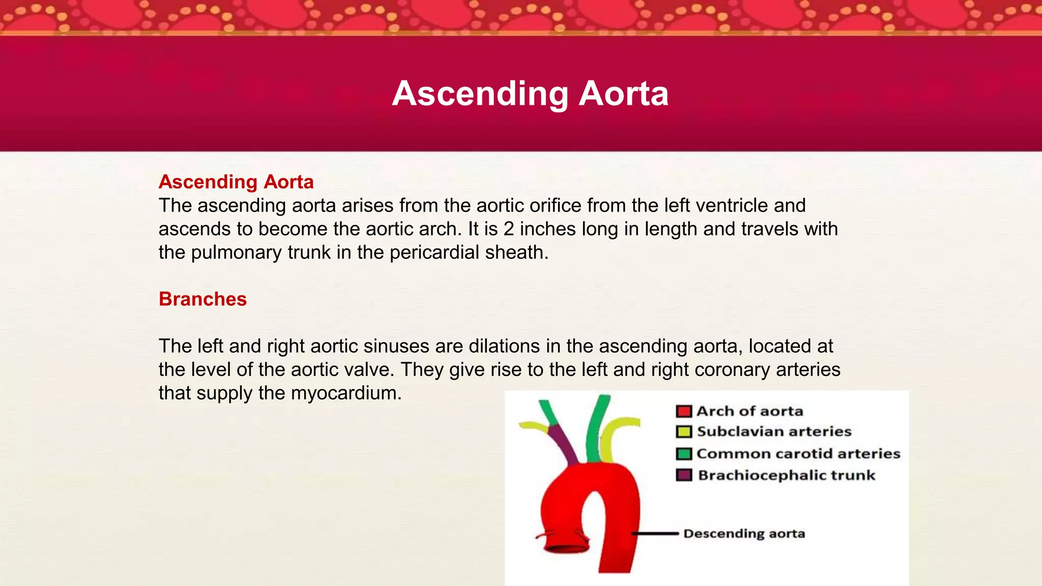
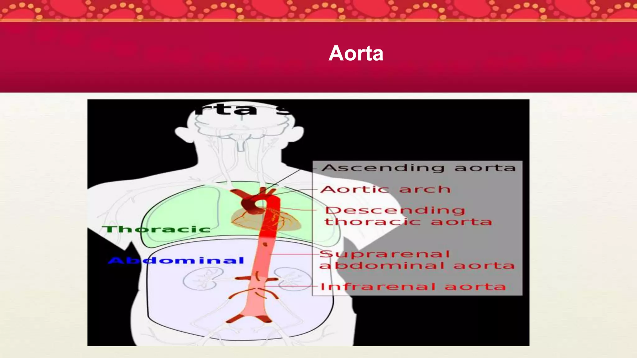
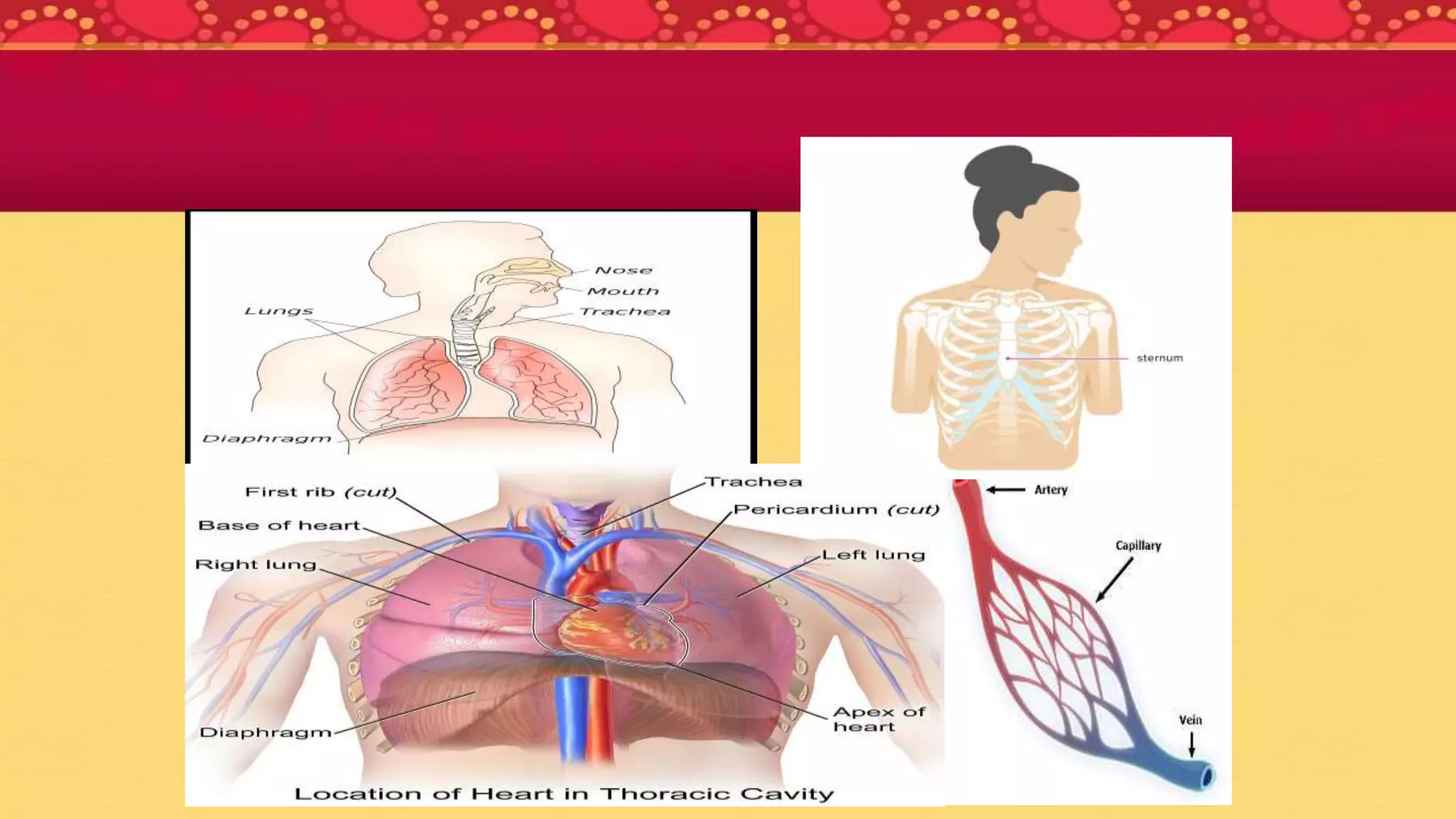
Submitted to,
As-Sazzad Mahmud
Lecturer
Department of Pharmacy
Dhaka International University
Submitted by,
Name: Anika Tabassum Shova
Roll: 04
Reg: PH-28-21-115828
Batch: 28th
Department of Pharmacy
Dhaka International University
Submission date: 22/01/2023
Course Title : Basic Anatomy
Course No : Pharm - 205
Cardiovascular
System
 The system that contains the heart and the
blood vessels and moves blood throughout
the body
The cardiovascular system consists of the
• heart,
• veins,
• arteries,
• and capillaries.
These components make up two circulatory systems:
• the systemic
• and pulmonary circulatory systems.
Two important circulation processes
Pulmonary circulation
Blood without oxygen comes into the right side of your heart and is sent to the
lungs to get oxygen and get rid of carbon dioxide. Then the oxygenated blood
comes back through the left side of your heart.
Systemic circulation
Blood that has just gotten oxygen from the lungs and returned through your heart’s
left side is pushed out to the rest of your body’s cells so they can receive oxygen
and nutrients. The cycle starts again when blood without oxygen goes to the right
side of our heart.
Heart
Location
The heart is located in the thoracic cavity
 Posterior to the sternum
 Superior to the diaphragm
 Between the lungs
 The tip of the heart is called the ‘apex’
Anatomy
The heart has:
3 layers
•
•
•
pericardium
endocardium
myocardium
4 chambers
• 2 atrium
• 2 ventricles
4 valves
•
•
•
•
Mitral
Aortic
Tricuspid
Pulmonary
Function
 The heart pumps oxygen and nutrient
rich blood to the organs, tissues and
cells of the body, and eliminates
waste products.
 Blood is carried from the heart to the
organs through arteries, arterioles and
capillaries.
 Blood returns to the heart through
venules and veins.
Layers of the Heart and function
Pericardium:
The heart is surrounded by a fibro serous
sac called the pericardium
The function of the pericardium is:
•To limit cardiac distension and restrict
excessive movement
•To protect and lubricate
The pericardium is composed of:
•
•
•
Visceral pericardium
Parietal pericardium
Pericardial cavity
Endocardium:
•
•
•
•
Innermost/deepest layer of the heart
Lines the heart chambers and the valves
Smooth thin lining to reduce friction of
blood flow through the chambers
Cardiac conduction system located in this
layer
Layers of the Heart
Myocardium:
 Middle, thickest layer of the heart
 Contains the muscle fibres which are
responsible for pumping
 Contraction of this layer allows blood
to be pumped through to the blood
vessels
Layers of the Heart
Chambers of the Heart
Heart is divided into four
chambers:
A: Right Atrium
B: Right Ventricle
C: Left Atrium
D: Left Ventricle
The upper chambers are:
• The atria
- Right
- Left
The right atrium:
Receives deoxygenated blood from the body
through the:
• superior vena cava (head and upper body)
• inferior vena cava (legs and lower torso)
The left atrium:
Receives oxygenated blood from the lungs
through the:
 pulmonary vein
Upper Chambers
The lower chambers are:
The ventricles
•Right
•Left
The right ventricle:
Receives de-oxygenated
blood as the right atrium
contracts
The left ventricle:
Receives oxygenated blood as
the left atrium contracts
Lower Chambers
LV
RV
Valves of the heart
The valves are located within the
chambers of the heart.
The function of the valves:
controls the direction of blood flow
allows one way flow of blood
-through chambers
-from the heart to the body
Valves of the heart
The four valves are known as:
• the tricuspid valve
• the pulmonic or pulmonary valve
• the mitral valve
• the aortic valve
The tricuspid valve:
• Is an atrioventricular valve, situated
between the atria and the ventricle
• Controls the opening between the right
atrium and the right ventricle
The mitral valve:
• Is an atrioventricular valve, situated
between the atria and the ventricle
• Controls the blood between the left
atrium and the left ventricle
Valves of the heart
The pulmonic or pulmonary valve:
• Is a semi lunar valve which controls
the blood leaving the heart
• Situated between the right ventricle
and the pulmonary valve
• Controls the flow of blood from the
right ventricle
• Prevents blood flow back to the right
ventricle, as it relaxes
Valves of the heart
The aortic valve:
•
• Is a semi lunar valve which
controls the blood leaving the
heart
Controls blood flow between the
left atrium and the aorta
Valves of the heart
The Aorta
 The Aorta is the largest artery of the
body which carries the oxygenated
blood from the left ventricle and
supplies it to all the parts of the
body.
 The aorta initially is one inch wide in
diameter.
 The aorta is classified as a large
elastic artery.
The Aorta
The aorta can be divided into four sections:
1. The ascending aorta,
2. The aortic arch,
3. The thoracic (descending) aorta,
4. The abdominal aorta.
The first 3 parts are confined to the thoracic cavity and together create the
thoracic aorta.
Thoracic aorta
Ascending Aorta
Ascending Aorta
The ascending aorta arises from the aortic orifice from the left ventricle and
ascends to become the aortic arch. It is 2 inches long in length and travels with
the pulmonary trunk in the pericardial sheath.
Branches
The left and right aortic sinuses are dilations in the ascending aorta, located at
the level of the aortic valve. They give rise to the left and right coronary arteries
that supply the myocardium.
Aortic Arch
Aortic Arch
The aortic arch is a continuation of the ascending aorta and begins at the level of the second
sternocostal joint. It arches superiorly, posteriorly and to the left before moving inferiorly.
The aortic arch ends at the level of the T4 vertebra. The arch is still connected to the pulmonary
trunk by the ligamentum arteriosum (remnant of the foetal ductus arteriosus).
Branches
There are three major branches arising from the aortic arch. Proximal to distal:
Brachiocephalic trunk: The first and largest branch that ascends laterally to split into the right
common carotid and right subclavian arteries. These arteries supply the right side of the head and
neck, and the right upper limb.
Left common carotid artery: Supplies the left side of the head and neck.
Left subclavian artery: Supplies the left upper limb.
Descending Thoracic Aorta
.
Descending thoracic aorta:
The thoracic descending aorta gives
rise to the intercostal and subcostal
arteries, as well as to the superior and
inferior left bronchial arteries and
variable branches to the esophagus,
mediastinum, and pericardium. Its
lowest pair of branches are the
superior phrenic arteries, which supply
the diaphragm, and the subcostal
arteries for the twelfth rib.
Abdominal aorta
Abdominal aorta
The abdominal aorta begins at the aortic
hiatus of the diaphragm at the level of the
twelfth thoracic vertebra. It gives rise to
lumbar and musculophrenic arteries, renal
and middle suprarenal arteries, and visceral
arteries (the celiac trunk, the superior
mesenteric artery and the inferior
mesenteric artery). It ends in a bifurcation
into the left and right common iliac arteries.
At the point of the bifurcation, there also
springs a smaller branch, the median sacral
artery.
Aorta
Most. Sumaiya Khatun Kali

Cardiovascular System

  • 1.
    Submitted to, As-Sazzad Mahmud Lecturer Departmentof Pharmacy Dhaka International University Submitted by, Name: Anika Tabassum Shova Roll: 04 Reg: PH-28-21-115828 Batch: 28th Department of Pharmacy Dhaka International University Submission date: 22/01/2023 Course Title : Basic Anatomy Course No : Pharm - 205
  • 2.
    Cardiovascular System  The systemthat contains the heart and the blood vessels and moves blood throughout the body The cardiovascular system consists of the • heart, • veins, • arteries, • and capillaries. These components make up two circulatory systems: • the systemic • and pulmonary circulatory systems.
  • 3.
    Two important circulationprocesses Pulmonary circulation Blood without oxygen comes into the right side of your heart and is sent to the lungs to get oxygen and get rid of carbon dioxide. Then the oxygenated blood comes back through the left side of your heart. Systemic circulation Blood that has just gotten oxygen from the lungs and returned through your heart’s left side is pushed out to the rest of your body’s cells so they can receive oxygen and nutrients. The cycle starts again when blood without oxygen goes to the right side of our heart.
  • 4.
  • 5.
    Location The heart islocated in the thoracic cavity  Posterior to the sternum  Superior to the diaphragm  Between the lungs  The tip of the heart is called the ‘apex’
  • 6.
    Anatomy The heart has: 3layers • • • pericardium endocardium myocardium 4 chambers • 2 atrium • 2 ventricles 4 valves • • • • Mitral Aortic Tricuspid Pulmonary
  • 7.
    Function  The heartpumps oxygen and nutrient rich blood to the organs, tissues and cells of the body, and eliminates waste products.  Blood is carried from the heart to the organs through arteries, arterioles and capillaries.  Blood returns to the heart through venules and veins.
  • 8.
    Layers of theHeart and function Pericardium: The heart is surrounded by a fibro serous sac called the pericardium The function of the pericardium is: •To limit cardiac distension and restrict excessive movement •To protect and lubricate The pericardium is composed of: • • • Visceral pericardium Parietal pericardium Pericardial cavity
  • 9.
    Endocardium: • • • • Innermost/deepest layer ofthe heart Lines the heart chambers and the valves Smooth thin lining to reduce friction of blood flow through the chambers Cardiac conduction system located in this layer Layers of the Heart
  • 10.
    Myocardium:  Middle, thickestlayer of the heart  Contains the muscle fibres which are responsible for pumping  Contraction of this layer allows blood to be pumped through to the blood vessels Layers of the Heart
  • 11.
    Chambers of theHeart Heart is divided into four chambers: A: Right Atrium B: Right Ventricle C: Left Atrium D: Left Ventricle The upper chambers are: • The atria - Right - Left
  • 12.
    The right atrium: Receivesdeoxygenated blood from the body through the: • superior vena cava (head and upper body) • inferior vena cava (legs and lower torso) The left atrium: Receives oxygenated blood from the lungs through the:  pulmonary vein Upper Chambers
  • 13.
    The lower chambersare: The ventricles •Right •Left The right ventricle: Receives de-oxygenated blood as the right atrium contracts The left ventricle: Receives oxygenated blood as the left atrium contracts Lower Chambers LV RV
  • 14.
    Valves of theheart The valves are located within the chambers of the heart. The function of the valves: controls the direction of blood flow allows one way flow of blood -through chambers -from the heart to the body
  • 15.
    Valves of theheart The four valves are known as: • the tricuspid valve • the pulmonic or pulmonary valve • the mitral valve • the aortic valve
  • 16.
    The tricuspid valve: •Is an atrioventricular valve, situated between the atria and the ventricle • Controls the opening between the right atrium and the right ventricle The mitral valve: • Is an atrioventricular valve, situated between the atria and the ventricle • Controls the blood between the left atrium and the left ventricle Valves of the heart
  • 17.
    The pulmonic orpulmonary valve: • Is a semi lunar valve which controls the blood leaving the heart • Situated between the right ventricle and the pulmonary valve • Controls the flow of blood from the right ventricle • Prevents blood flow back to the right ventricle, as it relaxes Valves of the heart
  • 18.
    The aortic valve: • •Is a semi lunar valve which controls the blood leaving the heart Controls blood flow between the left atrium and the aorta Valves of the heart
  • 19.
    The Aorta  TheAorta is the largest artery of the body which carries the oxygenated blood from the left ventricle and supplies it to all the parts of the body.  The aorta initially is one inch wide in diameter.  The aorta is classified as a large elastic artery.
  • 20.
    The Aorta The aortacan be divided into four sections: 1. The ascending aorta, 2. The aortic arch, 3. The thoracic (descending) aorta, 4. The abdominal aorta. The first 3 parts are confined to the thoracic cavity and together create the thoracic aorta. Thoracic aorta
  • 21.
    Ascending Aorta Ascending Aorta Theascending aorta arises from the aortic orifice from the left ventricle and ascends to become the aortic arch. It is 2 inches long in length and travels with the pulmonary trunk in the pericardial sheath. Branches The left and right aortic sinuses are dilations in the ascending aorta, located at the level of the aortic valve. They give rise to the left and right coronary arteries that supply the myocardium.
  • 22.
    Aortic Arch Aortic Arch Theaortic arch is a continuation of the ascending aorta and begins at the level of the second sternocostal joint. It arches superiorly, posteriorly and to the left before moving inferiorly. The aortic arch ends at the level of the T4 vertebra. The arch is still connected to the pulmonary trunk by the ligamentum arteriosum (remnant of the foetal ductus arteriosus). Branches There are three major branches arising from the aortic arch. Proximal to distal: Brachiocephalic trunk: The first and largest branch that ascends laterally to split into the right common carotid and right subclavian arteries. These arteries supply the right side of the head and neck, and the right upper limb. Left common carotid artery: Supplies the left side of the head and neck. Left subclavian artery: Supplies the left upper limb.
  • 23.
    Descending Thoracic Aorta . Descendingthoracic aorta: The thoracic descending aorta gives rise to the intercostal and subcostal arteries, as well as to the superior and inferior left bronchial arteries and variable branches to the esophagus, mediastinum, and pericardium. Its lowest pair of branches are the superior phrenic arteries, which supply the diaphragm, and the subcostal arteries for the twelfth rib.
  • 24.
    Abdominal aorta Abdominal aorta Theabdominal aorta begins at the aortic hiatus of the diaphragm at the level of the twelfth thoracic vertebra. It gives rise to lumbar and musculophrenic arteries, renal and middle suprarenal arteries, and visceral arteries (the celiac trunk, the superior mesenteric artery and the inferior mesenteric artery). It ends in a bifurcation into the left and right common iliac arteries. At the point of the bifurcation, there also springs a smaller branch, the median sacral artery.
  • 25.
  • 26.