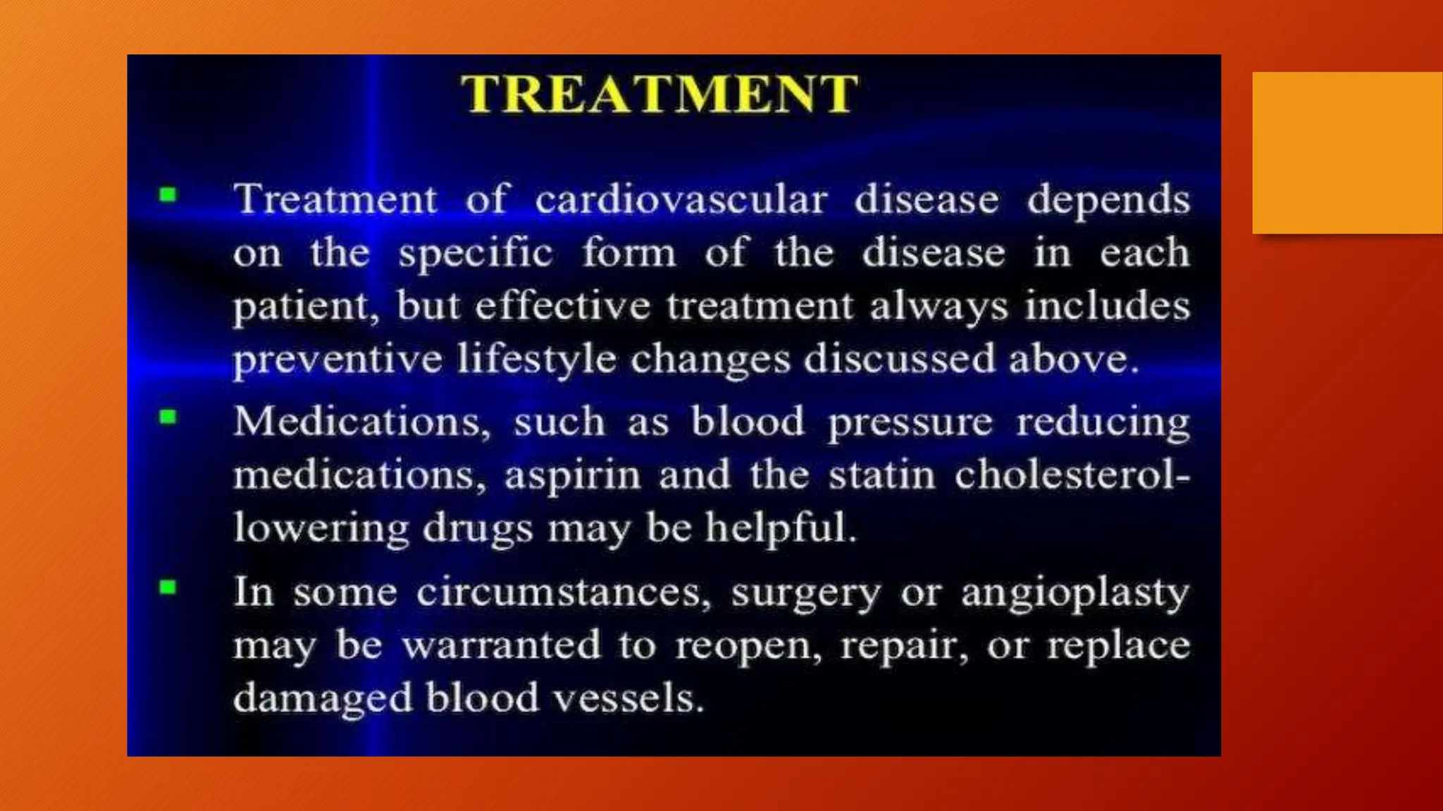
CARDIOVASCULAR
DISEASES
-By:Nisha D.
Barole
Post Graduate
Student
 The heart is divided by a septum into two
halves.
* Then halves divided into chambers.
* Upper two chambers of heart are called
atria and two lower chambers are called
ventricles .
* Valves allow blood flow in one direction
between the chambers of the heart.
What is CVD?
• Cardiovascular diseases (CVD) is a class of
diseases that involve the heart, the blood vessels
or the both.
• Cardiovascular disease refer to any disease
that effects the cardiovascular system,
principally cardiac disease, vascular disease of
the brain and kidney and peripheral disease.
▪︎ The diseases and the conditions affecting the heart
are collectively known as heart disease.
▪︎ Problems can be in any of the areas.
▪︎ Mainly according to WHO (World Health
Organization) this problem is common in women.
▪︎ 90% of women have one or more risk factors for
developing heart disease.
Mostcommon heartdiseasesare:
• Coronary artery disease • Hypertensive
heart disease
• Coronary heart disease • Inflammatory
heart disease
• Ischaemic heart disease • Valvular heart
disease
• Cardiovascular disease • Pericardial disease
Heart disease is an umbrella term for a
number of different diseases which
affect the heart.
◇ Coronary artery disease is most common forms of heart
attack (myocardial infarction) it develops when the major
blood vessels that supply your heart become damaged or
diseased. Cholesterol-containing deposits (plaques) or
condition called atherosclerosis in your coronary arteries.
◇ The coronary arteries supply blood, oxygen and nutrients
to your heart. A buildup of plaque can narrow these arteries,
decreasing blood flow to your heart. Eventually, the reduced
blood flow may cause chest pain (angina), shortness of
breath, or other coronary artery disease signs and symptoms.
Coronaryarterydisease
A disease of the heart itself caused by the accumulation of
atheromatous plaques within the walls of the arteries that
supply the myocardium.
Cardiovasculardisease
Sub – umbrella term for number of diseases that affect the
heart itself and the blood vessel system (veins/arteries).
Causes – include diabetes mellitus, hypertension,
hypercholesterolemia.
Coronaryheartdisease
Ischaemicheartdisease
A disease of heart itself, characterized by
reduced blood supply to the organ.
Pericardialdisease
Pericarditis may be caused by a viral infection or heart attack.
In many cases, the cause is unknown. A swelling and irritation
of the thin, sac-like membrane surrounding the heart
(pericardium). The most common symptom is sharp, stabbing
A failure of the right side of the heart. Pulmonary heart disease, also known
as cor pulmonale, is the enlargement and failure of the right ventricle of the
heart as a response to increased vascular resistance (such as from pulmonic
stenosis) or high blood pressure in the lungs.
Hereditaryheartdisease
Heart disease caused by unavoidable genetic factor.
Hypertensiveheartdisease
Heart disease caused by the high blood pressure .
Pulmonaryheartdisease
Heart disease that involves inflammation of the
heart muscle or/and tissue surrounding it .
Valvularheartdisease
Heart disease that affect the valves of the
heart.
Inflammatoryheart
disease
A congenital heart defect (CHD), also known as a congenital
heart anomaly and congenital heart disease, is a defect in the
structure of the heart or vessels that is present at birth. Signs and
symptoms depend on the specific type of defect.
HeartFailure
- Achronicconditioninwhichtheheartdoesn'tpumpbloodaswellasitshould.
-Heartfailurecanoccuriftheheartcannotpump(systolic)orfill (diastolic)adequately.
-Symptomsincludeshortnessofbreath,fatigue,swollenlegsandrapid heartbeat.
-Treatmentscaninclude eatinglesssalt,limitingfluidintakeandtakingprescription
Congenitalheartdisease
Sometypesofbloodvessel
disorders:
•Arteriosclerosis and
Atherosclerosis
•High blood pressure (
hypertension)
•Stroke
•Aneurysm
•Peripheral arterial disease and
• Vasculitis
• Venous incompetence
• Venous thrombosis
• Varicose veins
• Lymphedema
A build up of cholesterol plaque in the walls of arteries, causing
obstruction of blood flow. Plaques may rupture, causing acute
occlusion of the artery by clot.
* High BP (Hypertension)
A condition in which the force of the blood against the artery
walls is too high. Usually hypertension is defined as blood
pressure above 140/90, and is considered severe if the pressure
is above 180/120.
rteriosclerosis and Atherosclerosis -
* Stroke
Damagetothebrainfrominterruptionofitsbloodsupply.Astrokeisa
medicalemergency.
Symptomsofstrokeincludetroublewalking,speakingandunderstanding,as
wellasparalysisornumbnessoftheface,armorleg.
A ballooning and weakened area in an artery. A ruptured aneurysm
can result in internal bleeding and stroke. It can sometimes be fatal.
* Pericardial arterial disease and Claudication
Pain, commonly in the legs, caused by too little blood flow, usually
during exercise. Often indicates peripheral artery disease.
* Vasculitis
Aninflammationofthebloodvesselsthatcauses changesinthebloodvesselwalls.Vasculitiscan
cause vesselwalls tothickenandnarrow,cuttingoffvitalbloodsupplytotissuesandorgans.
Symptomsinclude fever,fatigue,weightlossandmuscle andjointpain.
Aneurysm-
The veins in your legs carry blood back to your heart.
* Venous thrombosis
Venous thrombosis is thrombosis in a vein, caused by a thrombus (blood clot).
The most common form of venous thrombosis is a Deep Vein
Thrombosis (DVT), when a blood clot forms in the deep veins of the leg.
* Varicose veins
Varicose veins are enlarged, swollen, and twisting veins, often appearing blue
or dark purple. They happen when faulty valves in the veins allow blood to
flow in the wrong direction or to pool. More than 23 % of all adults are thought
to be affected by by varicose. veins.
Venous
incompetence-
Swelling in an arm or leg caused by a lymphatic
system blockage.
Lymphedema-
Failure of conduction of impulses through the A.V. node
# Damage to S.A. node causes week impulses failing to reach
the ventricles .
Cardiac pacemaker established normal rhythm , it is small
, battery operated electronic device , it is inserted under the
skin . It lead that through a large vein to the heart, where the
wires are anchored, which send the electrical impulse to the
heart.
Arrhythmia/Dysrhythmia/Heartblock/Trio
ventricularblock
Atrial flutter : rapid , regular contraction of atria or
ventricle reaching upto 250- 300 beats per minute.
Fibrillation : an irregular , random , often rapid heart rate
/contraction that commonly causes poor blood flow reaching
upto 350- 400 beats per minute.
Defibrillation : is applied to the chest wall to help in
cardioversion. Defibrillation is a technique used to counter
the onset of ventricular fibrillation , a common cause of
cardiac arrest. Defibrillation is part of an advance cardiac life
support, it applies a controlled electric shock.
Cardiacarrest:Sudden, unexpected loss of heart
function, breathing and consciousness .
Palpitation : uncomfortablesensationinthechestassociated
witharrhythmia. This causes :
1. PAC- Premature Atrial Contraction.
2. PVC- Premature Ventricular
Contraction.
Myocardialinfarction
•Commonly known as heart attack.
•A blockage of blood flow the heart muscle.
Angina
•Commonly known as Ischemic chest pain.
•A type of chest pain caused by reduced blood
flow to the heart.
DIAGNOSIS :
•Blood test : laboratory test used to detect the risk
factors for heart diseases. These include detection of
the fats, cholesterol and lipids components of the
blood including LDL, HDL, triglycerides. Blood
sugar and glycosylated haemoglobin is measured for
detection of diabetes. C-reactive protein (CRP) and
other protein makers like Apolipoprotein A and B
are used to detect inflammation that may lead to
•Electrocardiogram : this is a simple and a
painless test that records the heart’s electrical
activity. The patient is strapped to the instrument
with several patches or leads placed over his/her
chest, wrists and ankles. A small portable
machine records the activity of the heart on a
stripe of graph paper, it shows how fast heart is
beating . An ECG can help to detect a heart ,
attacks of angina, arrhythmia.
•Stress testing : in this test patient is made to work
hard e.g. run on the treadmill or exercise which the
leads of ECG are placed over their body. Those
who cannot exercise are given pills to raise their
heart rate. This test detects the effects of the
exercise on the heart.
•Echocardiography : this test uses sound waves to
create a moving picture of the heart . This is also a
painless test where a probe is rolled over the chest and
the machine creates the image of the heart on the
monitor . This provides information on the shape, size,
working , valves and chambers of the heart .it may also
be combined with doppler to show the areas of poor
blood supply to the heart. It shows the areas of the heart
muscle that are not contacting normally and previous
injury to the heart muscle.
•Coronary angiography : this test is an invasive test .
A dye is injected into the veins to reach the coronary
arteries . This is done via coronary catheterisation .
Thereafter detailed picture of the blood vessels of the heart
are taken using special imaging methods . This is called
coronary angiography.
•Chest x ray : this is a test that shows the shape and
size of the heart lungs and major blood vessels . This
is a test seldom used in diagnosis of heart diseases at it
does not provide added information over
echocardiography and other imaging studies .
• Electron-beam computed tomography (EBCT) :
helps to detect the calcium deposits or calcification in the
walls of the coronary arteries . These are early markers of
atherosclerosis and coronary heart disease . This is not a
routine test for CHD.
•Cardiac MRI : MRI (Magnetic Resonance
Imaging) : that uses radio waves, magnets, and a
computer to create pictures of the heart . This gives a 3-D
image of the moving as well as pictures of the heart.
•Holter monitoring : A Holter monitor is a
portable device you wear to record a continuous ECG,
usually for 24 to 72 hours. Holter monitoring is used to
detect heart rhythm irregularities that aren’t found
during a regular ECG exam.
Treatment
Allopathic - angiotensin II receptor blocker , angiotensin-
converting enzyme (ACE) inhibitor, Beta blockers.
Cholesterol medications - consider the options
Nitrates - oral (synthetic), sub-lingual, chewable, or buccal
(systemic), topical (systematic).
Statins - are these cholesterol –lowering drug
Cardiac rehabilitation - building a better life after
heart disease.
Surgical - coronary angioplasty and stenting – opening
clogged arteries .
coronary artery bypass surgery.
Riskfactors:
• Age
• Absence of key nutritional elements, such as polyphenol
antioxidants.
• Diabetes Mellitus.
• Hypercholesterolemia and abnormal lipoprotein particle
profile.
• Tobacco smoking.
• Higher fibrinogen.
• Men have a higher rate of cardiovascular disease than
women, it is also the number one health problem for
women in industrialized countries.
• After menopause, the risk for women approaches that
of men.
• Hormone replacement therapy alleviates a number of
post – menopausal problems, but appears to increase
the risk Cardiovascular disease.
• Physical inactivity.
• Depression.
• Exposure to high levels of environmental noise.
Thank you

Cardiovascular Disease Power point presentation

  • 1.
  • 2.
     The heartis divided by a septum into two halves. * Then halves divided into chambers. * Upper two chambers of heart are called atria and two lower chambers are called ventricles . * Valves allow blood flow in one direction between the chambers of the heart.
  • 4.
    What is CVD? •Cardiovascular diseases (CVD) is a class of diseases that involve the heart, the blood vessels or the both. • Cardiovascular disease refer to any disease that effects the cardiovascular system, principally cardiac disease, vascular disease of the brain and kidney and peripheral disease.
  • 5.
    ▪︎ The diseasesand the conditions affecting the heart are collectively known as heart disease. ▪︎ Problems can be in any of the areas. ▪︎ Mainly according to WHO (World Health Organization) this problem is common in women. ▪︎ 90% of women have one or more risk factors for developing heart disease.
  • 6.
    Mostcommon heartdiseasesare: • Coronaryartery disease • Hypertensive heart disease • Coronary heart disease • Inflammatory heart disease • Ischaemic heart disease • Valvular heart disease • Cardiovascular disease • Pericardial disease Heart disease is an umbrella term for a number of different diseases which affect the heart.
  • 7.
    ◇ Coronary arterydisease is most common forms of heart attack (myocardial infarction) it develops when the major blood vessels that supply your heart become damaged or diseased. Cholesterol-containing deposits (plaques) or condition called atherosclerosis in your coronary arteries. ◇ The coronary arteries supply blood, oxygen and nutrients to your heart. A buildup of plaque can narrow these arteries, decreasing blood flow to your heart. Eventually, the reduced blood flow may cause chest pain (angina), shortness of breath, or other coronary artery disease signs and symptoms. Coronaryarterydisease
  • 8.
    A disease ofthe heart itself caused by the accumulation of atheromatous plaques within the walls of the arteries that supply the myocardium. Cardiovasculardisease Sub – umbrella term for number of diseases that affect the heart itself and the blood vessel system (veins/arteries). Causes – include diabetes mellitus, hypertension, hypercholesterolemia. Coronaryheartdisease
  • 9.
    Ischaemicheartdisease A disease ofheart itself, characterized by reduced blood supply to the organ. Pericardialdisease Pericarditis may be caused by a viral infection or heart attack. In many cases, the cause is unknown. A swelling and irritation of the thin, sac-like membrane surrounding the heart (pericardium). The most common symptom is sharp, stabbing
  • 10.
    A failure ofthe right side of the heart. Pulmonary heart disease, also known as cor pulmonale, is the enlargement and failure of the right ventricle of the heart as a response to increased vascular resistance (such as from pulmonic stenosis) or high blood pressure in the lungs. Hereditaryheartdisease Heart disease caused by unavoidable genetic factor. Hypertensiveheartdisease Heart disease caused by the high blood pressure . Pulmonaryheartdisease
  • 11.
    Heart disease thatinvolves inflammation of the heart muscle or/and tissue surrounding it . Valvularheartdisease Heart disease that affect the valves of the heart. Inflammatoryheart disease
  • 12.
    A congenital heartdefect (CHD), also known as a congenital heart anomaly and congenital heart disease, is a defect in the structure of the heart or vessels that is present at birth. Signs and symptoms depend on the specific type of defect. HeartFailure - Achronicconditioninwhichtheheartdoesn'tpumpbloodaswellasitshould. -Heartfailurecanoccuriftheheartcannotpump(systolic)orfill (diastolic)adequately. -Symptomsincludeshortnessofbreath,fatigue,swollenlegsandrapid heartbeat. -Treatmentscaninclude eatinglesssalt,limitingfluidintakeandtakingprescription Congenitalheartdisease
  • 15.
    Sometypesofbloodvessel disorders: •Arteriosclerosis and Atherosclerosis •High bloodpressure ( hypertension) •Stroke •Aneurysm •Peripheral arterial disease and • Vasculitis • Venous incompetence • Venous thrombosis • Varicose veins • Lymphedema
  • 16.
    A build upof cholesterol plaque in the walls of arteries, causing obstruction of blood flow. Plaques may rupture, causing acute occlusion of the artery by clot. * High BP (Hypertension) A condition in which the force of the blood against the artery walls is too high. Usually hypertension is defined as blood pressure above 140/90, and is considered severe if the pressure is above 180/120. rteriosclerosis and Atherosclerosis -
  • 17.
  • 18.
    A ballooning andweakened area in an artery. A ruptured aneurysm can result in internal bleeding and stroke. It can sometimes be fatal. * Pericardial arterial disease and Claudication Pain, commonly in the legs, caused by too little blood flow, usually during exercise. Often indicates peripheral artery disease. * Vasculitis Aninflammationofthebloodvesselsthatcauses changesinthebloodvesselwalls.Vasculitiscan cause vesselwalls tothickenandnarrow,cuttingoffvitalbloodsupplytotissuesandorgans. Symptomsinclude fever,fatigue,weightlossandmuscle andjointpain. Aneurysm-
  • 19.
    The veins inyour legs carry blood back to your heart. * Venous thrombosis Venous thrombosis is thrombosis in a vein, caused by a thrombus (blood clot). The most common form of venous thrombosis is a Deep Vein Thrombosis (DVT), when a blood clot forms in the deep veins of the leg. * Varicose veins Varicose veins are enlarged, swollen, and twisting veins, often appearing blue or dark purple. They happen when faulty valves in the veins allow blood to flow in the wrong direction or to pool. More than 23 % of all adults are thought to be affected by by varicose. veins. Venous incompetence-
  • 20.
    Swelling in anarm or leg caused by a lymphatic system blockage. Lymphedema-
  • 21.
    Failure of conductionof impulses through the A.V. node # Damage to S.A. node causes week impulses failing to reach the ventricles . Cardiac pacemaker established normal rhythm , it is small , battery operated electronic device , it is inserted under the skin . It lead that through a large vein to the heart, where the wires are anchored, which send the electrical impulse to the heart. Arrhythmia/Dysrhythmia/Heartblock/Trio ventricularblock
  • 23.
    Atrial flutter :rapid , regular contraction of atria or ventricle reaching upto 250- 300 beats per minute. Fibrillation : an irregular , random , often rapid heart rate /contraction that commonly causes poor blood flow reaching upto 350- 400 beats per minute. Defibrillation : is applied to the chest wall to help in cardioversion. Defibrillation is a technique used to counter the onset of ventricular fibrillation , a common cause of cardiac arrest. Defibrillation is part of an advance cardiac life support, it applies a controlled electric shock.
  • 24.
    Cardiacarrest:Sudden, unexpected lossof heart function, breathing and consciousness . Palpitation : uncomfortablesensationinthechestassociated witharrhythmia. This causes : 1. PAC- Premature Atrial Contraction. 2. PVC- Premature Ventricular Contraction.
  • 25.
    Myocardialinfarction •Commonly known asheart attack. •A blockage of blood flow the heart muscle.
  • 26.
    Angina •Commonly known asIschemic chest pain. •A type of chest pain caused by reduced blood flow to the heart.
  • 27.
    DIAGNOSIS : •Blood test: laboratory test used to detect the risk factors for heart diseases. These include detection of the fats, cholesterol and lipids components of the blood including LDL, HDL, triglycerides. Blood sugar and glycosylated haemoglobin is measured for detection of diabetes. C-reactive protein (CRP) and other protein makers like Apolipoprotein A and B are used to detect inflammation that may lead to
  • 28.
    •Electrocardiogram : thisis a simple and a painless test that records the heart’s electrical activity. The patient is strapped to the instrument with several patches or leads placed over his/her chest, wrists and ankles. A small portable machine records the activity of the heart on a stripe of graph paper, it shows how fast heart is beating . An ECG can help to detect a heart , attacks of angina, arrhythmia.
  • 30.
    •Stress testing :in this test patient is made to work hard e.g. run on the treadmill or exercise which the leads of ECG are placed over their body. Those who cannot exercise are given pills to raise their heart rate. This test detects the effects of the exercise on the heart.
  • 31.
    •Echocardiography : thistest uses sound waves to create a moving picture of the heart . This is also a painless test where a probe is rolled over the chest and the machine creates the image of the heart on the monitor . This provides information on the shape, size, working , valves and chambers of the heart .it may also be combined with doppler to show the areas of poor blood supply to the heart. It shows the areas of the heart muscle that are not contacting normally and previous injury to the heart muscle.
  • 32.
    •Coronary angiography :this test is an invasive test . A dye is injected into the veins to reach the coronary arteries . This is done via coronary catheterisation . Thereafter detailed picture of the blood vessels of the heart are taken using special imaging methods . This is called coronary angiography. •Chest x ray : this is a test that shows the shape and size of the heart lungs and major blood vessels . This is a test seldom used in diagnosis of heart diseases at it does not provide added information over echocardiography and other imaging studies .
  • 34.
    • Electron-beam computedtomography (EBCT) : helps to detect the calcium deposits or calcification in the walls of the coronary arteries . These are early markers of atherosclerosis and coronary heart disease . This is not a routine test for CHD. •Cardiac MRI : MRI (Magnetic Resonance Imaging) : that uses radio waves, magnets, and a computer to create pictures of the heart . This gives a 3-D image of the moving as well as pictures of the heart.
  • 36.
    •Holter monitoring :A Holter monitor is a portable device you wear to record a continuous ECG, usually for 24 to 72 hours. Holter monitoring is used to detect heart rhythm irregularities that aren’t found during a regular ECG exam.
  • 38.
    Treatment Allopathic - angiotensinII receptor blocker , angiotensin- converting enzyme (ACE) inhibitor, Beta blockers. Cholesterol medications - consider the options Nitrates - oral (synthetic), sub-lingual, chewable, or buccal (systemic), topical (systematic). Statins - are these cholesterol –lowering drug
  • 39.
    Cardiac rehabilitation -building a better life after heart disease. Surgical - coronary angioplasty and stenting – opening clogged arteries . coronary artery bypass surgery.
  • 41.
    Riskfactors: • Age • Absenceof key nutritional elements, such as polyphenol antioxidants. • Diabetes Mellitus. • Hypercholesterolemia and abnormal lipoprotein particle profile. • Tobacco smoking. • Higher fibrinogen. • Men have a higher rate of cardiovascular disease than women, it is also the number one health problem for women in industrialized countries.
  • 42.
    • After menopause,the risk for women approaches that of men. • Hormone replacement therapy alleviates a number of post – menopausal problems, but appears to increase the risk Cardiovascular disease. • Physical inactivity. • Depression. • Exposure to high levels of environmental noise.
  • 43.