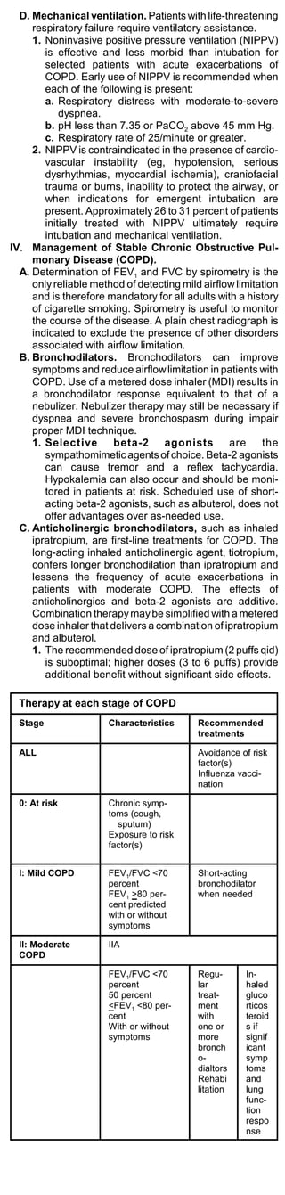
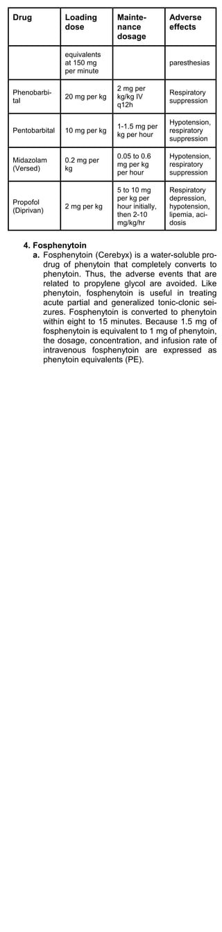
This document provides guidelines and templates for critical care documentation, including summaries of a patient's history and physical exam, daily progress notes, procedure notes, and discharge summaries. It outlines what information should be included in each section, such as vital signs, cardiac exam findings, lab results, assessments, and treatment plans by body system. It also provides guidelines for fluid and electrolyte replacement and blood component therapy.

![1.High-risk patients have a high-risk ACS if ST
segment depression (>0.05 mV [0.5 mm]) is present
in two or more contiguous leads and/or the TIMI risk
score is >5. This patient is admitted to an intensive
care unit, coronary care unit, or monitored cardiac
unit depending upon the persistence of symptoms
and evidence of hemodynamic compromise. Those
with persistent pain or hemodynamic compromise
generally undergo urgent angiography and
revascularization. Others with resolution of symp-
toms and stable hemodynamics are typically referred
for early elective angiography and revascularization
if appropriate.
a. If there is no ST segment elevation or depression
or new LBBB, regardless of the presence or
absence of Q waves, the patient with definite or
probable ACS should still be admitted to a moni-
tored care unit for further evaluation. Those
patients manifesting high-risk features either on
presentation or during their emergency room
course should be considered for early PCI.
2.Moderate-risk patient. Patients who have no ECG
changes and are at moderate risk for ACS can be
admitted to a chest pain observation unit, if available,
for further evaluation because a small percentage (2
to 4 percent) will have an ACS.
3.Low-risk patient. Patients with no ECG changes, a
TIMI risk score below 3, and no other concerning
features in their presentation can be considered for
early provocative testing or possible discharge with
outpatient follow-up. Patients at very low risk in
whom there is clear objective evidence for a non-
ischemic cause of their chest pain can be discharged
with outpatient follow-up.
V. Cardiac biomarkers (enzymes). Serial serum
biomarkers (also called cardiac enzymes) of acute
myocardial damage, such as troponin T and I, creatine
kinase (CK)-MB, and myoglobin, are essential for
confirming the diagnosis of infarction. The most com-
monly used are troponin T or I and CK-MB, which can
be measured by rapid bedside assay.
A. Sensitivity and specificity. An elevation in the
serum concentration of one or more of the above
markers is seen in virtually all patients with an acute
MI. However, the sensitivity of these tests is relatively
low until four to six hours after symptom onset. Thus,
a negative test in this time period does not exclude
infarction. Furthermore, some patients do not show
a biomarker elevation for as long as 12 hours.
B. Therefore, in patients who have an acute STEMI,
reperfusion therapy should not await the results of
cardiac biomarkers. In patients without diagnostic ST
segment elevation, serial biomarker testing is per-
formed after four or more hours if the initial values
are indeterminate, the ECG remains nondiagnostic,
and clinical suspicion remains high.
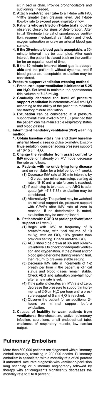
Common Markers for Acute Myocardial Infarction
Marker Initial Mean Time to
Elevation Time to Return to
After MI Peak Ele- Baseline
vations
Myoglobin 1-4 h 6-7 h 18-24 h
CTnl 3-12 h 10-24 h 3-10 d
CTnT 3-12 h 12-48 h 5-14 d
CKMB 4-12 h 10-24 h 48-72 h
CKMBiso 2-6 h 12 h 38 h
CTnI, CTnT = troponins of cardiac myofibrils; CPK-MB, MM =
tissue isoforms of creatine kinase.
C. Unstable angina. Patients with cardiac biomarker
elevations and unstable angina are considered to
have an NSTEMI and should be treated appropri-
ately.
D. Treadmill stress testing and echocardiography is
recommended for patients with a suspicion of coro-
nary ischemia.
Heart Failure Caused by Systolic
Left Ventricular Dysfunction
Over four million persons have HF in the United States.
The mortality rate is 50 percent at two years and 60 to 70
percent at three years. Heart failure (HF) can result from
any structural or functional cardiac disorder that impairs the](https://image.slidesharecdn.com/criticalcaremedicinebrenner-111115081447-phpapp02/85/Critical-care-medicine-brenner-15-320.jpg)
![(1) Carvedilol (Coreg), 3.125 mg BID and 25 to
50 mg BID (the higher dose being used in
subjects over 85 kg).
(2) Metoprolol (Toprol), 6.25 mg BID and 50 to
75 mg BID, and for extended-release
metoprolol, 12.5 or 25 mg daily and 200
mg/day.
(3) Bisoprolol (Ziac), 1.25 mg QD and 5 to 10
mg QD.
(4) The patient should weigh himself daily and
call the physician if there has been a 1 to 1.5
kg weight gain. Weight gain may be treated
with diuretics, but resistant edema or more
severe decompensation may require dose
reduction or cessation of the beta-blocker.
4. Digoxin is given to patients with HF and systolic
dysfunction to control symptoms (fatigue, dyspnea,
and exercise intolerance) and, in atrial fibrillation, to
control the ventricular rate. Digoxin should be
started in left ventricular systolic dysfunction (left
ventricular ejection fraction [LVEF] <40 percent)
who continue to have symptoms despite appropri-
ate therapy including an ACE inhibitor, beta-
blocker, and, if necessary for fluid control, a di-
uretic. The usual daily dose is 0.125 to 0.25 mg,
based upon renal function. The serum digoxin
concentration should be maintained between 0.5
and 0.8 ng/mL.
5. Diuretics. Sodium and water retention lead to
pulmonary and peripheral edema.
a. A loop diuretic should be given to control pulmo-
nary and/or peripheral edema. The most com-
monly used loop diuretic is furosemide (Lasix),
but some patients respond better to bumetanide
(Bumex) or torsemide (Demadex) because of
superior and more predictable absorption.
b. The usual starting dose in outpatients with HF is
20 to 40 mg of furosemide. In patients who are
volume overloaded, a reasonable goal is weight
reduction of 0.5 to 1.0 kg/day. If a patient does
not respond, the diuretic dose should initially be
increased.
c. In patients with HF and a normal glomerular
filtration rate, the maximum doses are 40 to 80
mg of furosemide, 2 to 3 mg of bumetanide, or
20 to 50 mg of torsemide. In patients with renal
insufficiency, a higher maximum dose of 160 to
200 mg of furosemide or its equivalent can be
given.
d. Intravenous diuretics (either as a bolus or a
continuous infusion) are more potent than their
equivalent oral doses and may be required for
unstable or severe disease. Thiazide diuretics
can be added for a synergistic effect.
e. A continuous infusion of a loop diuretic may
improve diuresis and reduce toxicity when
compared to intermittent bolus injections. Urine
output is significantly greater.
6. Aldosterone antagonists. Spironolactone and
eplerenone, which compete with aldosterone for
the mineralocorticoid receptor, prolong survival in
selected patients with HF. Eplerenone has greater
specificity for the mineralocorticoid receptor and a
lower incidence of endocrine side effects.
a. Therapy should be initiated with spironolactone,
and switch to eplerenone (25 to 50 mg/day) if
endocrine side effects occur. The serum potas-
sium should be monitored.
C. Drugs that are contraindicated in HF
1. Nonsteroidal anti-inflammatory drugs (NSAIDs)
can cause a worsening of pre-existing HF because
of systemic vasoconstriction. NSAID may blunt the
renal effects of diuretics and may reverse the effect
of angiotensin converting enzyme (ACE) inhibitors.
2. Thiazolidinediones are oral hypoglycemic agents
that increase insulin sensitivity. Drugs in this class
cause fluid retention, which may precipitate HF.
Patients with HF who are currently taking
thiazolidinediones should be carefully followed for
signs and symptoms of HF, and the agent should
be stopped if signs of fluid retention develop.
Thiazolidinediones should not be used in patients
with New York Heart Association class III or IV HF.
3. Metformin (Glucophage). Patients with HF who
take metformin are at increased risk of potentially
lethal lactic acidosis. Metformin is contraindicated
in patients with HF.
4. Cilostazol suppresses platelet aggregation and is
a direct arterial vasodilator. In patients with HF, oral
phosphodiesterase inhibitors has been associated
with increased mortality. HF of any severity is a
contraindication to the use of cilostazol.
5. Sildenafil (Viagra) is a phosphodiesterase inhibitor
that is used in the treatment of impotence. The
drug is a vasodilator that can lower systemic blood](https://image.slidesharecdn.com/criticalcaremedicinebrenner-111115081447-phpapp02/85/Critical-care-medicine-brenner-21-320.jpg)
![B. Hypertensive urgency is defined as diastolic blood
pressure >130 mm Hg without evidence of vascular
damage; the disorder is asymptomatic and no retinal
lesions are present.
C. Secondary hypertension includes renovascular
hypertension, pheochromocytoma, cocaine use,
withdrawal from alpha-2 stimulants, clonidine, beta-
blockers or alcohol, and noncompliance with
antihypertensive medications.
II. Initial assessment of severe hypertension
A. When severe hypertension is noted, the measure-
ment should be repeated in both arms to detect any
significant differences. Peripheral pulses should be
assessed for absence or delay, which suggests
dissecting aortic dissection. Evidence of pulmonary
edema should be sought.
B. Target organ damage is suggested by chest pain,
neurologic signs, altered mental status, profound
headache, dyspnea, abdominal pain, hematuria, focal
neurologic signs (paralysis or paresthesia), or hyper-
tensive retinopathy.
C. Prescription drug use should be assessed, including
missed doses of antihypertensives. History of recent
cocaine or amphetamine use should be sought.
D. If focal neurologic signs are present, a CT scan may
be required to differentiate hypertensive
encephalopathy from a stroke syndrome.
III. Laboratory evaluation
A. Complete blood cell count, urinalysis for protein,
glucose, and blood; urine sediment examination;
chemistry panel (SMA-18).
B. If chest pain is present, cardiac enzymes are ob-
tained.
C. If the history suggests a hyperadrenergic state, the
possibility of a pheochromocytoma should be ex-
cluded with a 24-hour urine for catecholamines. A
urine drug screen may be necessary to exclude illicit
drug use.
D. Electrocardiogram should be completed.
E. Suspected primary aldosteronism can be excluded
with a 24-hour urine potassium and an assessment of
plasma renin activity. Renal artery stenosis can be
excluded with captopril renography and intravenous
pyelography.
IV. Management of hypertensive emergencies
A. The patient should be hospitalized for intravenous
access, continuous intra-arterial blood pressure
monitoring, and electrocardiographic monitoring.
Volume status and urinary output should be moni-
tored. Rapid, uncontrolled reductions in blood pres-
sure should be avoided because coma, stroke,
myocardial infarction, acute renal failure, or death
may result.
B. The goal of initial therapy is to terminate ongoing
target organ damage. The mean arterial pressure
should be lowered not more than 20-25%, or to a
diastolic blood pressure of 100 mm Hg over 15 to 30
minutes. Blood pressure should be controlled over a
few hours.
V.Management of hypertensive urgencies
A. The initial goal in patients with severe asymptomatic
hypertension should be a reduction in blood pressure
to 160/110 over several hours with conventional oral
therapy.
B. If the patient is not volume depleted, furosemide
(Lasix) is given in a dosage of 20 mg if renal function
is normal, and higher if renal insufficiency is present.
A calcium channel blocker (isradipine ([DynaCirc], 5
mg or felodipine [Plendil], 5 mg) should be added. A
dose of captopril (Capoten)(12.5 mg) can be added if
the response is not adequate. This regimen should
lower the blood pressure to a safe level over three to
six hours and the patient can be discharged on a
regimen of once-a-day medications.
VI. Parenteral antihypertensive agents
A. Nitroprusside (Nipride)
1. Nitroprusside is the drug of choice in almost all
hypertensive emergencies (except myocardial
ischemia or renal impairment). It dilates both ar-
teries and veins, and it reduces afterload and
preload. Onset of action is nearly instantaneous,
and the effects disappear 1-2 minutes after discon-
tinuation.
2. The starting dosage is 0.25-0.5 mcg/kg/min by
continuous infusion with a range of 0.25-8.0
mcg/kg/min. Titrate dose to gradually reduce blood
pressure over minutes to hours.
3. When treatment is prolonged or when renal insuffi-
ciency is present, the risk of cyanide and
thiocyanate toxicity is increased. Signs of
thiocyanate toxicity include disorientation, fatigue,
hallucinations, nausea, toxic psychosis, and sei-
zures.](https://image.slidesharecdn.com/criticalcaremedicinebrenner-111115081447-phpapp02/85/Critical-care-medicine-brenner-29-320.jpg)
![Weight-based nomogram for intravenous heparin
infusion
Initial dose 80 U/kg bolus, then 18 U/kg per
hour
aPTT* <35 sec 80 U/kg bolus, then increase infu-
(<1.2 x control) sion rate by 4 U/kg per hour
aPTT 40 U/kg per hour, then increase
infusion by 2 U/kg per hour
aPTT No change
aPTT Decrease infusion rate by 2q U/kg
per hour
aPTT Hold infusion 1 hour, then de-
crease infusion rate by 3 U/kg per
hour
*aPTT = activated partial thromboplastin time
Asthma
Asthma is the most common chronic disease among
children. Asthma triggers include viral infections; environ-
mental pollutants, such as tobacco smoke; aspirin,
nonsteroidal anti-inflammatory drugs, and sustained
exercise, particularly in cold environments.
I. Diagnosis
A. Symptoms of asthma may include episodic com-
plaints of breathing difficulties, seasonal or nighttime
cough, prolonged shortness of breath after a respira-
tory infection, or difficulty sustaining exercise.
B. Wheezing does not always represent asthma.
Wheezing may persist for weeks after an acute
bronchitis episode. Patients with chronic obstructive
pulmonary disease may have a reversible component
superimposed on their fixed obstruction. Etiologic
clues include a personal history of allergic disease,
such as rhinitis or atopic dermatitis, and a family
history of allergic disease.
C. The frequency of daytime and nighttime symptoms,
duration of exacerbations and asthma triggers should
be assessed.
D. Physical examination. Hyperventilation, use of
accessory muscles of respiration, audible wheezing,
and a prolonged expiratory phase are common.
Increased nasal secretions or congestion, polyps, and
eczema may be present.
E. Measurement of lung function. An increase in the
forced expiratory volume in one second (FEV1) of
12% after treatment with an inhaled beta2 agonist is
sufficient to make the diagnosis of asthma. A 12%
change in peak expiratory flow rate (PEFR) measured
on a peak-flow meter is also diagnostic.
II. Treatment of asthma
A. Beta2 agonists
1. Inhaled short-acting beta2-adrenergic agonists are
the most effective drugs available for treatment of
acute bronchospasm and for prevention of
exercise-induced asthma. Levalbuterol (Xopenex),
the R-isomer of racemic albuterol, offers no signifi-
cant advantage over racemic albuterol.
2. Salmeterol (Serevent), a long-acting beta2 agonist,
has a relatively slow onset of action and a pro-
longed effect.
a. Salmeterol should not be used in the treatment
of acute bronchospasm. Patients taking
salmeterol should use a short-acting beta2
agonist as needed to control acute symptoms.
Twice-daily inhalation of salmeterol has been
effective for maintenance treatment in combina-
tion with inhaled corticosteroids.
b. Fluticasone/Salmeterol (Advair Diskus) is a
long-acting beta agonist and corticosteroid
combination; dry-powder inhaler [100, 250 or
500 g/puff],1 puff q12h.
3. Formoterol (Foradil) is a long-acting beta2 agonist
like salmeterol. It should only be used in patients
who already take an inhaled corticosteroid. Patients
taking formoterol should use a short-acting beta2
agonist as needed to control acute symptoms. For
maintenance treatment of asthma in adults and
children at least 5 years old, the recommended
dosage is 1 puff bid.
4. Adverse effects of beta2 agonists. Tachycardia,
palpitations, tremor and paradoxical bronchospasm
can occur. High doses can cause hypokalemia.](https://image.slidesharecdn.com/criticalcaremedicinebrenner-111115081447-phpapp02/85/Critical-care-medicine-brenner-39-320.jpg)
![B. Pharmacologic treatment
1. Inhaled beta-adrenergic agonists, such as
albuterol, are the mainstay of therapy for an acute
exacerbation of COPD because of their rapid onset
of action and efficacy in producing bronchodilation.
2. Typical doses of albuterol are 180 mcg (two puffs)
by metered dose inhaler, or 500 mcg by nebulizer,
given every one to two hours.
3. Anticholinergic bronchodilators, such as
ipratropium bromide, may be used in combination
with beta-adrenergic agonists to produce
bronchodilation in excess of that achieved by either
agent alone.
a. Ipratropium may be administered during acute
exacerbations either by nebulizer (500 mcg
every two to four hours), or via MDI (two puffs
[36 mcg] every two to four hours with a spacer).
Glycopyrrolate is available for nebulized use in
COPD (1 to 2 mg every two to four hours).
4. Parenteral corticosteroids are frequently used for
acute exacerbations of COPD. Methylprednisolone
(60 to 125 mg intravenously, two to four times
daily) commonly is given.
5. Antibiotics are recommended for acute exacerba-
tions of COPD characterized by increased volume
and purulence of secretions. A 10-day course of
amoxicillin, doxycycline, or trimethoprim-
sulfamethoxazole should be prescribed.
Choice of empirical antibiotic therapy for COPD
exacerbation
First-line treatment Dosage*
Amoxicillin (Amoxil, Trimox,
500 mg tid
Wymox)
Trimethoprim-sulfamethoxaz
1 tablet (80/400 mg) bid
ole (Bactrim, Cotrim, Septra)
Doxycycline 100 mg bid
Erythromycin 250-500 mg qid
Second-line treatment**
Amoxicillin and clavulanate
500-875 mg bid
(Augmentin)
Second- or third-generation
cephalosporin (eg, 250-500 mg bid
cefuroxime [Ceftin])
Macrolides
Clarithromycin (Biaxin) 250-500 mg bid
500 mg on day 1, then 250
Azithromycin (Zithromax)
mg qd X 4 days
Quinolones
Ciprofloxacin (Cipro) 500-750 mg bid
Levofloxacin (Levaquin)*** 500 mg qd
*May need adjustment in patients with renal or hepatic insuffi-
ciency.
**For patients in whom first-line therapy has failed and those
with moderate to severe disease or resistant or
gram-negative pathogens.
***Although the newer quinolones have better activity against
Streptococcus pneumoniae, ciprofloxacin may be preferable
in patients with gram-negative organisms.
6. Methylxanthines. Aminophylline and theophylline
are not recommended for the management of acute
exacerbations of COPD.
C. Oxygen therapy
1. Supplemental oxygen should achieve a target
PaO2 of 60 to 65 mm Hg, with a hemoglobin satura-
tion exceeding 90 percent.
2. Venturi masks are the preferred means of oxygen
delivery because they permit a precise delivered
fraction of inspired oxygen (FiO2).
3. Nasal cannulae can provide flow rates up to 6
L/min with an associated FiO2 of approximately 44
percent. Nasal cannulae are also more comfortable
for the patient and permit oral feedings.
4. Facemasks can be used when higher inspired
concentrations of oxygen are needed. Simple
facemasks using flow rates of 6 to 10 L/min provide
an FiO2 up to 55 percent.
5. Non-rebreather masks with a reservoir, one-way
valves, and a tight face seal can deliver an inspired
oxygen concentration up to 90 percent.](https://image.slidesharecdn.com/criticalcaremedicinebrenner-111115081447-phpapp02/85/Critical-care-medicine-brenner-45-320.jpg)

![patients who are recovering from influenza (post-
influenza pneumonia).
3. Legionella accounts for 2 to 8 percent of cases of
CAP. Among patients with pneumonia who can be
treated as outpatients, the frequency of Legion-
naires’ disease is less than 1 percent. Legionella
is much more frequent in hospitalized patients,
especially those admitted to intensive care units.
4. Gram-negative bacilli, especially P. aeruginosa,
are an uncommon cause of CAP except in pa-
tients with neutropenia, cystic fibrosis, late stage
HIV infection and bronchiectasis.
5. Other bacteria that can cause CAP include
Neisseria meningitidis, Moraxella catarrhalis, and
Streptococcus pyogenes.
6. Anaerobic organisms may be the cause of aspira-
tion pneumonia and lung abscess.
II.Diagnostic approach
A. Clinical evaluation. CAP caused by pyogenic
organisms presents with the sudden onset of rigors,
fever, pleuritic chest pain, and cough productive of
purulent sputum. Chest pain occurs in 30 percent of
cases, chills in 40 to 50 percent, and rigors in 15
percent.
B. On physical examination, 80 percent are febrile,
although this finding is frequently absent in older
patients. A respiratory rate above 24 breaths/minute
is noted in 45 to 70 percent of patients; tachycardia
is also common. Chest examination reveals audible
rales in most patients, while one-third have consoli-
dation.
C. The major blood test abnormality is leukocytosis
(15,000 and 30,000 per mm3) with a leftward shift.
Leukopenia can occur.
D. Chest radiographs. The presence of an infiltrate on
plain chest radiograph is the “gold standard” for
diagnosing pneumonia. The radiographic appear-
ances include lobar consolidation, interstitial infil-
trates, and cavitation.
E. Sputum examination. An etiologic agent is found in
51 percent of patients. A specimen that has greater
than 25 PMN and less than 10 epithelial cells per low
power field represents a purulent specimen. Gram
stain and culture may identify the cause of the
pneumonia.
F. Blood cultures should be obtained in patients who
require hospitalization.
G. Urinary antigen testing for pneumococcal cell wall
components has a sensitivity of 70 to 90 percent;
specificity is 80 to 100 percent. Urinary antigen
testing is also available for Legionella species and is
highly sensitive and specific and inexpensive. Urinary
antigen testing is recommended for both S.
pneumoniae and Legionella, particularly in patients
with risk factors for Legionella infection (eg, smoking,
chronic lung disease, immunosuppression, and CAP
requiring hospitalization).
Causes of Community-acquired Pneumonia
Etiology Prevalence (percent)
Streptococcus 20-60
pneumoniae 3-10
Hemophilus influenzae 3-5
Staphylococcus aureus 3-10
Gram-negative bacilli 6-10
Aspiration 3-5
Miscellaneous 2-8
Legionella sp. 1-6
Mycoplasma pneumoniae 4-6
Chlamydia pneumoniae 2-15
Viruses
III. Treatment of Community-Acquired Pneumonia
A. Empiric regimens. For uncomplicated pneumonia
in patients who do not require hospitalization,
macrolide therapy is recommended. Erythromycin
is the least expensive macrolide but is associated
with gastrointestinal upset in many patients.
Azithromycin (Zithromax [500 mg PO QD]) is
recommended because it causes less gastrointesti-
nal upset; if macrolide resistance in the community
is high, doxycycline (100 mg PO twice a day)
should be considered. Telithromycin (Ketek) or a
quinolone is also reasonable if macrolide resis-
tance is prevalent. Quinolones are not recom-
mended for patients with CAP because achievable
tissue concentrations of these agents will be close
to the minimum inhibitory concentration for
pneumococcus and resistance may emerge with
overuse.
B. Duration of therapy. The usual recommended
duration of therapy is 7 to 14 days. Three days of](https://image.slidesharecdn.com/criticalcaremedicinebrenner-111115081447-phpapp02/85/Critical-care-medicine-brenner-62-320.jpg)
![azithromycin may be as effective as longer course
of antibiotics. For a hospitalized patient, ceftriaxone
(Rocephin [2 g IV QD]) with or without azithromycin
(depending upon the likelihood of an atypical
organism) is recommended. For more severely ill
patients who might have an atypical pneumonia or
patients admitted to an ICU, therapy consists of a
quinolone, such as levofloxacin (Levaquin [500 mg
IV QD]) or azithromycin to treat legionella infection
as well as mycoplasma and chlamydia. Consider-
ation should be given to adding a second agent for
pneumococcus to levofloxacin. Ceftriaxone should
be given with azithromycin in these sicker patients.
Recommended Empiric Drug Therapy for Pa-
tients with Community-Acquired Pneumonia
Clinical Situa- Primary Treat- Alternative(s)
tion ment
Younger (<60 Macrolide anti- Levofloxacin or
yr) outpatients biotics doxycycline
without under- (azithromycin,
lying disease clarithromycin,
dirithromycin,
or
erythromycin)
Older (>60 yr) Levofloxacin or Beta-lactamase in-
outpatients with cefuroxime or hibitor (with
underlying dis- Trimethoprim- macrolide if
ease sulfa- legionella infection
methoxazole suspected)
Add
vancomycin in
severe, life-
threatening
pneumonias
Gross aspira- Clindamycin IV Cefotetan,
tion suspected ampicillin/sulbactam
Common Antimicrobial Agents for Community-
Acquired Pneumonia in Adults
Type Agent Dosage
Oral therapy
Macrolides Erythromycin 500 mg PO qid
Clarithromycin 500 mg PO bid
(Biaxin) 500 mg PO on day
Azithromycin 1, then 250 mg qd
(Zithromax) x 4 days
Beta- Amoxicillin- 500 mg tid or 875
lactam/beta- clavulanate mg PO bid
lactamase in- (Augmentin)
hibitor Augmentin XR 2 tabs q12h
Quinolones Ciprofloxacin 500 mg PO bid
(Cipro) 500 mg PO qd
Levofloxacin 400 mg PO bid
(Levaquin)
Ofloxacin (Floxin)
Tetracycline Doxycycline 100 m g PO bid
Sulfonamide Trimethoprim- 160 mg/800 mg
sulfamethoxazole (DS) PO bid
Intravenous Therapy
Cephalosporin
s Cefuroxime 0.75-1.5 g IV q8h
Second-gen- (Kefurox, Zinacef)
eration Ceftizoxime 1-2 g IV q8h
(Cefizox) 1-2 g IV q8h
Third-genera- Ceftazidime 1-2 g IV q8h
tion (anti- (Fortaz)
Pseudomo- Cefoperazone
nas (Cefobid)
aeruginosa)
Beta- Ampicillin- 1.5 g IV q6h
lactam/beta- sulbactam
lactamase in- (Unasyn) 3.375 g IV q6h
hibitors Piperacillin/tazoba
ctam (Zosyn) 3.1 g IV q6h
Ticarcillin-
clavulanate
(Timentin)
Quinolones Ciprofloxacin 400 mg IV q12h
(Cipro) 500 mg IV q24h
Levofloxacin 400 mg IV q12h
(Levaquin)
Ofloxacin (Floxin)](https://image.slidesharecdn.com/criticalcaremedicinebrenner-111115081447-phpapp02/85/Critical-care-medicine-brenner-63-320.jpg)
![diagnostic procedure is required to confirm the
diagnosis of PCP.
B. Transbronchial biopsy and bronchoalveolar
lavage. The sensitivity of transbronchial biopsy for
PCP is 98%. The sensitivity of bronchoalveolar is
90%.
C. Open-lung biopsy should be reserved for patients
with progressive pulmonary disease in whom the less
invasive procedures are nondiagnostic.
IV. Diagnostic algorithm
A. If the chest radiograph of a symptomatic patient
appears normal, a DLCO should be performed.
Patients with significant symptoms, a normal-appear-
ing chest radiograph, and a normal DLCO should
undergo high-resolution CT. Patients with abnormal
findings at any of these steps should proceed to
sputum induction or bronchoscopy. Sputum speci-
mens collected by induction that reveal P. carinii
should also be stained for acid-fast organisms and
fungi, and the specimen should be cultured for
mycobacteria and fungi.
B. Patients whose sputum examinations do not show P.
carinii or another pathogen should undergo bron-
choscopy.
C. Lavage fluid is stained for P. carinii, acid-fast organ-
isms, and fungi. Also, lavage fluid is cultured for
mycobacteria and fungi and inoculated onto cell
culture for viral isolation. Touch imprints are made
from tissue specimens and stained for P. carinii. Fluid
is cultured for mycobacteria and fungi, and stained
for P. carinii, acid-fast organisms, and fungi. If all
procedures are nondiagnostic and the lung disease
is progressive, open-lung biopsy may be considered.
V. Treatment of Pneumocystic carinii pneumonia
A. Trimethoprim-sulfamethoxazole DS (Bactrim DS,
Septra DS) is the recommended initial therapy for
PCP. Dosage is 15-20 mg/kg/day of TMP IV divided
q6h for 14-21 days. Adverse effects include rash
(33%), elevation of liver enzymes (44%), nausea and
vomiting (50%), anemia (40%), creatinine elevation
(33%), and hyponatremia (94%).
B. Pentamidine is an alternative in patients who have
adverse reactions or fail to respond to TMP-SMX.
The dosage is 4 mg/kg/day IV for 14-21 days. Ad-
verse effects include anemia (33%), creatinine
elevation (60%), LFT elevation (63%), and
hyponatremia (56%). Pancreatitis, hypoglycemia, and
hyperglycemia are common side effects.
C. Corticosteroids. Adjunctive corticosteroid treatment
is beneficial with anti-PCP therapy in patients with a
partial pressure of oxygen (PaO2) less than 70 mm
Hg, (A-a)DO2 greater than 35 mm Hg, or oxygen
saturation less than 90% on room air. Contraindica-
tions include suspected tuberculosis or disseminated
fungal infection. Treatment with methylprednisolone
(SoluMedrol) should begin at the same time as anti-
PCP therapy. The dosage is 30 mg IV q12h x 5 days,
then 30 mg IV qd x 5 days, then 15 mg qd x 11 days
OR prednisone, 40 mg twice daily for 5 days, then 40
mg daily for 5 days, and then 20 mg daily until day 21
of therapy.
VI. Prophylaxis
A. HIV-infected patients who have CD4 counts less than
200 cells/mcL should receive prophylaxis against
PCP. If CD4 count increases to greater than 200
cells/mcL after receiving antiretroviral therapy, PCP
prophylaxis can be safely discontinued.
B. Trimethoprim-sulfamethoxazole (once daily to
three times weekly) is the preferred regimen for PCP
prophylaxis.
C. Dapsone (100 mg daily or twice weekly) is a prophy-
lactic regimen for patients who can not tolerate TMP-
SMX.
D. Aerosolized pentamidine (NebuPent) 300 mg in 6
mL water nebulized over 20 min q4 weeks is another
alternative.
Antiretroviral Therapy and Opportu-
nistic Infections in AIDS
I. Antiretroviral therapy
A. A combination of three agents is recommended as
initial therapy. The preferred options are 2 nucleo-
sides plus 1 protease inhibitor or 1 non-nucleoside.
Alternative options are 2 protease inhibitors plus 1
nucleoside or 1 non-nucleoside. Combinations of 1
nucleoside, 1 non-nucleoside, and 1 protease inhibi-
tor are also effective.
B. Nucleoside analogs
1. Abacavir (Ziagen) 300 mg PO bid [300 mg].
2. Didanosine (Videx) 200 mg PO bid [chewable
tabs: 25, 50, 100, 150 mg]; oral ulcers discourage
common usage.
3. Lamivudine (Epivir) 150 mg PO bid [tab: 150 mg].](https://image.slidesharecdn.com/criticalcaremedicinebrenner-111115081447-phpapp02/85/Critical-care-medicine-brenner-65-320.jpg)
![4. Stavudine (Zerit) 40 mg PO bid [cap: 15, 20, 30,
40 mg].
5. Zalcitabine (Hivid) 0.75 mg PO tid [tab: 0.375, 0.75
mg].
6. Zidovudine (Retrovir, AZT) 200 mg PO tid or 300
mg PO bid [cap: 100, 300 mg].
7. Zidovudine 300 mg/lamivudine 150 mg (Combivir)
1 tab PO bid.
C. Protease inhibitors
1. Amprenavir (Agenerase) 1200 mg PO bid [50, 150
mg]
2. Indinavir (Crixivan) 800 mg PO tid [cap: 200, 400
mg].
3. Nelfinavir (Viracept) 750 mg PO tid [tab: 250 mg]
4. Ritonavir (Norvir) 600 mg PO bid [cap: 100 mg].
5. Saquinavir ( Invirase) 600 mg PO tid [cap: 200
mg].
D. Non-nucleoside analogs
1. Delavirdine (Rescriptor) 400 mg PO tid [tab: 100
mg]
2. Efavirenz (Sustiva) 600 mg qhs [50, 100, 200 mg]
3. Nevirapine (Viramune) 200 mg PO bid [tab: 200
mg]
II. Oral candidiasis
A. Fluconazole (Diflucan), acute: 200 mg PO x 1, then
100 mg qd x 5 days OR
B. Ketoconazole (Nizoral), acute: 400 mg po qd 1-2
weeks or until resolved OR
C. Clotrimazole (Mycelex) troches 10 mg dissolved
slowly in mouth 5 times/d.
III. Candida esophagitis
A. Fluconazole (Diflucan) 200 mg PO x 1, then 100 mg
PO qd until improved.
B. Ketoconazole (Nizoral) 200 mg po bid.
IV. Primary or recurrent mucocutaneous HSV.
Acyclovir (Zovirax), 200-400 mg PO 5 times a day for
10 days, or 5 mg/kg IV q8h; or in cases of acyclovir
resistance, foscarnet 40 mg/kg IV q8h for 21 days.
V. Herpes simplex encephalitis. Acyclovir 10 mg/kg IV
q8h x 10-21 days.
VI. Herpes varicella zoster
A. Acyclovir (Zovirax) 10 mg/kg IV over 60 min q8h OR
B. Valacyclovir (Valtrex) 1000 mg PO tid x 7 days
[caplet: 500 mg].
VII.Cytomegalovirus infections
A. Ganciclovir (Cytovene) 5 mg/kg IV (dilute in 100 mL
D5W over 60 min) q12h x 14-21 days (concurrent
use with zidovudine increases hematological toxic-
ity).
B. Suppressive treatment for CMV: Ganciclovir
(Cytovene) 5 mg/kg IV qd, or 6 mg/kg IV 5 times/wk,
or 1000 mg orally tid with food.
VIII. Toxoplasmosis
A. Pyrimethamine 200 mg PO loading dose, then 50-
75 mg qd plus leucovorin calcium (folinic acid) 10-
20 mg PO qd for 6-8 weeks for acute therapy AND
B. Sulfadiazine (1.0-1.5 gm PO q6h) or clindamycin
450 mg PO qid/600-900 mg IV q6h.
C. Suppressive treatment for toxoplasmosis
1. Pyrimethamine 25-50 mg PO qd with or without
sulfadiazine 0.5-1.0 gm PO q6h; and folinic acid 5-
10 mg PO qd OR
2. Pyrimethamine 50 mg PO qd; and clindamycin
300 mg PO q6h; and folinic acid 5-10 mg PO qd.
IX. Cryptococcus neoformans meningitis
A. Amphotericin B at 0.7 mg/kg/d IV for 14 days or until
clinically stable, followed by fluconazole (Diflucan)
400 mg qd to complete 10 weeks of therapy, fol-
lowed by suppressive therapy with fluconazole
(Diflucan) 200 mg PO qd indefinitely.
B. Amphotericin B lipid complex (Abelcet) may be used
in place of non-liposomal amphotericin B if the
patient is intolerant to non-liposomal amphotericin
B. The dosage is 5 mg/kg IV q24h.
X. Active tuberculosis
A. Isoniazid (INH) 300 mg PO qd; and rifabutin 300 mg
PO qd; and pyrazinamide 15-25 mg/kg PO qd (500
mg PO bid-tid); and ethambutol 15-25 mg/kg PO qd
(400 mg PO bid-tid).
B. All four drugs are continued for 2 months; isoniazid
and rifabutin (depending on susceptibility testing)
are continued for a period of at least 9 months and
at least 6 months after the last negative cultures.
C. Pyridoxine (vitamin B6) 50 mg PO qd, concurrent
with INH.
XI. Disseminated mycobacterium avium complex
(MAC)
A. Azithromycin (Zithromax) 500-1000 mg PO qd or
clarithromycin (Biaxin) 500 mg PO bid; AND
B. Ethambutol 15-25 mg/kg PO qd (400 mg bid-tid)
AND
C. Rifabutin 300 mg/d (two 150 mg tablets qd).
D. Prophylaxis for MAC
1. Clarithromycin (Biaxin) 500 mg PO bid OR
2. Rifabutin (Mycobutin) 300 mg PO qd or 150 mg
PO bid.](https://image.slidesharecdn.com/criticalcaremedicinebrenner-111115081447-phpapp02/85/Critical-care-medicine-brenner-66-320.jpg)
![2. Dopamine (Intropin) traditionally has been used
as the initial therapy in hypotension, primarily
because it is thought to increase systemic blood
pressure. However, dopamine is a relatively weak
vasoconstrictor in septic shock.
Hemodynamic effects of vasoactive agents
Agent Dose Effect
CO MAP SVR
Dopamine 5-20
2+ 1+ 3+
(Intropin) mcg/kg/min
Norepin-
ephrine 0.05-0.5
-/0/+ 2+ 4+
(Levophed mcg/kg/min
)
Dobutamin
10 -/0/+
e 2+ -/0
mcg/kg/min
(Dobutrex)
Epineph- 0.05-2
3+ 2+ 4+
rine mcg/kg/min
Phenyleph
rine 2-10
0 2+ 4+
(Neo-Syne mcg/kg/min
phrine)
3. Norepinephrine (Levophed) is superior to
dopamine in the treatment of hypotension associ-
ated with septic shock. Norepinephrine is the
agent of choice for treatment of hypotension
related to septic shock.
4. Dobutamine (Dobutrex) should be reserved for
patients with a persistently low cardiac index or
underlying left ventricular dysfunction.
E. Antibiotics should be administered within 2 hours
of the recognition of sepsis. Use of vancomycin
should be restricted to settings in which the caus-
ative agent is most likely resistant Enterococcus,
methicillin-resistant Staphylococcus aureus, or
high-level penicillin-resistant Streptococcus
pneumoniae.
Recommended Antibiotics in Septic Shock
Suspected
source Recommended antibiotics
Pneumonia Third or 4th-generation cephalosporin
(cefepime, ceftazidime, cefotaxime,
ceftizoxime) plus macrolide
(antipseudomonal beta lactam plus
aminoglycoside if hospital-acquired) + an-
aerobic coverage with metronidazole or
clindamycin.
Urinary tract Ampicillin plus gentamicin (Garamycin) or
third-generation cephalosporin (ceftazidime,
cefotaxime, ceftizoxime) or a quinolone
(ciprofloxacin, levofloxacin).
Skin or soft Nafcillin (add metronidazole [Flagyl] or
tissue clindamycin if anaerobic infection suspected)
Meningitis Third-generation cephalosporin (ceftazidime,
cefotaxime, ceftizoxime)
Intra-abdomin Third-generation cephalosporin (ceftazidime,
al cefotaxime, ceftizoxime) plus metronidazole
or clindamycin
Primary Ticarcillin/clavulanate (Timentin) or
bacteremia piperacillin/tazobactam(Zosyn)
Dosages of Antibiotics Used in Sepsis
Agent Dosage
Cefepime (Maxipime) 2 gm IV q12h; if neutropenic,
use 2 gm q8h
Ceftizoxime (Cefizox) 2 gm IV q8h
Ceftazidime (Fortaz) 2 g IV q8h
Cefotaxime (Claforan) 2 gm q4-6h
Cefuroxime (Kefurox, 1.5 g IV q8h
Zinacef)
Cefoxitin (Mefoxin) 2 gm q6h](https://image.slidesharecdn.com/criticalcaremedicinebrenner-111115081447-phpapp02/85/Critical-care-medicine-brenner-69-320.jpg)
![Agent Dosage
Cefotetan (Cefotan) 2 gm IV q12h
Piperacillin/tazobactam 3.375-4.5 gm IV q6h
(Zosyn)
Ticarcillin/clavulanate 3.1 gm IV q4-6h (200-300
(Timentin) mg/kg/d)
Ampicillin 1-3.0 gm IV q6h
Ampicillin/sulbactam 3.0 gm IV q6h
(Unasyn)
Nafcillin (Nafcil) 2 gm IV q4-6h
Piperacillin, ticarcillin, 3 gm IV q4-6h
mezlocillin
Meropenem (Merrem) 1 gm IV q8h
Imipenem/cilastatin 1.0 gm IV q6h
(Primaxin)
Gentamicin or 2 mg/kg IV loading dose, then
tobramycin 1.7 mg/kg IV q8h
Amikacin (Amikin) 7.5 mg/kg IV loading dose, then
5 mg/kg IV q8h
Vancomycin 1 gm IV q12h
Metronidazole (Flagyl) 500 mg IV q6-8h
Clindamycin (Cleocin) 600-900 mg IV q8h
Linezolid (Zyvox) 600 mg IV/PO q12h
Quinupristin/dalfopristin 7.5 mg/kg IV q8h
(Synercid)
1. Initial treatment of life-threatening sepsis
usually consists of a third or 4th-generation
cephalosporin (cefepime, ceftazidime, cefotaxime,
ceftizoxime) or piperacillin/tazobactam (Zosyn).
An aminoglycoside (gentamicin, tobramycin, or
amikacin) should also be included.
Antipseudomonal coverage is important for
hospital- or institutional-acquired infections.
Appropriate choices include an antipseudomonal
penicillin, cephalosporin, or an aminoglycoside.
2. Methicillin-resistant staphylococci. If line
sepsis or an infected implanted device is a possi-
bility, vancomycin should be added to the regimen
to cover for methicillin-resistant Staph aureus and
methicillin-resistant Staph epidermidis.
3. Vancomycin-resistant enterococcus (VRE):
An increasing number of enterococcal strains are
resistant to ampicillin and gentamicin. The inci-
dence of vancomycin-resistant enterococcus
(VRE) is rapidly increasing.
a. Linezolid (Zyvox) is an oral or parenteral
agent active against vancomycin-resistant
enterococci, including E. faecium and E.
faecalis. Linezolid is also active against
methicillin-resistant staphylococcus aureus.
b. Quinupristin/dalfopristin (Synercid) is a
parenteral agent active against strains of
vancomycin-resistant enterococcus faecium,
but not enterococcus faecalis. Most strains of
VRE are enterococcus faecium.
F. Other therapies
1. Hydrocortisone (100 mg every 8 hours) in
patients with refractory shock significantly im-
proves hemodynamics and survival rates.
Corticosteroids may be beneficial in patients with
refractory shock caused by an Addison’s crisis.
2. Activated protein C (drotrecogin alfa [Xigris])
has antithrombotic, profibrinolytic, and
anti-inflammatory properties. Activated protein C
reduces the risk of death by 20%. Activated
protein C is approved for treatment of patients
with severe sepsis who are at high risk of death.
Drotrecogin alfa is administered as 24 mcg/kg/hr
for 96 hours. There is a small risk of bleeding.
Contraindications are thrombocytopenia,](https://image.slidesharecdn.com/criticalcaremedicinebrenner-111115081447-phpapp02/85/Critical-care-medicine-brenner-70-320.jpg)
![Hepatoportal sclerosis
Schistosomiasis
Sinusoidal: Cirrhosis, alcoholic hepatitis
Postsinusoidal
Budd-Chiari syndrome (hepatic vein thrombosis)
Veno-occlusive disease
Severe congestive heart failure
Restrictive heart disease
I. Pathophysiology
A. Varices develop annually in 5% to 15% of patients
with cirrhosis, and varices enlarge by 4% to 10%
each year. Each episode of variceal hemorrhage
carries a 20% to 30% risk of death.
B. After an acute variceal hemorrhage, bleeding re-
solves spontaneously in 50% of patients. Bleeding is
least likely to stop in patients with large varices and
a Child-Pugh class C cirrhotic liver.
II. Management of variceal hemorrhage
A. Primary prophylaxis
1. All patients with cirrhosis should undergo endos-
copy to screen for varices every 2 to 3 years.
2. Propranolol (Inderal) and nadolol (Corgard) re-
duce portal pressure through beta blockade.
2
Beta-blockade reduces the risk of bleeding by
45% and bleeding-related death by 50%. The
beta-blocker dose is adjusted to decrease the
resting heart rate by 25% from its baseline, but not
to less than 55 to 60 beats/min.
3. Propranolol (Inderal) is given at 10 to 480 mg
daily, in divided doses, or nadolol (Corgard) 40 to
320 mg daily in a single dose.
B. Treatment of acute hemorrhage
1. Variceal bleeding should be considered in any
patient who presents with significant upper gastro-
intestinal bleeding. Signs of cirrhosis may include
spider angiomas, palmar erythema, leukonychia,
clubbing, parotid enlargement, and Dupuytren's
contracture. Jaundice, lower extremity edema and
ascites are indicative of decompensated liver
disease.
2. The severity of the bleeding episode can be
assessed on the basis of orthostatic changes (eg,
resting tachycardia, postural hypotension), which
indicates one-third or more of blood volume loss.
3. Blood should be replaced as soon as possible.
While blood for transfusion is being made avail-
able, intravascular volume should be replenished
with normal saline solution. Once euvolemia is
established, the intravenous infusion should be
changed to solutions with a lower sodium content
(5% dextrose with 1/2 or 1/4 normal saline). Blood
should be transfused to maintain a hematocrit of
at least 30%. Serial hematocrit estimations should
be obtained during continued bleeding.
4. Fresh frozen plasma is administered to patients
who have been given massive transfusions. Each
3 units of PRBC should be accompanied by
CaCL2 1 gm IV over 30 min. Clotting factors
should be assessed. Platelet transfusions are
reserved for counts below 50,000/mL in an ac-
tively bleeding patient.
5. If the patient's sensorium is altered because of
hepatic encephalopathy, the risk of aspiration
mandates endotracheal intubation. Placement of
a large-caliber nasogastric tube (22 F or 24 F)
permits tap water lavage for removal of blood and
clots in preparation for endoscopy.
6. Octreotide acetate (Sandostatin) is a synthetic,
analogue of somatostatin, which causes
splanchnic vasoconstriction. Octreotide is the drug
of choice in the pharmacologic management of
acute variceal bleeding. Octreotide infusion should
be started with a loading dose of 50 micrograms,
followed by an infusion of 50 micrograms/hr.
Treatment is continued until hemorrhage sub-
sides. Definitive endoscopic therapy is performed
shortly after hemostasis is achieved.
7. Endoscopic therapy
a. A sclerosant (eg, morrhuate [Scleromate]) is
injected into each varix. Complications include
bleeding ulcers, dysphagia due to strictures,
and pleural effusions.
b. Endoscopic variceal ligation with elastic bands
is an alternative to sclerotherapy because of
fewer complications and similar efficacy.
c. If bleeding persists (or recurs within 48 hours
of the initial episode) despite pharmacologic
therapy and two endoscopic therapeutic at-
tempts at least 24 hours apart, patients should
be considered for salvage therapy with TIPS or
surgical treatment (transection of esophageal
varices and devascularization of the stomach,
portacaval shunt, or liver transplantation).
8. Transjugular intrahepatic portosystemic shunt
(TIPS) consists of the angiographic creation of a](https://image.slidesharecdn.com/criticalcaremedicinebrenner-111115081447-phpapp02/85/Critical-care-medicine-brenner-74-320.jpg)
![E. Bladder catheterization is useful to rule out sus-
pected bladder outlet obstruction. A residual volume
of more than 100 mL suggests bladder outlet obstruc-
tion.
F. Central venous monitoring is used to measure
cardiac output and left ventricular filling pressure if
prerenal failure is suspected.
V. Laboratory evaluation
A. Spot urine sodium concentration
1. Spot urine sodium can help distinguish between
prerenal azotemia and acute tubular necrosis.
2. Prerenal failure causes increased reabsorption of
salt and water and will manifest as a low spot urine
sodium concentration <20 mEq/L and a low frac-
tional sodium excretion <1%, and a urine/plasma
creatinine ration of >40. Fractional excretion of
sodium (%) = ([urine sodium/plasma sodium] ÷
[urine creatinine/plasma creatinine] x 100).
3. If tubular necrosis is the cause, the spot urine
concentration will be >40 mEq/L, and fractional
excretion of sodium will be >1%.
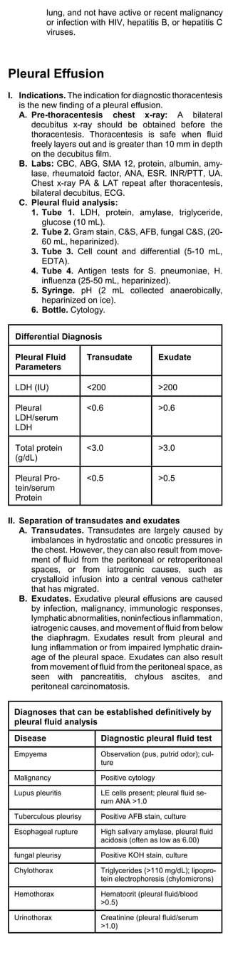
B. Urinalysis
1. Normal urine sediment is a strong indicator of
prerenal azotemia or may be an indicator of
obstructive uropathy.
2. Hematuria, pyuria, or crystals may be asso-
ciated with postrenal obstructive azotemia.
3. Abundant cells, casts, or protein suggests an
intrarenal disorder.
4. Red cells alone may indicate vascular disorders.
RBC casts and abundant protein suggest glomeru-
lar disease (glomerulonephritis).
5. White cell casts and eosinophilic casts indicate
interstitial nephritis.
6. Renal epithelial cell casts and pigmented
granular casts are associated with acute tubular
necrosis.
C. Ultrasound is useful for evaluation of suspected
postrenal obstruction (nephrolithiasis). The presence
of small (<10 cm in length), scarred kidneys is diag-
nostic of chronic renal insufficiency.
VI. Management of acute renal failure
A. Reversible disorders, such as obstruction, should be
excluded, and hypovolemia should be corrected with
volume replacement. Cardiac output should be
maintained. In critically ill patients, a pulmonary artery
catheter should be used for evaluation and monitor-
ing.
B. Extracellular fluid volume expansion. Infusion of a
1-2 liter crystalloid fluid bolus may confirm suspected
volume depletion.
C. If the patient remains oliguric despite euvolemia, IV
diuretics may be administered. A large single dose of
furosemide (100-200 mg) may be administered
intravenously to promote diuresis. If urine flow is not
improved, the dose of furosemide may be doubled.
Furosemide may be repeated in 2 hours, or a contin-
uous IV infusion of 10-40 mg/hr (max 1000 mg/day)
may be used.
D. The dosage or dosing intervals of renally excreted
drugs should be modified.
E. Hyperkalemia is the most immediately life-threaten-
ing complication of renal failure. Serum potassium
values greater than 6.5 mEq/L may lead to
arrhythmias and cardiac arrest. Potassium should be
removed from IV solutions. Hyperkalemia may be
treated with sodium polystyrene sulfonate
(Kayexalate), 30-60 gm PO/PR every 4-6 hours.
F. Hyperphosphatemia can be controlled with alu-
minum hydroxide antacids (eg, Amphojel or Basaljel),
15-30 ml or one to three capsules PO with meals,
should be used.
G. Fluids. After normal volume has been restored, fluid
intake should be reduced to an amount equal to uri-
nary and other losses plus insensible losses of 300-
500 mL/day. In oliguric patients, daily fluid intake may
need to be restricted to less than 1 L.
H. Nutritional therapy. A renal diet consisting of daily
high biologic value protein intake of 0.5 gm/kg/d,
sodium 2 g, potassium 40-60 mg/day, and at least 35
kcal/kg of nonprotein calories is recommended.
Phosphorus should be restricted to 800 mg/day
I. Dialysis. Indications for dialysis include uremic
pericarditis, severe hyperkalemia, pulmonary edema,
persistent severe metabolic acidosis (pH less than
7.2), and symptomatic uremia.
Hyperkalemia
Body potassium is 98% intracellular. Only 2% of total body
potassium, about 70 mEq, is in the extracellular fluid, with
the normal concentration of 3.5-5 mEq/L.](https://image.slidesharecdn.com/criticalcaremedicinebrenner-111115081447-phpapp02/85/Critical-care-medicine-brenner-99-320.jpg)
![II.Treatment of hypermagnesemia
A. Asymptomatic, hemodynamically stable patients.
Moderate hypermagnesemia can be managed by
elimination of intake.
B. Severe hypermagnesemia
1. Furosemide 20-40 mg IV q3-4h should be given as
needed. Saline diuresis should be initiated with
0.9% saline, infused at 120 cc/h to replace urine
loss.
2. If ECG abnormalities (peaked T waves, loss of P
waves, or widened QRS complexes) or if respira-
tory depression is present, IV calcium gluconate
should be given as 1-3 ampules (10% solution, 1
gm per 10 mL amp), added to saline infusate.
Calcium gluconate can be infused to reverse acute
cardiovascular toxicity or respiratory failure as 15
mg/kg over a 4-hour period.
3. Parenteral insulin and glucose can be given to shift
magnesium into cells. Dialysis is necessary for
patients who have severe hypermagnesemia.
Disorders of Water and Sodium Bal-
ance
I. Pathophysiology of water and sodium balance
A. Volitional intake of water is regulated by thirst. Mainte-
nance intake of water is the amount of water sufficient
to offset obligatory losses.
B. Maintenance water needs
= 100 mL/kg for first 10 kg of body weight
+ 50 mL/kg for next 10 kg
+ 20 mL/kg for weight greater than 20 kg
C. Clinical signs of hyponatremia. Confusion, agita-
tion, lethargy, seizures, and coma.
D. Pseudohyponatremia
1. Elevation of blood glucose may creates an osmotic
gradient that pulls water from cells into the
extracellular fluid, diluting the extracellular sodium.
The contribution of hyperglycemia to hyponatremia
can be estimated using the following formula:
Expected change in serum sodium = (serum
glucose - 100) x 0.016
2. Marked elevation of plasma lipids or protein can
also result in erroneous hyponatremia because of
laboratory inaccuracy. The percentage of plasma
water can be estimated with the following formula:
% plasma water = 100 - [0.01 x lipids (mg/dL)] -
[0.73 x protein (g/dL)]
II.Diagnostic evaluation of hyponatremia
A. Pseudohyponatremia should be excluded by repeat
testing. The cause of the hyponatremia should be
determined based on history, physical exam, urine
osmolality, serum osmolality, urine sodium and
chloride. An assessment of volume status should
determine if the patient is volume contracted, normal
volume, or volume expanded.
B. Classification of hyponatremic patients based on
urine osmolality
1. Low-urine osmolality (50-180 mOsm/L) indicates
primary excessive water intake (psychogenic water
drinking).
2. High-urine osmolality (urine osmolality >serum
osmolality)
a. High-urine sodium (>40 mEq/L) and volume
contraction indicates a renal source of sodium
loss and fluid loss (excessive diuretic use, salt-
wasting nephropathy, Addison's disease,
osmotic diuresis).
b. High-urine sodium (>40 mEq/L) and normal
volume is most likely caused by water reten-
tion due to a drug effect, hypothyroidism, or the
syndrome of inappropriate antidiuretic hor-
mone secretion. In SIADH, the urine sodium
level is usually high. SIADH is found in the
presence of a malignant tumor or a disorder of
the pulmonary or central nervous system.
c. Low-urine sodium (<20 mEq/L) and volume
contraction, dry mucous membranes, de-
creased skin turgor, and orthostatic
hypotension indicate an extrarenal source of
fluid loss (gastrointestinal disease, burns).
d. Low-urine sodium (<20 mEq/L) and volume-
expansion, and edema is caused by conges-
tive heart failure, cirrhosis with ascites, or
nephrotic syndrome. Effective arterial blood
volume is decreased. Decreased renal perfu-
sion causes increased reabsorption of water.](https://image.slidesharecdn.com/criticalcaremedicinebrenner-111115081447-phpapp02/85/Critical-care-medicine-brenner-104-320.jpg)
![Drugs Associated with SIADH
Acetaminophen Isoproterenol
Barbiturates Prostaglandin E1
Carbamazepine Meperidine
Chlorpropamide Nicotine
Clofibrate Tolbutamide
Cyclophosphamide Vincristine
Indomethacin
III. Treatment of water excess hyponatremia
A. Determine the volume of water excess
Water excess = total body water x ([140/measured
sodium] -1)
B. Treatment of asymptomatic hyponatremia. Water
intake should be restricted to 1,000 mL/day. Food
alone in the diet contains this much water, so no
liquids should be consumed. If an intravenous
solution is needed, an isotonic solution of 0.9%
sodium chloride (normal saline) should be used.
Dextrose should not be used in the infusion because
the dextrose is metabolized into water.
C. Treatment of symptomatic hyponatremia
1. If neurologic symptoms of hyponatremia are
present, the serum sodium level should be cor-
rected with hypertonic saline. Excessively rapid
correction of sodium may result in a syndrome of
central pontine demyelination.
2. The serum sodium should be raised at a rate of 1
mEq/L per hour. If hyponatremia has been
chronic, the rate should be limited to 0.5 mEq/L
per hour. The goal of initial therapy is a serum
sodium of 125-130 mEq/L, then water restriction
should be continued until the level normalizes.
3. The amount of hypertonic saline needed is esti-
mated using the following formula:
Sodium needed (mEq) = 0.6 x wt in kg x (desired sodium -
measured sodium)
4. Hypertonic 3% sodium chloride contains 513
mEq/L of sodium. The calculated volume required
should be administered over the period required
to raise the serum sodium level at a rate of 0.5-1
mEq/L per hour. Concomitant administration of
furosemide may be required to lessen the risk of
fluid overload.
IV.Hypernatremia
A. Clinical manifestations of hypernatremia: Clinical
manifestations include tremulousness, irritability,
ataxia, spasticity, mental confusion, seizures, and
coma.
B. Causes of hypernatremia
1. Net sodium gain or net water loss will cause
hypernatremia
2. Failure to replace obligate water losses may
cause hypernatremia, as in patients unable to
obtain water because of an altered mental status
or severe debilitating disease.
3. Diabetes insipidus: If urine volume is high but
urine osmolality is low, diabetes insipidus is the
most likely cause.
Drugs Associated with Diabetes Insipidus
Ethanol Glyburide
Phenytoin Amphotericin B
Chlorpromazine Colchicine
Lithium Vinblastine
C. Diagnosis of hypernatremia
1. Assessment of urine volume and osmolality are
essential in the evaluation of hyperosmolality. The
usual renal response to hypernatremia is the
excretion of the minimum volume (<500 mL/day)
of maximally concentrated urine (urine osmolality
>800 mOsm/kg). These findings suggest
extrarenal water loss.
2. Diabetes insipidus generally presents with
polyuria and hypotonic urine (urine osmolality
<250 mOsm/kg).
V. Management of hypernatremia
A. If there is evidence of hemodynamic compromise
(eg, orthostatic hypotension, marked oliguria), fluid
deficits should be corrected initially with isotonic
saline. Once hemodynamic stability is achieved, the
remaining free water deficit should be corrected with
5% dextrose water or 0.45% NaCl.
B. The water deficit can be estimated using the follow-
ing formula:
Water deficit = 0.6 x wt in kg x (1 - [140/measured
sodium]).
C. The change in sodium concentration should not
exceed 1 mEq/liter/hour. One-half of the calculated
water deficit can be administered in the first 24
hours, followed by correction of the remaining deficit
over the next 1-2 days. The serum sodium concen-](https://image.slidesharecdn.com/criticalcaremedicinebrenner-111115081447-phpapp02/85/Critical-care-medicine-brenner-105-320.jpg)
![tration and ECF volume status should be evaluated
every 6 hours. Excessively rapid correction of
hypernatremia may lead to lethargy and seizures
secondary to cerebral edema.
D. Maintenance fluid needs from ongoing renal and
insensible losses must also be provided. If the
patient is conscious and able to drink, water should
be given orally or by nasogastric tube.
E. Treatment of diabetes insipidus
1. Vasopressin (Pitressin) 5-10 U IV/SQ q6h; fast
onset of action with short duration.
2. Desmopressin (DDAVP) 2-4 mcg IV/SQ q12h;
slow onset of action with long duration of effect.
VI.Mixed disorders
A. Water excess and saline deficit occurs when
severe vomiting and diarrhea occur in a patient who
is given only water. Clinical signs of volume contrac-
tion and a low serum sodium are present. Saline
deficit is replaced and free water intake restricted
until the serum sodium level has normalized.
B. Water and saline excess often occurs with heart
failure, manifesting as edema and a low serum
sodium. An increase in the extracellular fluid volume,
as evidenced by edema, is a saline excess. A
marked excess of free water expands the
extracellular fluid volume, causing apparent hypo-
natremia. However, the important derangement in
edema is an excess of sodium. Sodium and water
restriction and use of furosemide are usually indi-
cated in addition to treatment of the underlying
disorder.
C. Water and saline deficit is frequently caused by
vomiting and high fever and is characterized by signs
of volume contraction and an elevated serum so-
dium. Saline and free water should be replaced in
addition to maintenance amounts of water.
Hypercalcemic Crisis
Hypercalcemic crisis is defined as an elevation in serum
calcium that is associated with volume depletion, mental
status changes, and life-threatening cardiac arrhythmias.
Hypercalcemic crisis is most commonly caused by malig-
nancy-associated bone resorption.
I. Diagnosis
A. Hypercalcemic crisis is often complicated by nausea,
vomiting, hypovolemia, mental status changes, and
hypotension.
B. A correction for the low albumin level must be made
because ionized calcium is the physiologically impor-
tant form of calcium.
Corrected serum calcium (mg/dL) = serum calcium +
0.8 x (4.0 - albumin [g/dL])
C. Most patients in hypercalcemic crisis have a cor-
rected serum calcium level greater than 13 mg/dL.
D. The ECG often demonstrates a short QT interval.
Bradyarrhythmias, heart blocks, and cardiac arrest
may also occur.
II. Treatment of hypercalcemic crisis
A. Normal saline should be administered until the
patient is normovolemic. If signs of fluid overload
develop, furosemide (Lasix) can be given to promote
sodium and calcium diuresis. Thiazide diuretics,
vitamin D supplements and antacids containing
sodium bicarbonate should be discontinued.
B. Pamidronate disodium (Aredia) is the agent of choice
for long-term treatment of hypercalcemia. A single
dose of 90-mg infused IV over 24 hours should
normalize calcium levels in 4 to 7 days. The
pamidronate dose of 30- to 90-mg IV infusion may be
repeated 7 days after the initial dose. Smaller doses
(30 or 60 mg IV over 4 hours) are given every few
weeks to maintain normal calcium levels.
C. Calcitonin (Calcimar, Miacalcin) has the advantage of
decreasing serum calcium levels within hours; 4 to 8
U/kg SQ/IM q12h. Calcitonin should be used in
conjunction with pamidronate in severely
hypercalcemic patients.
Hypophosphatemia
Clinical manifestations of hypophosphatemia include heart
failure, muscle weakness, tremor, ataxia, seizures, coma,
respiratory failure, delayed weaning from ventilator,
hemolysis, and rhabdomyolysis.
I. Differential diagnosis of hypophosphatemia
A. Increased urinary excretion: Hyperparathyroidism,
renal tubular defects, diuretics.
B. Decrease in GI absorption: Malnutrition,](https://image.slidesharecdn.com/criticalcaremedicinebrenner-111115081447-phpapp02/85/Critical-care-medicine-brenner-106-320.jpg)


![PERI-PROSTHETIC FRACTURE NAIL-PLATE CONSTRUCT [NPC].pptx](https://cdn.slidesharecdn.com/ss_thumbnails/drarunkumardrmohamedashrafperiprostheticfrasturenail-plateconstructnpc-260209164459-7e9d15a1-thumbnail.jpg?width=640&height=640&fit=bounds)


