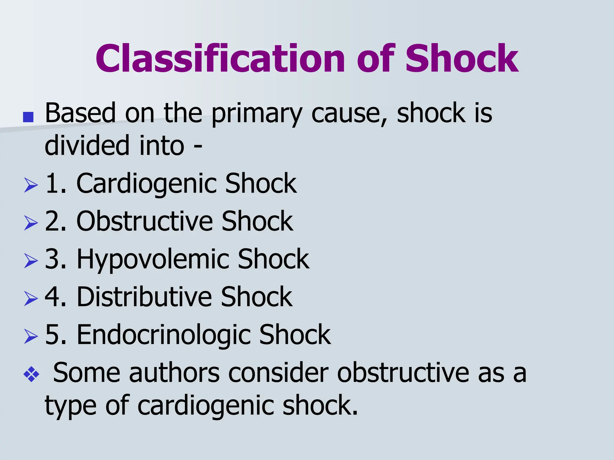
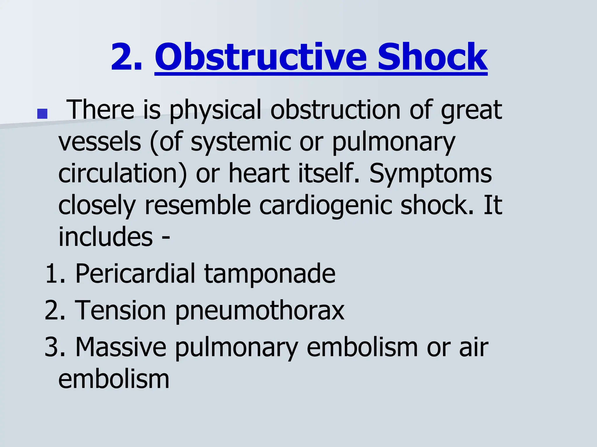
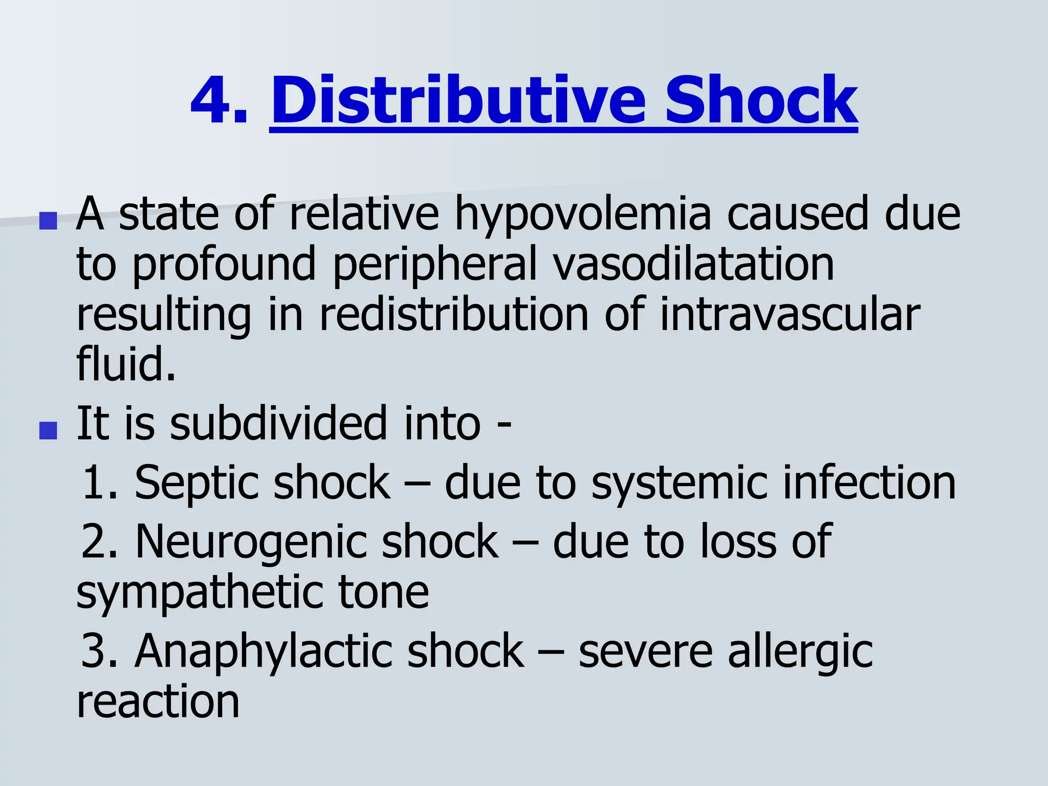
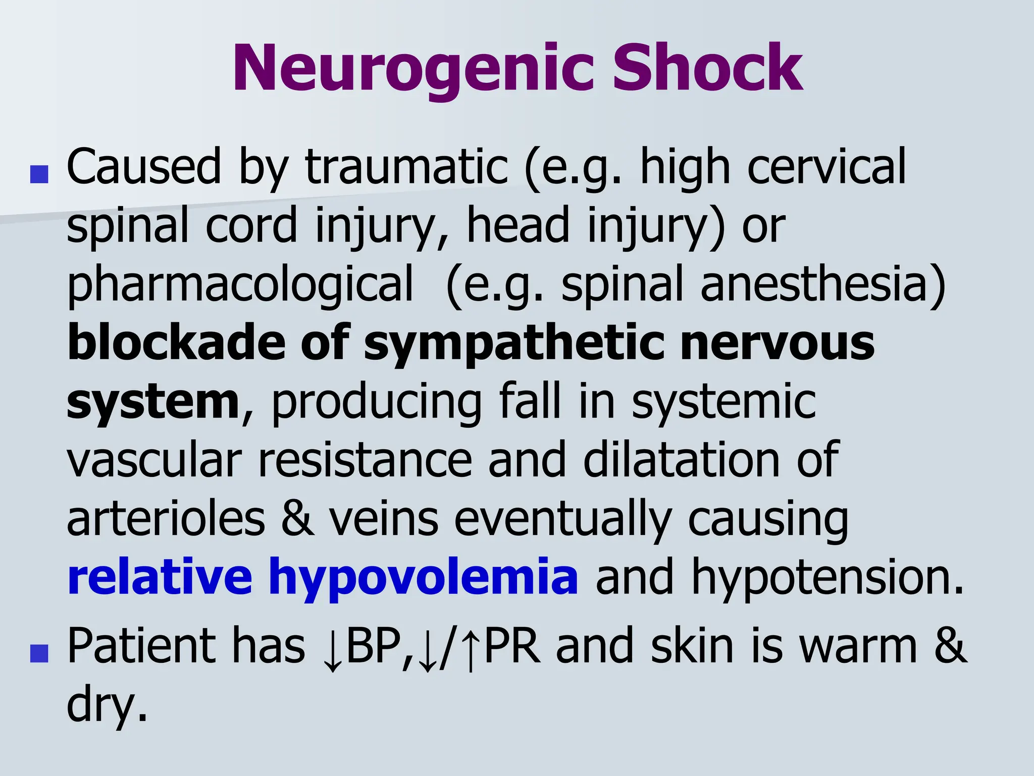
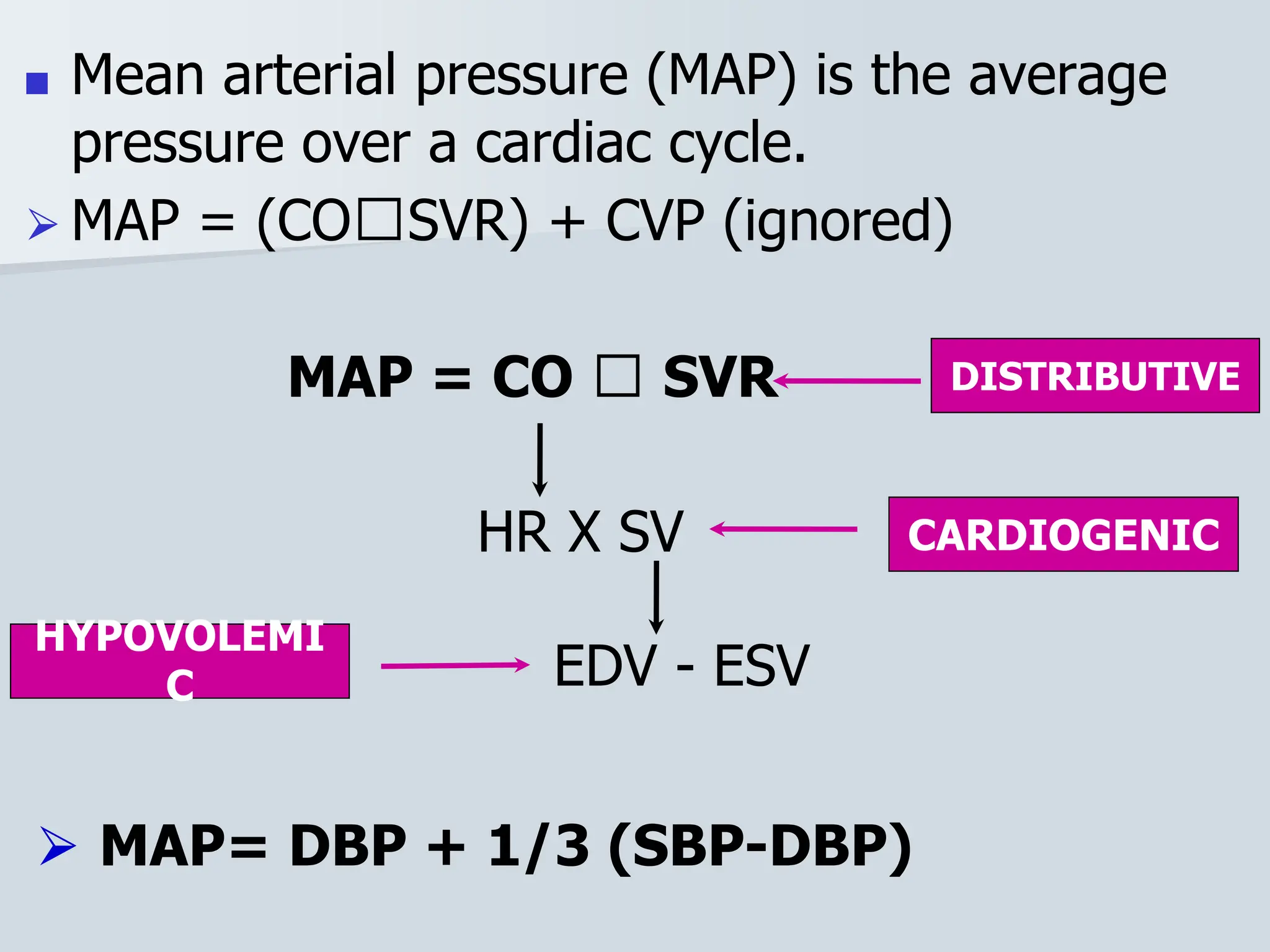
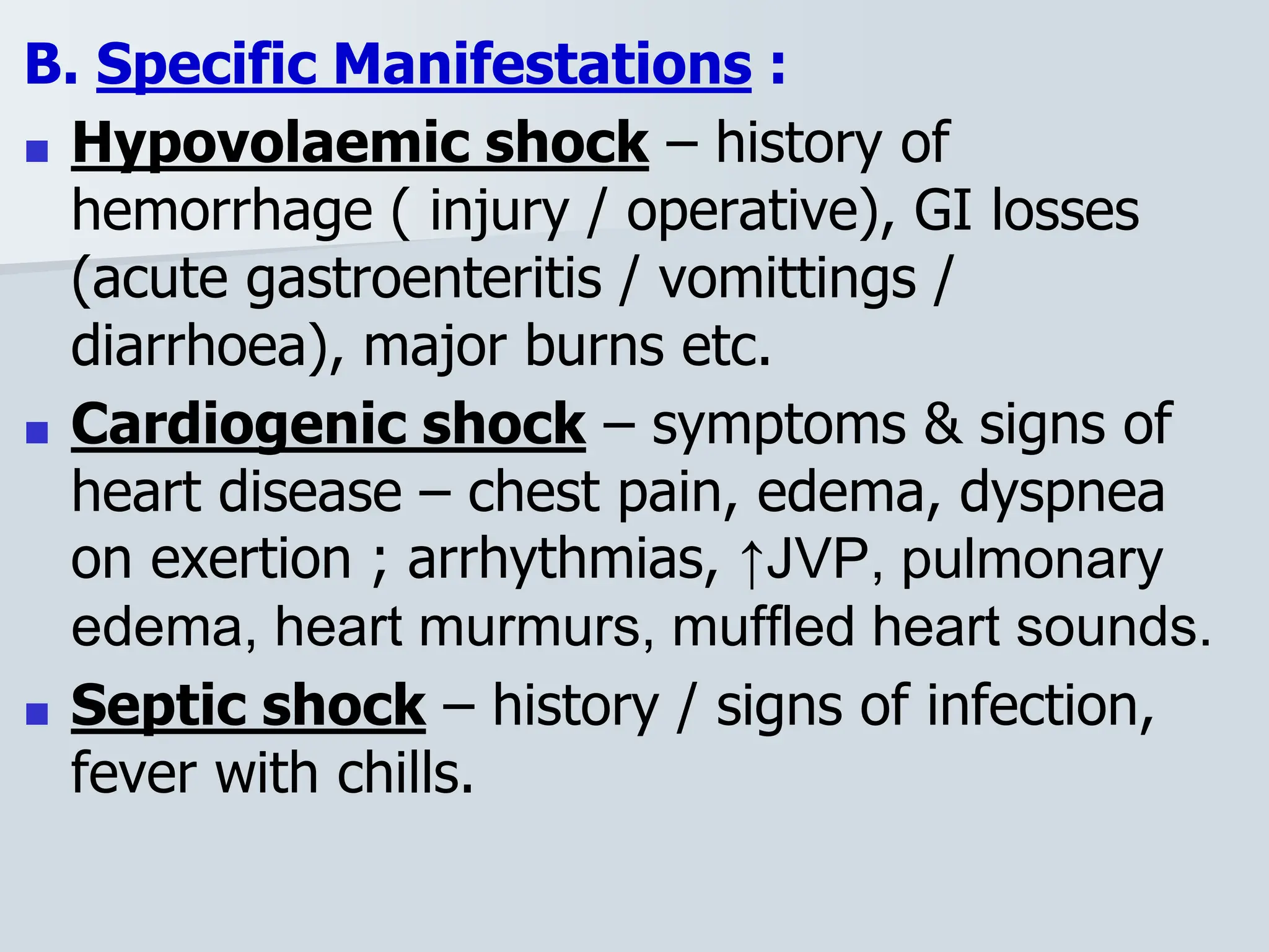
2. Objectives
■ Definition
■ To understand cardiovascular
physiology
■ Types of shock, their
pathophysiology and manifestations
■ Management of shock
3. Introduction
■ Definition :- A state of failure of
circulatory system to maintain adequate
cellular perfusion, resulting in widespread
reduction in delivery of oxygen and other
nutrients to tissues.
■ Diagnosed with presence of hypotension
(SBP < 90mmHg)
■ Uncorrected it eventually results in death of
cells, tissues, organs and ultimately the
patient.
4. Circulatory Homeostasis
■ Depends on :-
A) Preload – means end-diastolic volume
of heart.
- Greater the preload, greater is the
output of heart
- Preload depends on – changes in
position, intrathoracic pressure,
intrapericardial pressure and the blood
volume.
5. B) Ventricular contraction – greater the
force of contraction, greater is the stroke
volume.
- Myocardial injury, valve dysfunction and
cardiac hypertrophy lead to impaired
contractility.
C) Afterload – describes the pressure that
the heart has to overcome, during every
beat, to send blood into the aorta.
- Greater the afterload (e.g. HT), lesser
the cardiac output.
■ SV α Preload, Contractility, 1/Afterload
6. Classification of Shock
■ Based on the primary cause, shock is
divided into -
⮚ 1. Cardiogenic Shock
⮚ 2. Obstructive Shock
⮚ 3. Hypovolemic Shock
⮚ 4. Distributive Shock
⮚ 5. Endocrinologic Shock
❖ Some authors consider obstructive as a
type of cardiogenic shock.
7. 1. Cardiogenic Shock
■ Results from failure of heart to pump blood to
tissues -
a. Myocardial problems – Acute MI, CHF,
cardiomyopathies, myocarditis, drug induced &
blunt trauma to heart.
b. Rhythm problems – severe brady or
tachyarrhythmias.
c. Mechanical causes – include advanced
valvular heart diseases like stenosis,
regurgitation etc.
8. 2. Obstructive Shock
■ There is physical obstruction of great
vessels (of systemic or pulmonary
circulation) or heart itself. Symptoms
closely resemble cardiogenic shock. It
includes -
1. Pericardial tamponade
2. Tension pneumothorax
3. Massive pulmonary embolism or air
embolism
9.
10. 3. Hypovolemic Shock
■ Most common type of shock.
■ It results from loss of circulating blood
volume (usually >20%) & occurs due to -
1. Hemorrhage (also called Hemorrhagic
shock) – trauma, medical causes
2. Non-hemorrhagic causes – burns, vomiting,
diarrhea, urinary losses or pancreatitis etc.
11. 4. Distributive Shock
■ A state of relative hypovolemia caused due
to profound peripheral vasodilatation
resulting in redistribution of intravascular
fluid.
■ It is subdivided into -
1. Septic shock – due to systemic infection
2. Neurogenic shock – due to loss of
sympathetic tone
3. Anaphylactic shock – severe allergic
reaction
12. 5. Endocrinologic shock
■ Occurs due to severe hormonal imbalance
and may present as a combination of
features of cardiogenic, hypovolemic and
distributive shock. Causes include –
1. Severe adrenal insufficiency
2. Hypo or hyperthyroidism.
13. Neurogenic Shock
■ Caused by traumatic (e.g. high cervical
spinal cord injury, head injury) or
pharmacological (e.g. spinal anesthesia)
blockade of sympathetic nervous
system, producing fall in systemic
vascular resistance and dilatation of
arterioles & veins eventually causing
relative hypovolemia and hypotension.
■ Patient has ↓BP,↓/↑PR and skin is warm &
dry.
14.
15. Anaphylactic Shock
■ Anaphylaxis is a severe, life threatening,
generalized or systemic hypersensitivity
reaction mediated by IgE antibody
against an antigen e.g. insect venom, drugs
and certain foods.
■ There is activation and release of
inflammatory mediators such as cytokines,
histamine, prostaglandins etc. These lead
to vasodilatation, increased capillary
permeability, bronchospasm, airway edema
and circulatory collapse.
19. ■ Mean arterial pressure (MAP) is the average
pressure over a cardiac cycle.
⮚ MAP = (CO🞪SVR) + CVP (ignored)
MAP = CO 🞪 SVR
HR X SV
EDV - ESV
HYPOVOLEMI
C
CARDIOGENIC
DISTRIBUTIVE
⮚ MAP= DBP + 1/3 (SBP-DBP)
21. VASOVAGAL SHOCK
• A patient may faint due to emotional
distress, fear, or pain during a dental
procedure.
• Signs and Symptoms:
• Pallor
• sweating,
• nausea,
• bradycardia
• loss of consciousness.
22. INSULIN SHOCK
■ Insulin shock is a form of severe
hypoglycemia, when blood sugar falls to
dangerously low levels. If left untreated
for too long, it can lead to loss of
consciousness and even death.
■ Insulin shock can occur in people with
type 1 or type 2 diabetes if they are using
certain medications.
23. Stages of Shock
■ Typically shock passes through 4 stages :-
1. Initial : Hypoperfusion causes hypoxia which
causes anaerobic metabolism leading to metabolic
acidosis.
2. Compensatory : The body employs several
neural, hormonal & biochemical mechanisms -
■ Tachypnea – to correct acidosis.
■ Release of catecholamines – causing
vasoconstriction and tachycardia.
■ Activation of Renin Angiotensin system & ADH
release – to retain Na and water.
24. 3. Progressive (de-compensated )shock
: The mechanisms begin to fail –
■ Metabolic acidosis worsens, causing cardiac
depression & stasis of blood in the capillaries. The
pressure within the capillaries increases. This,
combined with membrane dysfunction and
histamine release, leads to fluid loss into the
interstitial spaces.
■ Prolonged vasoconstriction & DIC cause
compromise of vital organs.
■ Bacterial translocation occurs in gut due to
ischemia, can cause systemic infection.
REFERENCE : Shock, a clinical syndrome: an update. Part 2. The stages of shock - PubMed (nih.gov)
25. 4. Refractory : Vital organs fail
and the shock can no longer be
reversed. Death is imminent.
Also called as Irreversible de-
compensated shock
27. Clinical Manifestations
A. Common to all forms of Shock:
1. Hypotension – mean arterial pressure
<60mm Hg, or systolic BP <90mm Hg
in previously normotensive patient, or
fall >40mmHg from baseline BP of an
individual.
2. Tachycardia, tachypnea
3. Oliguria, anuria
4. Clouded sensorium
5. Cool mottled extremities (warm pink
in neurogenic & early septic shock)
28. B. Specific Manifestations :
■ Hypovolaemic shock – history of
hemorrhage ( injury / operative), GI losses
(acute gastroenteritis / vomittings /
diarrhoea), major burns etc.
■ Cardiogenic shock – symptoms & signs of
heart disease – chest pain, edema, dyspnea
on exertion ; arrhythmias, ↑JVP, pulmonary
edema, heart murmurs, muffled heart sounds.
■ Septic shock – history / signs of infection,
fever with chills.
29. ■ Anaphylaxis is likely when all 3 criteria are
met :-
1. Sudden onset & rapid progression of
symptoms.
2. Life threatening Airway &/or Breathing
&/or Circulation problems.
3. Skin &/or mucosal problems – flushing,
urticaria, angioedema.
■ The diagnosis is supported by history of
exposure to a known allergen for the patient.
30. DIAGNOSIS OF SHOCK
■ The diagnosis of shock is based on identifying a
mechanism for shock, the patient’s symptoms,
and the patient’s vital signs. A significant drop in
blood pressure is usually a late finding, Stage
III, and don’t delay care waiting for blood
pressure to drop below normal. Extremely low
urine output, measured at the hospital or skilled
nursing facility, is a possible indicator of shock
as the patient’s body is working to maintain
adequate fluid volume. Blood test can diagnosis
infection causing sepsis.
31. Investigations
■ Diagnosis of shock is clinical.
Investigations are done to know the
cause, extent of damage, for
assessment of patient’s condition &
management.
1. Hematological : Hb, TLC, DLC,
platelet count, blood grouping, BT, CT,
PT.
2. RBS.
3. Urine R/M.
32. 4. Kidney function tests : blood urea,
serum creatinine, serum sodium &
potassium.
5. Liver function tests : serum bilirubin,
SGOT, SGPT etc.
6. ECG , Echocardiography
7. Blood C/S, Pus C/S : in septic shock
8. X-ray chest : for pneumothorax,
Pericardial tamponade, ARDS.
9. Arterial blood gases
10. X-ray, USG, CT
33. Management
■ Prognosis depends on duration and
degree of shock, so prompt &
aggressive management is essential.
■ Management is initiated immediately,
side-by-side history is taken, patient
examined and investigations sent, so
as to make proper diagnosis.
55. Septic Shock
■ Restore “ ORDER ”.
- O – Oxygenation
- R – Resuscitation
- D – Drugs
- E – Evaluate response to therapy
- R – Removal of infection
56. Septic Shock Management
■ Early phase treatment: ionotropic
agents
■ Early onset neonatal sepsis: ampicillin
and gentamicin, cefotaxime
■ First line of drugs: Dopamine
■ Treatment if renal failure: gentamicin
57. Neurogenic Shock
■ Immobilization
■ IV Fluids
■ If Blood Pressure is low:
vasopressors like – norepinephrine,
epinephrine, dopamine
■ If slower heart rhythm: atropine
■ High dose steroids
■ Prevent blood pooling
■ Elevation of head
58. Cardiogenic Shock
A. Supplemental oxygen
B. Inotropic agents : Dopamine,
Dobutamine
C. Mechanical support : Intra-aortic
balloon pulsation device
D. Extra-cardiac cause : treat it.
59. Anaphylactic Shock
A. Discontinue the cause / agent.
B. Oxygenation with 100% oxygen. Intubate
if necessary.
C. Intra-venous fluids to manage
hypotension.
D. Adrenaline is the drug of choice. 0.3-0.5
ml of 1:1000 adrenaline IM repeated every
5-10 mins. according to patient response. It
reverses vasodilatation, dilates bronchial
airways and increases cAMP in leucocytes &
mast cells thus decreasing histamine release.
60. ■ E. Secondary measures :
1. Antihistaminics – Inj. Avil IV.
2. Steroids – Inj. Hydrocortisone IV
3. Bronchodilators – Salbutamol (inhaled /
IV), or Aminophylline IV
62. INSULIN SHOCK MANAGEMENT
■ If the individual is conscious, treatment is with rapidly
absorbed sugars. It is recommended to follow the “15-
15 rule,” where an individual consumes 15 grams of
sugars, then blood glucose is rechecked with a
fingerstick in 15 minutes.
■ If blood glucose is still less than 70 mg/dL, repeat the
same steps until the blood glucose normalizes.
■ The following contain 15 grams of sugar:
• Juice, 4 ounces (half a standard drinking glass or a
quarter of a pint glass)
• Regular soda, 5-6 ounces (about half a regular can)
• Granulated sugar, about 4 teaspoons
• Glucose tablets, 3-4 tabs (follow instructions)
• Glucose gel, 1 tube (follow instructions)
REFERENCE: Insulin Shock: Causes, Warning Signs, Treatment & Prevention (healthcentral.com)
63. ■ Avoid chocolate or other fat-
containing foods, since this slows
the absorption of sugars and
therefore delays the normalization of
blood sugar.
■ If the individual is unconscious or
unable to consume sugars orally,
treatment is through the
administration of glucagon.
Glucagon is a hormone that
increases blood glucose levels.
■ It comes in two forms: a nasal spray
(brand name Baqsimi) or an
intramuscular (IM) injection. The IM
injection comes in a prefilled auto-
injector (like an EpiPen) or as a
powder with a separate fluid that the
64. Patient Monitoring
1. Pulse – rate and volume
2. Blood pressure
3. Respiratory rate, SPO2
4. Central venous pressure (CVP)
5. Pulmonary capillary wedge pressure in
severe shock & doubtful diagnosis.
6. Urine output of ≥ 1 ml/Kg/hr.
7. Serial blood gases and serum lactate &
electrolytes
65. MANAGEMENT OF VASOVAGAL
SHOCK
■ Place the patient in a supine
position
■ Ensure a patent airway
■ monitor vital signs
■ Reassure the patient and provide
appropriate care.
71. Summary
■ A state of cardiovascular collapse with failure to
maintain adequate systemic perfusion and
diagnosed with presence of SBP<90mmHg.
■ Types - cardiogenic, obstructive, hypovolemic,
distributive and endocrinologic shock.
■ As patient’s condition worsens each type
contributes irrespective of initial cause.
■ Multiple organ involvement & failure.
■ Diagnosis is clinical
■ Aggressive management is the key. Prognosis
depends on duration and severity of shock.
72. REFERENCES & SUGGESTED READING
■ Bailey & Love’s Short Practice of Surgery
■ Harrison’s Principles of Internal Medicine
■ Schwartz Textbook of surgery