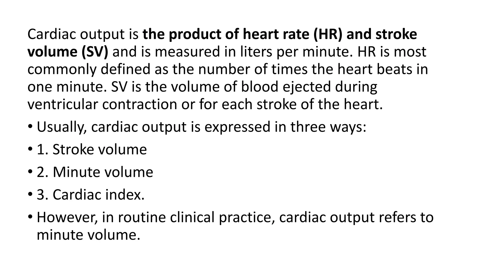
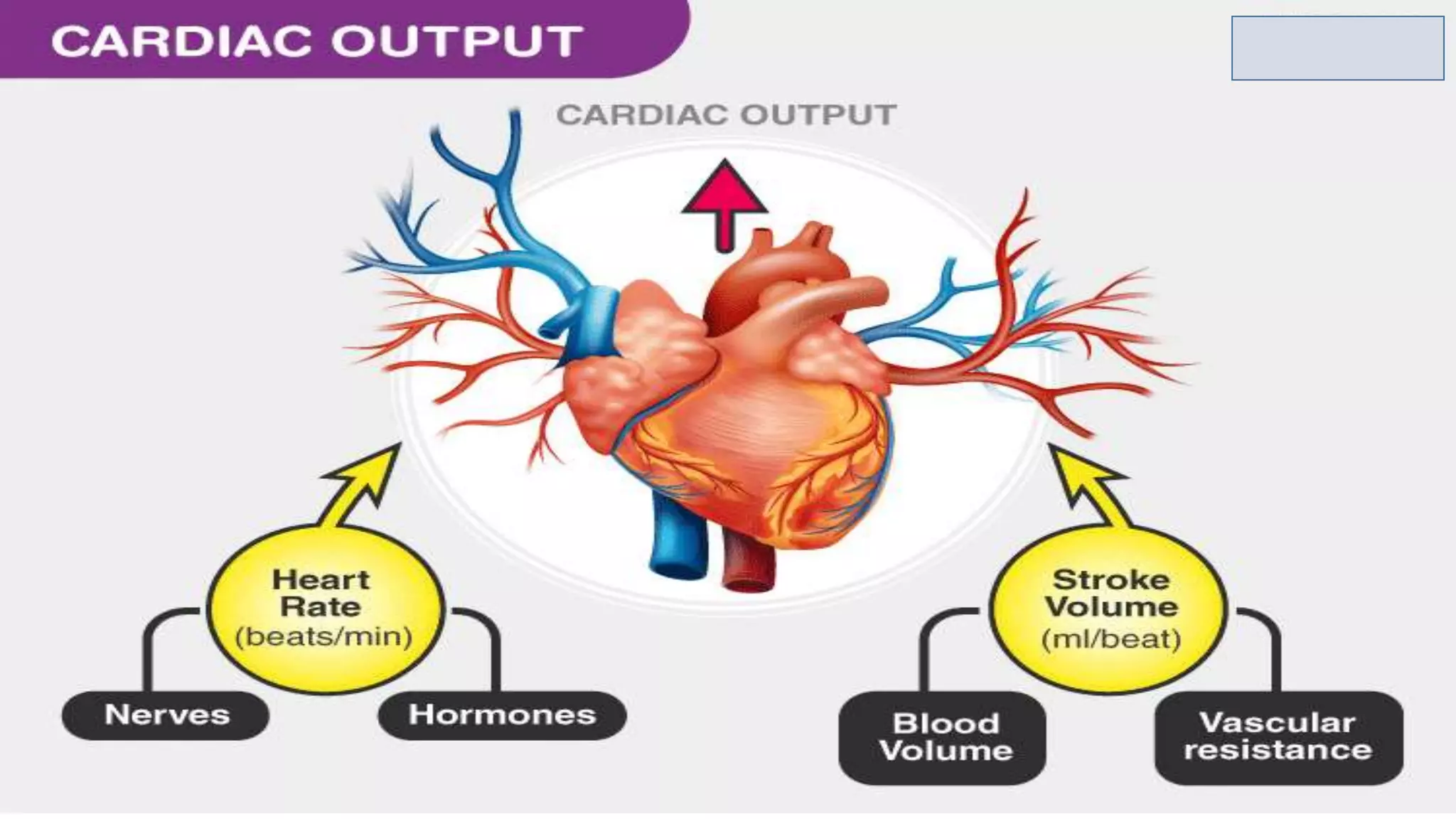
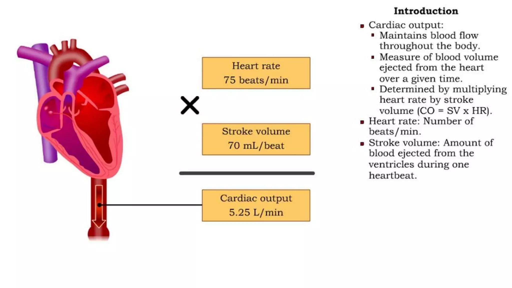
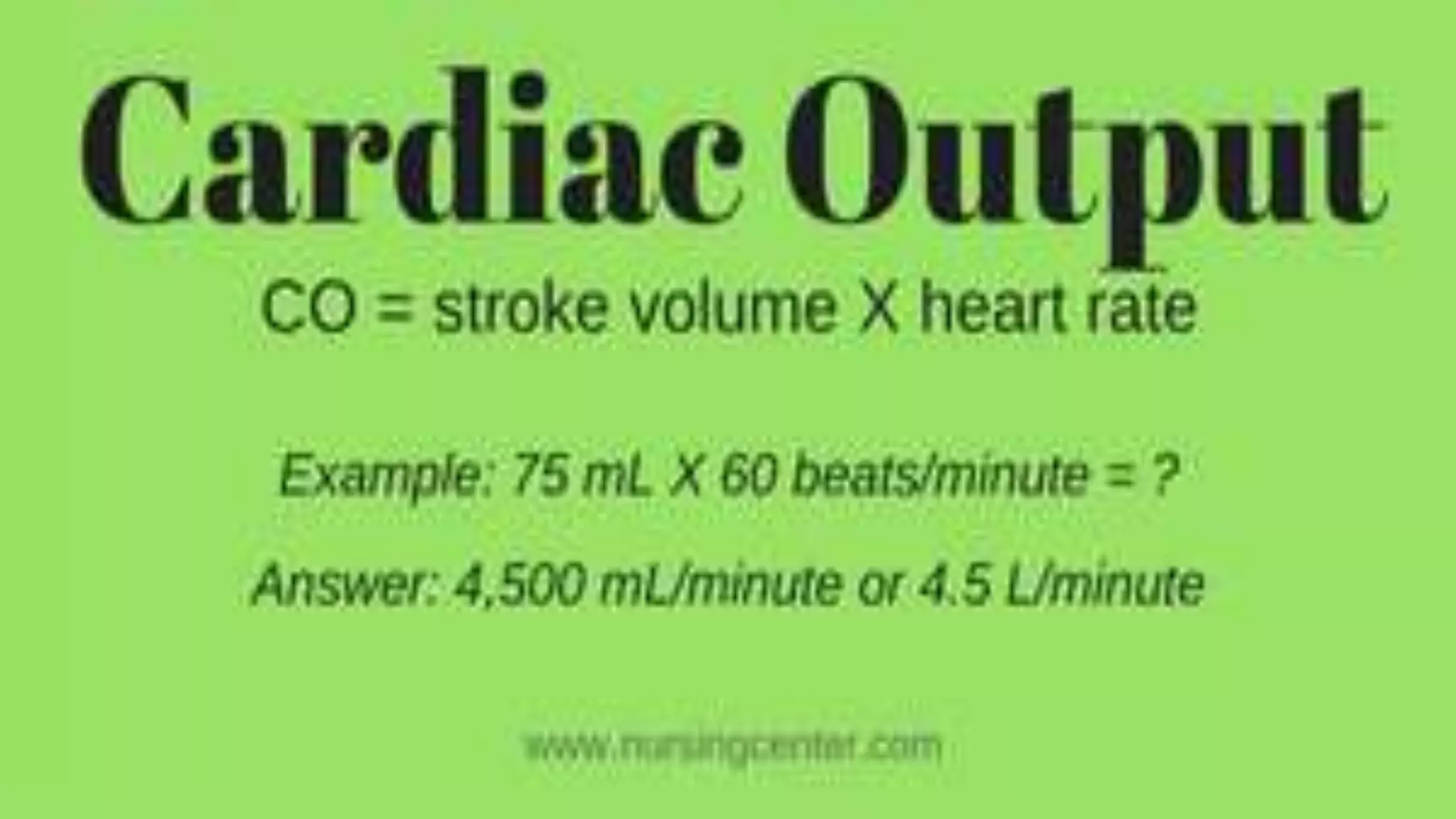
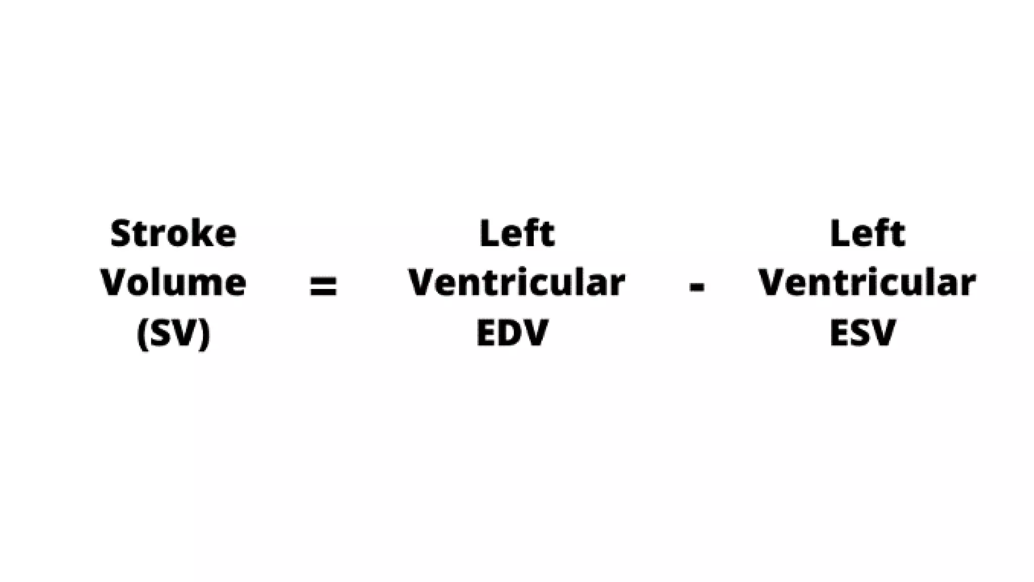
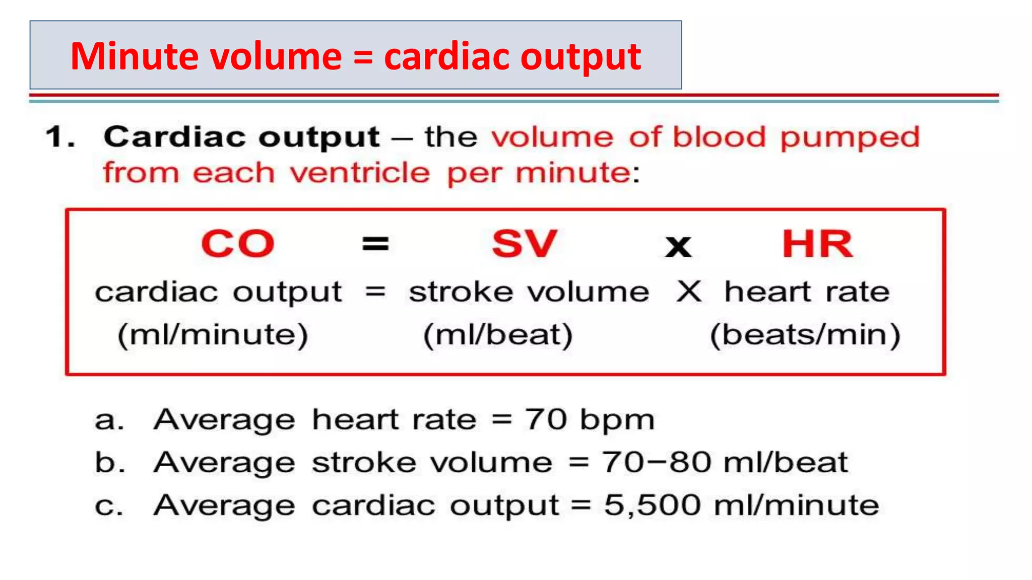
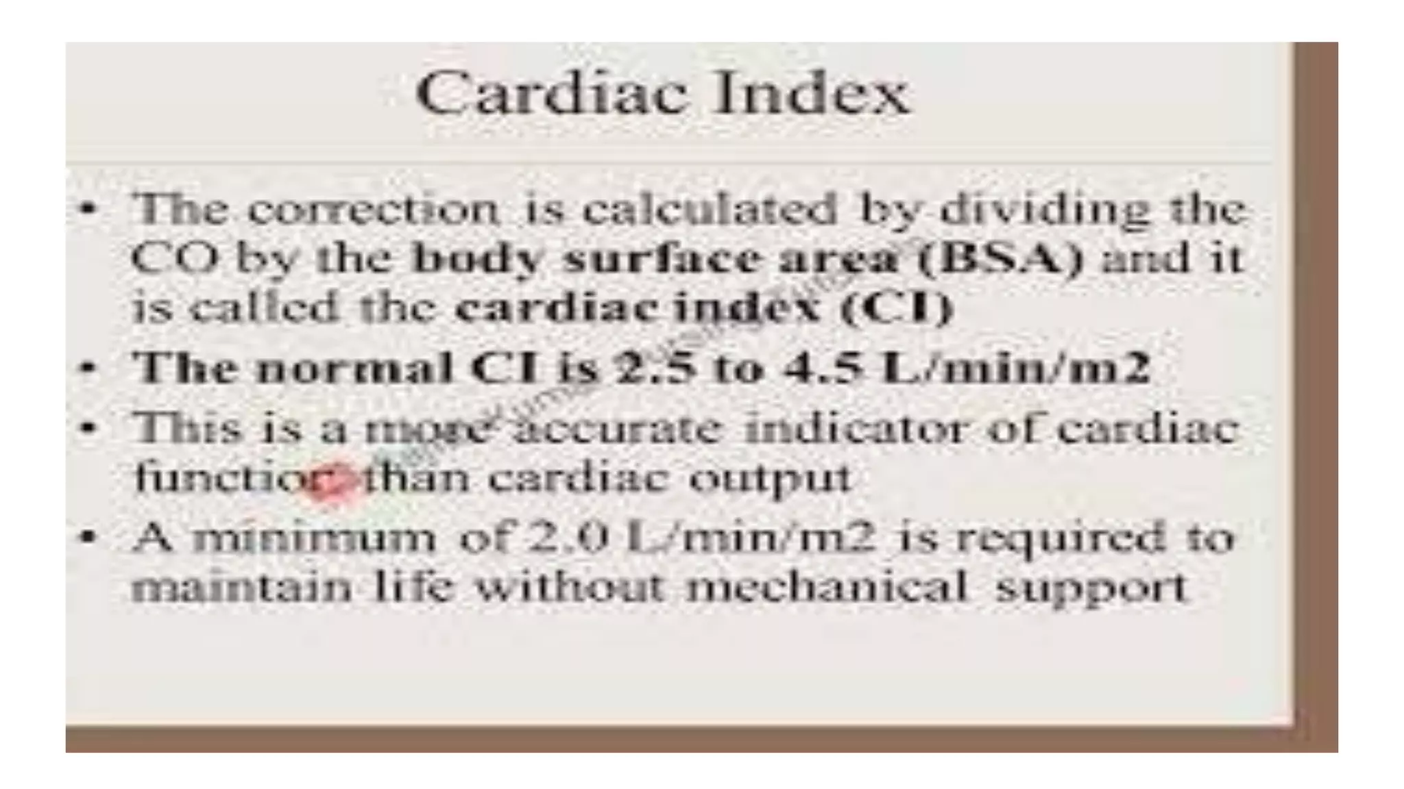
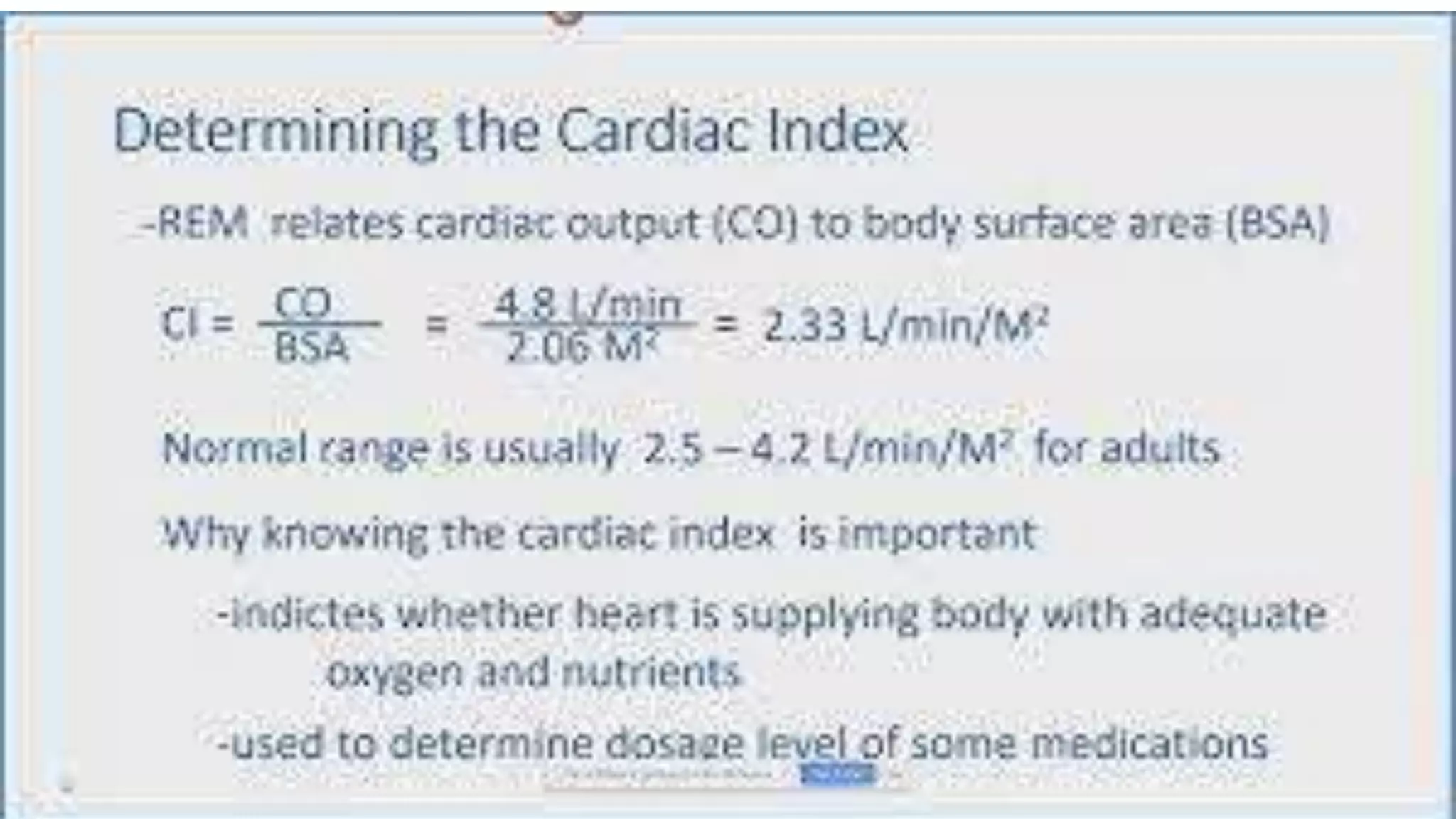
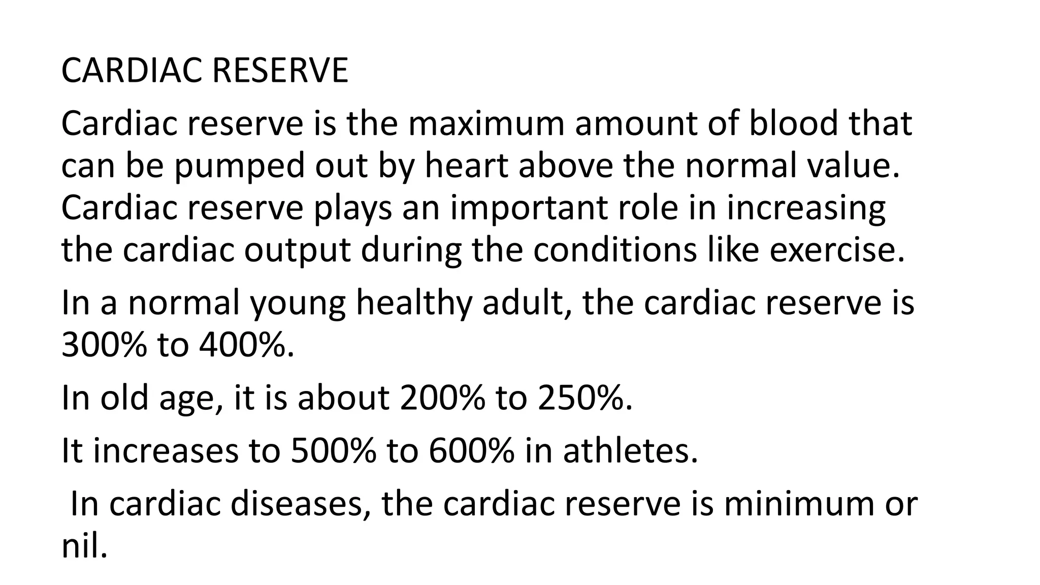
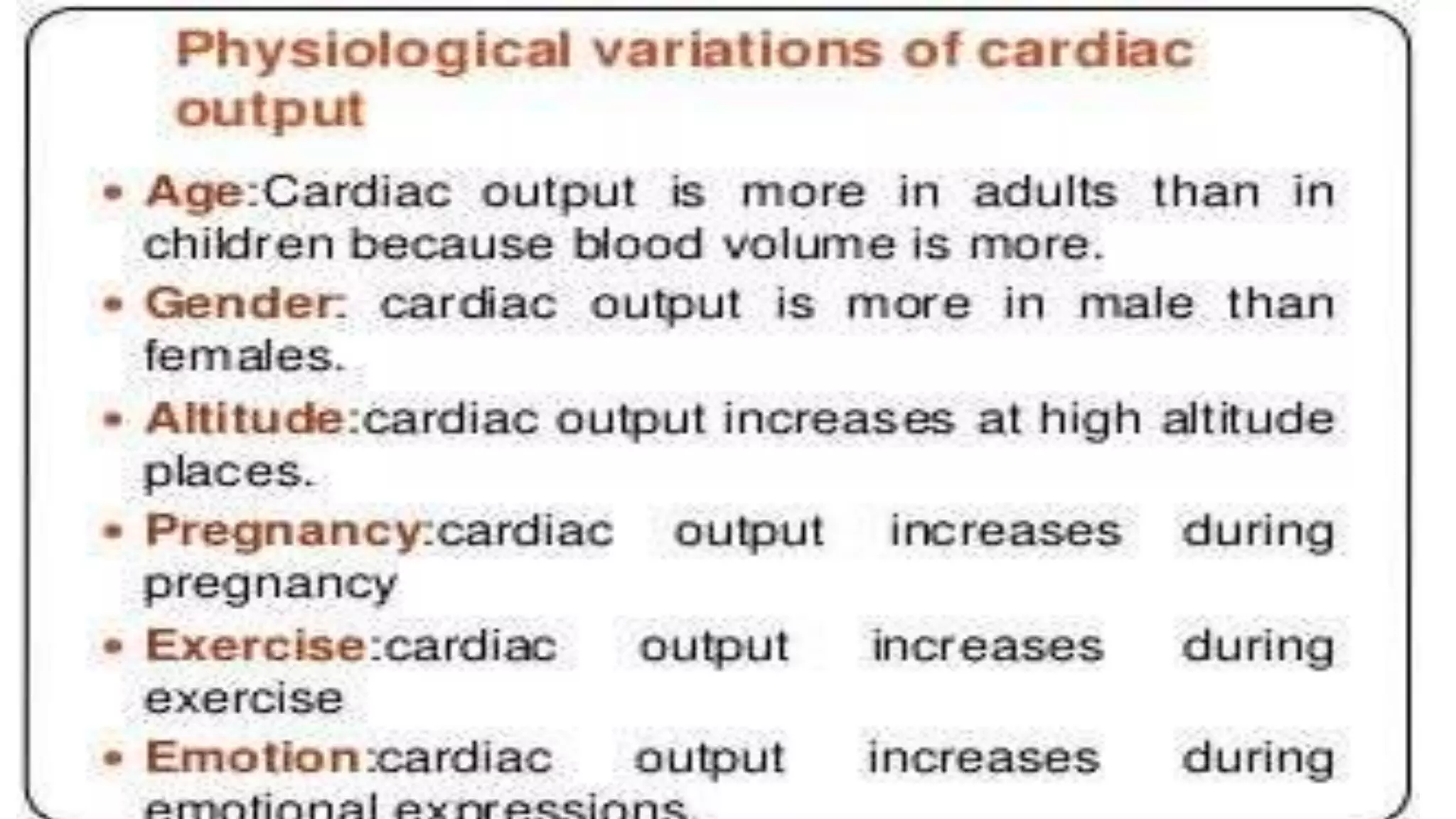
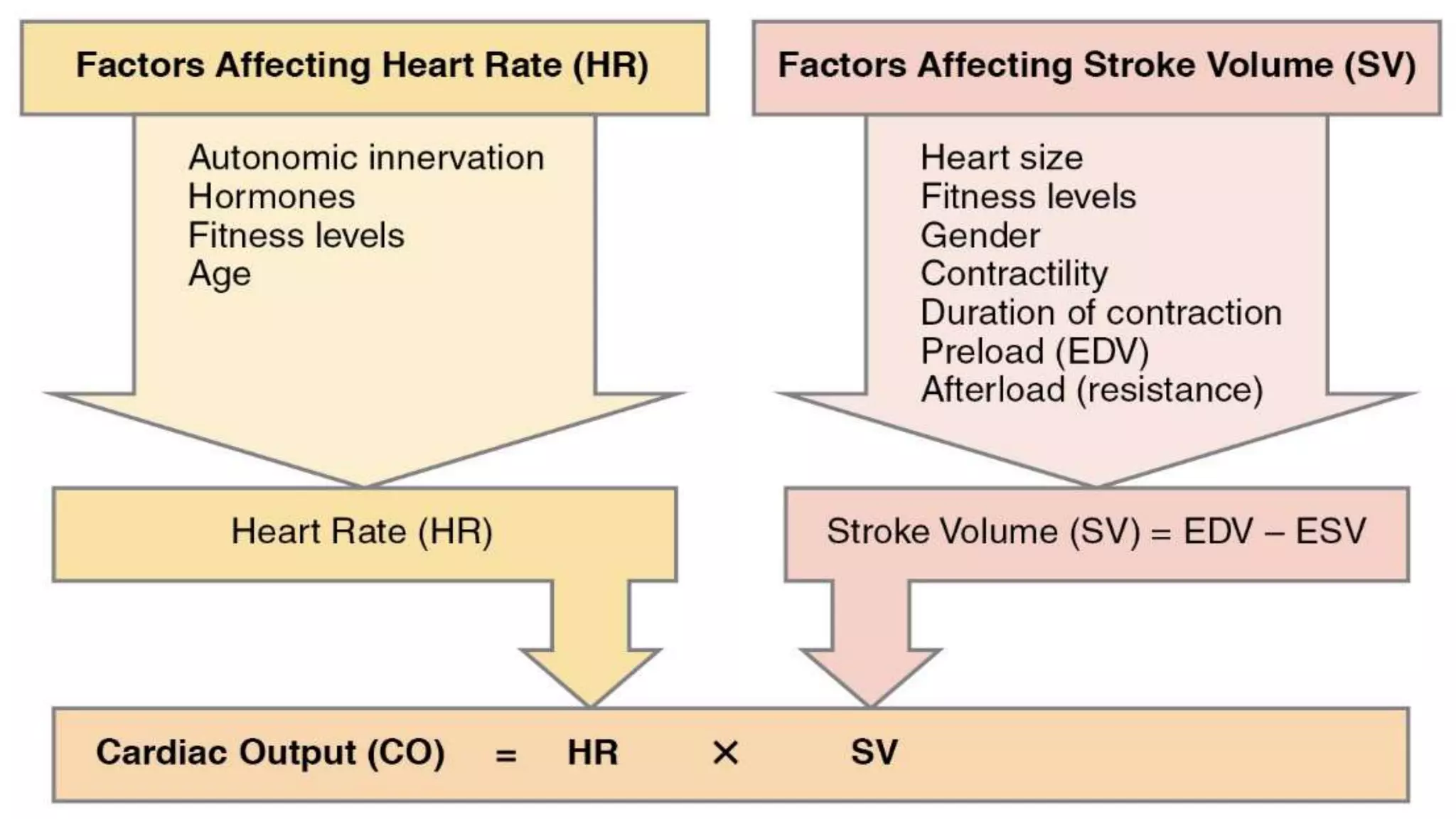
Cardiac output is the volume of blood pumped by the heart per minute and is equal to heart rate multiplied by stroke volume. Cardiac output can be expressed in three ways: stroke volume, minute volume, or cardiac index. Minute volume and cardiac output refer to the same thing in clinical practice. Stroke volume is the volume of blood pumped out of the left ventricle per contraction. Ejection fraction is the percentage of blood ejected from the ventricles during each contraction, normally 60-65%. Cardiac reserve is the maximum increase in cardiac output above normal levels, important for conditions like exercise.
“Cardiac output refers to the volume of blood pumped out per ventricle per minute.”
Cardiac output is the function of heart rate and stroke volume.
STROKE VOLUME:
The amount of blood pumped by the left ventricle in one compression is called the stroke volume.
Heart Rate
The cardiac output increases with the increase in heart rate.
Right Atrium of human heart
This PPT help to understand the external and internal structures of right atrium.
sulcus terminalis on external surface of rt atrium,
crista terminalis on internal side of rt. atrium,
interior is divided into rough anterior part and smooth posterior part ( sinus venarum)
superior and inferior venae cavae drains deoxygenated blood into rt. atrim
there is Eustachian valve to guard the opening of IVC and Thebesian valve to guard the opening of coronary sinus
septal wall presents fossa ovalis with its border limbus fossa ovalis
This presentation is an overview of the description of the 4 stages of the cardiac cycle (atrial diastole, atrial systole, ventricular systole, ventricular diastole) as well as explaining the mechanism of the cardiac cycle.
Cardiac output (The Guyton and Hall Physiology)Maryam Fida
The volume of blood pumped by each ventricle per minute is called cardiac output
Cardiac output = Stroke Volume X Heart Rate
Normal value = 5 Liters /Minute
Cardiac output = Stroke Volume X Heart Rate
The factors which regulate stroke volume and Heart rate are basically regulating Cardiac output
Volume of blood ejected by each ventricle in single systole; Normal Value = 70 ml/beat
Stroke Volume = End diastolic Volume – End Systolic Volume
So stroke volume is mainly controlled by
EDV
ESV
VENOUS RETURN: What ever blood volume returns to the heart, same is pumped forward through the Frank’s Starlings Law. According to this law 13- 15 liters of blood volume can be pumped out without cardiac stimulation.
DURATION OF DIASTOLE OR FILLING TIME: ventricular filling occurs during diastole, so there must be adequate ventricular filling time.
DISTENSIBILITY OF THE VENTRICLES: Normally ventricles are distensible to accommodate adequate blood volume. Infarction decreases the distensibility which decreases the EDV.
ATRIAL CONTRACTION: There must be adequate atrial contraction to have adequate EDV. If atrial function is not adequate then EDV will decrease.
E.S.V is basically CONTROLLED BY MYOCARDIAL CONTRACTION
FORCE OF MYOCARDIAL CONTRACTION: It depends upon the initial length of muscle fibers according to frank’s starlings law.
PRELOAD: The effect of EDV on initial length is called preload. So EDV also effects the ESV.
AFTER LOAD: Force of contraction is also dependant upon the resistance against which the ventricles have to pump
CONDITION OF THE MYOCARDIUM : It also effects the force of contraction.
AUTONOMIC NERVES : Sympathetic stimulation increases and parasympathetic stimulation decreases force of contraction
HORMONES: Catecholamines, thyroxine, glucagon, digitalis, calcium, increased temp, caffeine, theophyline increase the force.
Force decreases by hypoxia, acidosis, barniturates, procainamide and quinidine decrease the force of contraction.
HEART RATE
REGULATION OF HEART RATE
VASOMOTOR CENTER – CARDIAC CENTER
MOTOR (EFFERENT) NERVE FIBERS TO HEART
FACTORS AFFECTING VASOMOTOR CENTER
for all medical & health care students
overview of heart and its disease
The heart is a powerful muscle that pumps
blood throughout the body by means of a
coordinated contraction.
The contraction is generated by an
electrical activation, which is spread by a
wave of bioelectricity that propagates in a
coordinated manner throughout the
heart.
It is located in thoracic cavity,
posterior to the sternum ,superior
to the diaphragm between the
lungs.
A human heart beats an average of
100,000 times per day. During that
time, it pumps more than 4,300
gallons of blood throughout
the entire body.
EPICARDIUM:
It is the outer layer of the wall of the heart which is composed of
connective tissue covered by epithelium. It is also known as
visceral pericardium.
2) MYOCARDIUM:
It is the muscular middle layer of the wall of the heart. It is
composed of spontaneously contracting cardiac muscle fibers
which allow the heart to contract. It stimulates heart contraction
to pump blood from the ventricles and relaxes the heart to allow
the arteries to receive blood.
ENDOCARDIUM:
It is the inner layer of the heart which consist of epithelial
tissue and connective tissue.
Hey, these are the slides me n my friends made... Use them if u want to... for viewing the videos used click on the links given ahead.
http://www.youtube.com/watch?v=jzOti_MtmBk
http://www.youtube.com/watch?v=N9MARqmqSf4
http://www.youtube.com/watch?v=yokcKhqq48c
http://www.youtube.com/watch?v=rJZVFRJmc9M
Presentation on:
Describe the gross anatomy of heart under following: external features, surface markings and areas of auscultation.
List the components of examination of Cardiovascular System.
Define cardiac cycle and its phases.
Define venous return, stroke volume, cardiac output, preload, afterload and ejection fraction with their normal range.
Cardiac output as you know is made up of heart rate and stroke volume. At rest, these are relatively constant however with exercise the heart beats faster, and more blood is pumped out with each beat. These factors both contribute to a rise in BP, as would any other factor that caused the heart to speed up
Anti ulcer drugs and their Advance pharmacology ||
Anti-ulcer drugs are medications used to prevent and treat ulcers in the stomach and upper part of the small intestine (duodenal ulcers). These ulcers are often caused by an imbalance between stomach acid and the mucosal lining, which protects the stomach lining.
||Scope: Overview of various classes of anti-ulcer drugs, their mechanisms of action, indications, side effects, and clinical considerations.
Flu Vaccine Alert in Bangalore Karnatakaaddon Scans
As flu season approaches, health officials in Bangalore, Karnataka, are urging residents to get their flu vaccinations. The seasonal flu, while common, can lead to severe health complications, particularly for vulnerable populations such as young children, the elderly, and those with underlying health conditions.
Dr. Vidisha Kumari, a leading epidemiologist in Bangalore, emphasizes the importance of getting vaccinated. "The flu vaccine is our best defense against the influenza virus. It not only protects individuals but also helps prevent the spread of the virus in our communities," he says.
This year, the flu season is expected to coincide with a potential increase in other respiratory illnesses. The Karnataka Health Department has launched an awareness campaign highlighting the significance of flu vaccinations. They have set up multiple vaccination centers across Bangalore, making it convenient for residents to receive their shots.
To encourage widespread vaccination, the government is also collaborating with local schools, workplaces, and community centers to facilitate vaccination drives. Special attention is being given to ensuring that the vaccine is accessible to all, including marginalized communities who may have limited access to healthcare.
Residents are reminded that the flu vaccine is safe and effective. Common side effects are mild and may include soreness at the injection site, mild fever, or muscle aches. These side effects are generally short-lived and far less severe than the flu itself.
Healthcare providers are also stressing the importance of continuing COVID-19 precautions. Wearing masks, practicing good hand hygiene, and maintaining social distancing are still crucial, especially in crowded places.
Protect yourself and your loved ones by getting vaccinated. Together, we can help keep Bangalore healthy and safe this flu season. For more information on vaccination centers and schedules, residents can visit the Karnataka Health Department’s official website or follow their social media pages.
Stay informed, stay safe, and get your flu shot today!
Title: Sense of Smell
Presenter: Dr. Faiza, Assistant Professor of Physiology
Qualifications:
MBBS (Best Graduate, AIMC Lahore)
FCPS Physiology
ICMT, CHPE, DHPE (STMU)
MPH (GC University, Faisalabad)
MBA (Virtual University of Pakistan)
Learning Objectives:
Describe the primary categories of smells and the concept of odor blindness.
Explain the structure and location of the olfactory membrane and mucosa, including the types and roles of cells involved in olfaction.
Describe the pathway and mechanisms of olfactory signal transmission from the olfactory receptors to the brain.
Illustrate the biochemical cascade triggered by odorant binding to olfactory receptors, including the role of G-proteins and second messengers in generating an action potential.
Identify different types of olfactory disorders such as anosmia, hyposmia, hyperosmia, and dysosmia, including their potential causes.
Key Topics:
Olfactory Genes:
3% of the human genome accounts for olfactory genes.
400 genes for odorant receptors.
Olfactory Membrane:
Located in the superior part of the nasal cavity.
Medially: Folds downward along the superior septum.
Laterally: Folds over the superior turbinate and upper surface of the middle turbinate.
Total surface area: 5-10 square centimeters.
Olfactory Mucosa:
Olfactory Cells: Bipolar nerve cells derived from the CNS (100 million), with 4-25 olfactory cilia per cell.
Sustentacular Cells: Produce mucus and maintain ionic and molecular environment.
Basal Cells: Replace worn-out olfactory cells with an average lifespan of 1-2 months.
Bowman’s Gland: Secretes mucus.
Stimulation of Olfactory Cells:
Odorant dissolves in mucus and attaches to receptors on olfactory cilia.
Involves a cascade effect through G-proteins and second messengers, leading to depolarization and action potential generation in the olfactory nerve.
Quality of a Good Odorant:
Small (3-20 Carbon atoms), volatile, water-soluble, and lipid-soluble.
Facilitated by odorant-binding proteins in mucus.
Membrane Potential and Action Potential:
Resting membrane potential: -55mV.
Action potential frequency in the olfactory nerve increases with odorant strength.
Adaptation Towards the Sense of Smell:
Rapid adaptation within the first second, with further slow adaptation.
Psychological adaptation greater than receptor adaptation, involving feedback inhibition from the central nervous system.
Primary Sensations of Smell:
Camphoraceous, Musky, Floral, Pepperminty, Ethereal, Pungent, Putrid.
Odor Detection Threshold:
Examples: Hydrogen sulfide (0.0005 ppm), Methyl-mercaptan (0.002 ppm).
Some toxic substances are odorless at lethal concentrations.
Characteristics of Smell:
Odor blindness for single substances due to lack of appropriate receptor protein.
Behavioral and emotional influences of smell.
Transmission of Olfactory Signals:
From olfactory cells to glomeruli in the olfactory bulb, involving lateral inhibition.
Primitive, less old, and new olfactory systems with different path
These simplified slides by Dr. Sidra Arshad present an overview of the non-respiratory functions of the respiratory tract.
Learning objectives:
1. Enlist the non-respiratory functions of the respiratory tract
2. Briefly explain how these functions are carried out
3. Discuss the significance of dead space
4. Differentiate between minute ventilation and alveolar ventilation
5. Describe the cough and sneeze reflexes
Study Resources:
1. Chapter 39, Guyton and Hall Textbook of Medical Physiology, 14th edition
2. Chapter 34, Ganong’s Review of Medical Physiology, 26th edition
3. Chapter 17, Human Physiology by Lauralee Sherwood, 9th edition
4. Non-respiratory functions of the lungs https://academic.oup.com/bjaed/article/13/3/98/278874
Explore natural remedies for syphilis treatment in Singapore. Discover alternative therapies, herbal remedies, and lifestyle changes that may complement conventional treatments. Learn about holistic approaches to managing syphilis symptoms and supporting overall health.
These lecture slides, by Dr Sidra Arshad, offer a quick overview of physiological basis of a normal electrocardiogram.
Learning objectives:
1. Define an electrocardiogram (ECG) and electrocardiography
2. Describe how dipoles generated by the heart produce the waveforms of the ECG
3. Describe the components of a normal electrocardiogram of a typical bipolar leads (limb II)
4. Differentiate between intervals and segments
5. Enlist some common indications for obtaining an ECG
Study Resources:
1. Chapter 11, Guyton and Hall Textbook of Medical Physiology, 14th edition
2. Chapter 9, Human Physiology - From Cells to Systems, Lauralee Sherwood, 9th edition
3. Chapter 29, Ganong’s Review of Medical Physiology, 26th edition
4. Electrocardiogram, StatPearls - https://www.ncbi.nlm.nih.gov/books/NBK549803/
5. ECG in Medical Practice by ABM Abdullah, 4th edition
6. ECG Basics, http://www.nataliescasebook.com/tag/e-c-g-basics
Tom Selleck Health: A Comprehensive Look at the Iconic Actor’s Wellness Journeygreendigital
Tom Selleck, an enduring figure in Hollywood. has captivated audiences for decades with his rugged charm, iconic moustache. and memorable roles in television and film. From his breakout role as Thomas Magnum in Magnum P.I. to his current portrayal of Frank Reagan in Blue Bloods. Selleck's career has spanned over 50 years. But beyond his professional achievements. fans have often been curious about Tom Selleck Health. especially as he has aged in the public eye.
Follow us on: Pinterest
Introduction
Many have been interested in Tom Selleck health. not only because of his enduring presence on screen but also because of the challenges. and lifestyle choices he has faced and made over the years. This article delves into the various aspects of Tom Selleck health. exploring his fitness regimen, diet, mental health. and the challenges he has encountered as he ages. We'll look at how he maintains his well-being. the health issues he has faced, and his approach to ageing .
Early Life and Career
Childhood and Athletic Beginnings
Tom Selleck was born on January 29, 1945, in Detroit, Michigan, and grew up in Sherman Oaks, California. From an early age, he was involved in sports, particularly basketball. which played a significant role in his physical development. His athletic pursuits continued into college. where he attended the University of Southern California (USC) on a basketball scholarship. This early involvement in sports laid a strong foundation for his physical health and disciplined lifestyle.
Transition to Acting
Selleck's transition from an athlete to an actor came with its physical demands. His first significant role in "Magnum P.I." required him to perform various stunts and maintain a fit appearance. This role, which he played from 1980 to 1988. necessitated a rigorous fitness routine to meet the show's demands. setting the stage for his long-term commitment to health and wellness.
Fitness Regimen
Workout Routine
Tom Selleck health and fitness regimen has evolved. adapting to his changing roles and age. During his "Magnum, P.I." days. Selleck's workouts were intense and focused on building and maintaining muscle mass. His routine included weightlifting, cardiovascular exercises. and specific training for the stunts he performed on the show.
Selleck adjusted his fitness routine as he aged to suit his body's needs. Today, his workouts focus on maintaining flexibility, strength, and cardiovascular health. He incorporates low-impact exercises such as swimming, walking, and light weightlifting. This balanced approach helps him stay fit without putting undue strain on his joints and muscles.
Importance of Flexibility and Mobility
In recent years, Selleck has emphasized the importance of flexibility and mobility in his fitness regimen. Understanding the natural decline in muscle mass and joint flexibility with age. he includes stretching and yoga in his routine. These practices help prevent injuries, improve posture, and maintain mobilit
HOT NEW PRODUCT! BIG SALES FAST SHIPPING NOW FROM CHINA!! EU KU DB BK substit...GL Anaacs
Contact us if you are interested:
Email / Skype : kefaya1771@gmail.com
Threema: PXHY5PDH
New BATCH Ku !!! MUCH IN DEMAND FAST SALE EVERY BATCH HAPPY GOOD EFFECT BIG BATCH !
Contact me on Threema or skype to start big business!!
Hot-sale products:
NEW HOT EUTYLONE WHITE CRYSTAL!!
5cl-adba precursor (semi finished )
5cl-adba raw materials
ADBB precursor (semi finished )
ADBB raw materials
APVP powder
5fadb/4f-adb
Jwh018 / Jwh210
Eutylone crystal
Protonitazene (hydrochloride) CAS: 119276-01-6
Flubrotizolam CAS: 57801-95-3
Metonitazene CAS: 14680-51-4
Payment terms: Western Union,MoneyGram,Bitcoin or USDT.
Deliver Time: Usually 7-15days
Shipping method: FedEx, TNT, DHL,UPS etc.Our deliveries are 100% safe, fast, reliable and discreet.
Samples will be sent for your evaluation!If you are interested in, please contact me, let's talk details.
We specializes in exporting high quality Research chemical, medical intermediate, Pharmaceutical chemicals and so on. Products are exported to USA, Canada, France, Korea, Japan,Russia, Southeast Asia and other countries.
Couples presenting to the infertility clinic- Do they really have infertility...Sujoy Dasgupta
Dr Sujoy Dasgupta presented the study on "Couples presenting to the infertility clinic- Do they really have infertility? – The unexplored stories of non-consummation" in the 13th Congress of the Asia Pacific Initiative on Reproduction (ASPIRE 2024) at Manila on 24 May, 2024.
Ozempic: Preoperative Management of Patients on GLP-1 Receptor Agonists Saeid Safari
Preoperative Management of Patients on GLP-1 Receptor Agonists like Ozempic and Semiglutide
ASA GUIDELINE
NYSORA Guideline
2 Case Reports of Gastric Ultrasound
New Directions in Targeted Therapeutic Approaches for Older Adults With Mantl...i3 Health
i3 Health is pleased to make the speaker slides from this activity available for use as a non-accredited self-study or teaching resource.
This slide deck presented by Dr. Kami Maddocks, Professor-Clinical in the Division of Hematology and
Associate Division Director for Ambulatory Operations
The Ohio State University Comprehensive Cancer Center, will provide insight into new directions in targeted therapeutic approaches for older adults with mantle cell lymphoma.
STATEMENT OF NEED
Mantle cell lymphoma (MCL) is a rare, aggressive B-cell non-Hodgkin lymphoma (NHL) accounting for 5% to 7% of all lymphomas. Its prognosis ranges from indolent disease that does not require treatment for years to very aggressive disease, which is associated with poor survival (Silkenstedt et al, 2021). Typically, MCL is diagnosed at advanced stage and in older patients who cannot tolerate intensive therapy (NCCN, 2022). Although recent advances have slightly increased remission rates, recurrence and relapse remain very common, leading to a median overall survival between 3 and 6 years (LLS, 2021). Though there are several effective options, progress is still needed towards establishing an accepted frontline approach for MCL (Castellino et al, 2022). Treatment selection and management of MCL are complicated by the heterogeneity of prognosis, advanced age and comorbidities of patients, and lack of an established standard approach for treatment, making it vital that clinicians be familiar with the latest research and advances in this area. In this activity chaired by Michael Wang, MD, Professor in the Department of Lymphoma & Myeloma at MD Anderson Cancer Center, expert faculty will discuss prognostic factors informing treatment, the promising results of recent trials in new therapeutic approaches, and the implications of treatment resistance in therapeutic selection for MCL.
Target Audience
Hematology/oncology fellows, attending faculty, and other health care professionals involved in the treatment of patients with mantle cell lymphoma (MCL).
Learning Objectives
1.) Identify clinical and biological prognostic factors that can guide treatment decision making for older adults with MCL
2.) Evaluate emerging data on targeted therapeutic approaches for treatment-naive and relapsed/refractory MCL and their applicability to older adults
3.) Assess mechanisms of resistance to targeted therapies for MCL and their implications for treatment selection
- Video recording of this lecture in English language: https://youtu.be/lK81BzxMqdo
- Video recording of this lecture in Arabic language: https://youtu.be/Ve4P0COk9OI
- Link to download the book free: https://nephrotube.blogspot.com/p/nephrotube-nephrology-books.html
- Link to NephroTube website: www.NephroTube.com
- Link to NephroTube social media accounts: https://nephrotube.blogspot.com/p/join-nephrotube-on-social-media.html
Title: Sense of Taste
Presenter: Dr. Faiza, Assistant Professor of Physiology
Qualifications:
MBBS (Best Graduate, AIMC Lahore)
FCPS Physiology
ICMT, CHPE, DHPE (STMU)
MPH (GC University, Faisalabad)
MBA (Virtual University of Pakistan)
Learning Objectives:
Describe the structure and function of taste buds.
Describe the relationship between the taste threshold and taste index of common substances.
Explain the chemical basis and signal transduction of taste perception for each type of primary taste sensation.
Recognize different abnormalities of taste perception and their causes.
Key Topics:
Significance of Taste Sensation:
Differentiation between pleasant and harmful food
Influence on behavior
Selection of food based on metabolic needs
Receptors of Taste:
Taste buds on the tongue
Influence of sense of smell, texture of food, and pain stimulation (e.g., by pepper)
Primary and Secondary Taste Sensations:
Primary taste sensations: Sweet, Sour, Salty, Bitter, Umami
Chemical basis and signal transduction mechanisms for each taste
Taste Threshold and Index:
Taste threshold values for Sweet (sucrose), Salty (NaCl), Sour (HCl), and Bitter (Quinine)
Taste index relationship: Inversely proportional to taste threshold
Taste Blindness:
Inability to taste certain substances, particularly thiourea compounds
Example: Phenylthiocarbamide
Structure and Function of Taste Buds:
Composition: Epithelial cells, Sustentacular/Supporting cells, Taste cells, Basal cells
Features: Taste pores, Taste hairs/microvilli, and Taste nerve fibers
Location of Taste Buds:
Found in papillae of the tongue (Fungiform, Circumvallate, Foliate)
Also present on the palate, tonsillar pillars, epiglottis, and proximal esophagus
Mechanism of Taste Stimulation:
Interaction of taste substances with receptors on microvilli
Signal transduction pathways for Umami, Sweet, Bitter, Sour, and Salty tastes
Taste Sensitivity and Adaptation:
Decrease in sensitivity with age
Rapid adaptation of taste sensation
Role of Saliva in Taste:
Dissolution of tastants to reach receptors
Washing away the stimulus
Taste Preferences and Aversions:
Mechanisms behind taste preference and aversion
Influence of receptors and neural pathways
Impact of Sensory Nerve Damage:
Degeneration of taste buds if the sensory nerve fiber is cut
Abnormalities of Taste Detection:
Conditions: Ageusia, Hypogeusia, Dysgeusia (parageusia)
Causes: Nerve damage, neurological disorders, infections, poor oral hygiene, adverse drug effects, deficiencies, aging, tobacco use, altered neurotransmitter levels
Neurotransmitters and Taste Threshold:
Effects of serotonin (5-HT) and norepinephrine (NE) on taste sensitivity
Supertasters:
25% of the population with heightened sensitivity to taste, especially bitterness
Increased number of fungiform papillae
2. Cardiac output is the product of heart rate (HR) and stroke
volume (SV) and is measured in liters per minute. HR is most
commonly defined as the number of times the heart beats in
one minute. SV is the volume of blood ejected during
ventricular contraction or for each stroke of the heart.
• Usually, cardiac output is expressed in three ways:
• 1. Stroke volume
• 2. Minute volume
• 3. Cardiac index.
• However, in routine clinical practice, cardiac output refers to
minute volume.
3.
4.
5.
6. Stroke volume
he definition of stroke volume is the volume of blood pumped out of
the left ventricle of the heart during each systolic cardiac contraction.
12. EJECTION FRACTION
Ejection fraction is the fraction of end diastolic
volume that is ejected out by each ventricle. Normal
ejection fraction is 60% to 65%.
13. CARDIAC RESERVE
Cardiac reserve is the maximum amount of blood that
can be pumped out by heart above the normal value.
Cardiac reserve plays an important role in increasing
the cardiac output during the conditions like exercise.
In a normal young healthy adult, the cardiac reserve is
300% to 400%.
In old age, it is about 200% to 250%.
It increases to 500% to 600% in athletes.
In cardiac diseases, the cardiac reserve is minimum or
nil.
18. Venous return
Venous return in turn, depends upon five factors:
i. Respiratory pump: Respiratory pump is the respiratory activity that helps the
return of blood, to heart during inspiration. During inspiration, it increases the
diameter of inferior vena cava, resulting in increased venous return.
ii. Muscle pump: Muscle pump is the muscular activity that helps in return of the
blood to heart. During muscular activities, the veins are compressed or squeezed.
Due to the presence of valves in veins, during compression the blood is moved
towards the heart.
iii. Gravity
iv. Venous pressure
v. Sympathetic tone.
19. Peripheral resistance.
• Peripheral vascular resistance (systemic vascular resistance, SVR)
is the resistance in the circulatory system that is used to create
blood pressure, the flow of blood and is also a component of cardiac
function. When blood vessels constrict (vasoconstriction) this leads to
an increase in SVR.