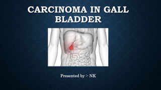
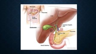
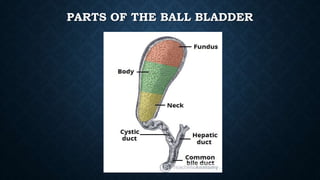
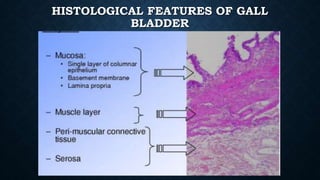
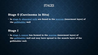
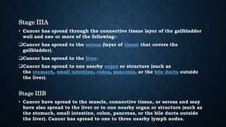
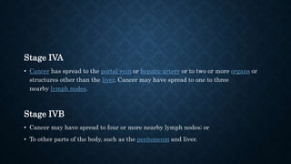
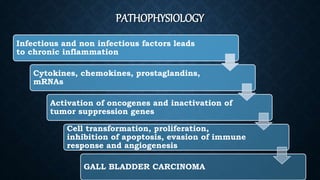
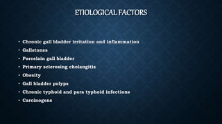
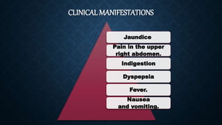
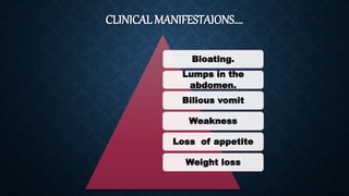
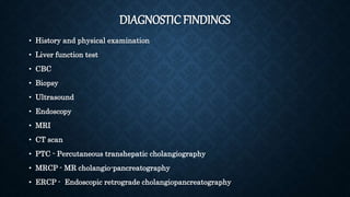
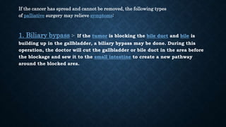
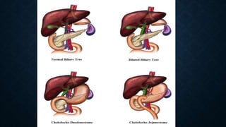
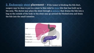
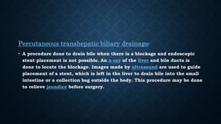
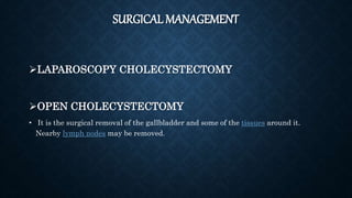
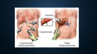
CARCINOMA IN GALL
BLADDER
Presented by :- NK
OBJECTIVES
• Anatomy and Physiology of the Gall Bladder
• Definition of the Carcinoma of Gall Bladder
• Types and Stages of the Carcinoma of Gall Bladder
• Pathophysiology of the Carcinoma of Gall Bladder
• Etiology of the Carcinoma of Gall Bladder
• Clinical manifestations of the Carcinoma of Gall Bladder
• Risk factors of the Carcinoma of Gall Bladder
• Diagnostic findings of the Carcinoma of Gall Bladder
• Medical management of the Carcinoma of Gall Bladder
• Nursing management
• Health education
• Rehabilitition
ANATOMY
• Gall bladder is a small hollow organ where bile is stored and concentrated before it
is released into the small intestine.
• It measures approximately 7 to 10 cm in length and 4 cm in diameter when fully
distended. It has a capacity of 50 ml.
• It is pear shaped and its tip opens into the cystic duct.
• It is divided into three sections :- fundus, body and neck.
• It is attached to liver on segment IVB and V.
• Cystic duct unites with the common hepatic duct to become a common bile duct.
PARTS OF THE BALL BLADDER
HISTOLOGICAL FEATURES OF GALL
BLADDER
PHYSIOLOGY
• The main function of gall bladder is to store bile needed for the digestion
of fats in food.
• Bile consists primarily of water and bile salts and also acts as a means of
eliminating bilirubin , a product of hemoglobin metabolism from the body.
• The bile emulsifies fats in partly digested food, thereby assisting their
absorption.
DEFINITION
• Gallbladder cancer is a disease in which malignant (cancer) cells form in
the tissues of the gallbladder.
• It is a rare disease that is thought to be related to gallstones building up,
which also can lead to calcification of the gallbladder, a condition known
as porcelain gallbladder.
TYPES
The gall bladder carcinoma is of following types based on the type of cells
affected :-
1. Adenocarcinoma :- it is a cancer that forms in mucous secreting gland
cells in the lining of the gall bladder.
2. Squamous cell carcinoma :- It develops from the skin like cells that form
the lining of the gallbladder, along with the gland cells.
3. Adenosquamous carcinomas :- These are cancers that have both
squamous cancer cells and glandular cancer cells.
STAGES
Stage 0 (Carcinoma in Situ)
• In stage 0, abnormal cells are found in the mucosa (innermost layer) of
the gallbladder wall
Stage I
• In stage I, cancer has formed in the mucosa (innermost layer) of
the gallbladder wall and may have spread to the muscle layer of the
gallbladder wall.
Stage IIA
• Cancer has spread through the muscle layer to the connective tissue layer of the
gallbladder wall on the side of the gallbladder that is not near the liver.
Stage IIB
• Cancer has spread through the muscle layer to the connective tissue layer of the
gallbladder wall on the same side as the liver. Cancer has not spread to the liver
Stage IIIA
• Cancer has spread through the connective tissue layer of the gallbladder
wall and one or more of the following:-
Cancer has spread to the serosa (layer of tissue that covers the
gallbladder).
Cancer has spread to the liver.
Cancer has spread to one nearby organ or structure (such as
the stomach, small intestine, colon, pancreas, or the bile ducts outside
the liver).
Stage IIIB
• Cancer have spread to the muscle, connective tissue, or serosa and may
have also spread to the liver or to one nearby organ or structure (such as
the stomach, small intestine, colon, pancreas, or the bile ducts outside
the liver). Cancer has spread to one to three nearby lymph nodes.
Stage IVA
• Cancer has spread to the portal vein or hepatic artery or to two or more organs or
structures other than the liver. Cancer may have spread to one to three
nearby lymph nodes.
Stage IVB
• Cancer may have spread to four or more nearby lymph nodes; or
• To other parts of the body, such as the peritoneum and liver.
PATHOPHYSIOLOGY
Infectious and non infectious factors leads
to chronic inflammation
Cytokines, chemokines, prostaglandins,
mRNAs
Activation of oncogenes and inactivation of
tumor suppression genes
Cell transformation, proliferation,
inhibition of apoptosis, evasion of immune
response and angiogenesis
GALL BLADDER CARCINOMA
ETIOLOGICAL FACTORS
• Chronic gall bladder irritation and inflammation
• Gallstones
• Porcelain gall bladder
• Primary sclerosing cholangitis
• Obesity
• Gall bladder polyps
• Chronic typhoid and para typhoid infections
• Carcinogens
CLINICAL MANIFESTATIONS
Jaundice
Pain in the upper
right abdomen.
Indigestion
Dyspepsia
Fever.
Nausea
and vomiting.
CLINICAL MANIFESTAIONS….
Bloating.
Lumps in the
abdomen.
Bilious vomit
Weakness
Loss of appetite
Weight loss
RISK FACTORS
• Female
• Obesity
• Chronic cholecystitis and cholelithiasis
• Primary sclerosing cholangitis
DIAGNOSTICFINDINGS
• History and physical examination
• Liver function test
• CBC
• Biopsy
• Ultrasound
• Endoscopy
• MRI
• CT scan
• PTC - Percutaneous transhepatic cholangiography
• MRCP - MR cholangio-pancreatography
• ERCP - Endoscopic retrograde cholangiopancreatography
MEDICAL MANAGEMENT
• CHEMOTHERAPY
• Gemcitabine
• Cisplatin
• Methotrexate
• 5FU
• RADIOTHERAPY :- It is a cancer treatment that uses high-energy x-rays or other
types of radiation to kill cancer cells or keep them from growing.
If the cancer has spread and cannot be removed, the following types
of palliative surgery may relieve symptoms:
1. Biliary bypass :- If the tumor is blocking the bile duct and bile is
building up in the gallbladder, a biliary bypass may be done. During this
operation, the doctor will cut the gallbladder or bile duct in the area before
the blockage and sew it to the small intestine to create a new pathway
around the blocked area.
2. Endoscopic stent placement :- If the tumor is blocking the bile duct,
surgery may be done to put in a stent (a thin tube) to drain bile that has built up in
the area. The doctor may place the stent through a catheter that drains the bile into a
bag on the outside of the body or the stent may go around the blocked area and drain
the bile into the small intestine.
Percutaneous transhepatic biliary drainage:
• A procedure done to drain bile when there is a blockage and endoscopic
stent placement is not possible. An x-ray of the liver and bile ducts is
done to locate the blockage. Images made by ultrasound are used to guide
placement of a stent, which is left in the liver to drain bile into the small
intestine or a collection bag outside the body. This procedure may be done
to relieve jaundice before surgery.
SURGICAL MANAGEMENT
LAPAROSCOPY CHOLECYSTECTOMY
OPEN CHOLECYSTECTOMY
• It is the surgical removal of the gallbladder and some of the tissues around it.
Nearby lymph nodes may be removed.
NURSING MANAGEMENT
• Monitor vital signs
• Monitor intake output
• Provide high protein and carbohydrate diet :- fresh fruits and
vegetables, fruit juice, low-fat dairy products, whole grains.
• Provide plenty of fluids orally – 2-3 liters of fluids.
HEALTH EDUCATION
 Teach the patient about the effects of chemotherapy and how to overcome it.
 Instruct the patient about regular follow up for further cycles of chemotherapy.
 Tell the patient to avoid :-
• soft drinks, refined grains
• baked potato
• pickled food
• Fried foods (fried chicken, French fries, potato chips)
• High fat dairy products (milk, butter, cheese, ice cream)
• Fatty meats (beef, pork)
• Processed meats (bacon, ham, sausage)
• Alcohol.
HEALTHEDUCATION
Tell the patient to have protein and carbohydrate rich diet which includes fresh
fruits and vegetables, fruit juice, low-fat dairy products, whole grains, etc.
Tell the patient to take frequent and small meals
Provide adequate rest to the patient
Tell the patient to do the following :-
• Proper hand washing
• Maintain personal hygiene
• Keep skin clean and free from trauma.
REHABILITATION
• Patient needs special care and attention from the time she is being diagnosed.
• Encourage the patient to maintain the family and social interactions.
• Encourage the patient to continue with the activities of daily living.
• Encourage the patient to get involve in diversional activities like yoga, meditation,
art etc.
• Provide psychological support to the patient and family before, during and after
chemotherapy.
• Inform the patient about the side effects of chemotherapy and radiotherapy before
the therapy starts so that patient will not go in sudden shock.
• Tell the patient how to overcome the side effects of the chemotherapy like hair loss
by covering the head by scarves or using wigs.
THANK YOU

Carcinoma in gall bladder

  • 1.
  • 2.
    OBJECTIVES • Anatomy andPhysiology of the Gall Bladder • Definition of the Carcinoma of Gall Bladder • Types and Stages of the Carcinoma of Gall Bladder • Pathophysiology of the Carcinoma of Gall Bladder • Etiology of the Carcinoma of Gall Bladder • Clinical manifestations of the Carcinoma of Gall Bladder • Risk factors of the Carcinoma of Gall Bladder • Diagnostic findings of the Carcinoma of Gall Bladder • Medical management of the Carcinoma of Gall Bladder • Nursing management • Health education • Rehabilitition
  • 3.
    ANATOMY • Gall bladderis a small hollow organ where bile is stored and concentrated before it is released into the small intestine. • It measures approximately 7 to 10 cm in length and 4 cm in diameter when fully distended. It has a capacity of 50 ml. • It is pear shaped and its tip opens into the cystic duct. • It is divided into three sections :- fundus, body and neck. • It is attached to liver on segment IVB and V. • Cystic duct unites with the common hepatic duct to become a common bile duct.
  • 5.
    PARTS OF THEBALL BLADDER
  • 6.
  • 7.
    PHYSIOLOGY • The mainfunction of gall bladder is to store bile needed for the digestion of fats in food. • Bile consists primarily of water and bile salts and also acts as a means of eliminating bilirubin , a product of hemoglobin metabolism from the body. • The bile emulsifies fats in partly digested food, thereby assisting their absorption.
  • 8.
    DEFINITION • Gallbladder canceris a disease in which malignant (cancer) cells form in the tissues of the gallbladder. • It is a rare disease that is thought to be related to gallstones building up, which also can lead to calcification of the gallbladder, a condition known as porcelain gallbladder.
  • 9.
    TYPES The gall bladdercarcinoma is of following types based on the type of cells affected :- 1. Adenocarcinoma :- it is a cancer that forms in mucous secreting gland cells in the lining of the gall bladder. 2. Squamous cell carcinoma :- It develops from the skin like cells that form the lining of the gallbladder, along with the gland cells. 3. Adenosquamous carcinomas :- These are cancers that have both squamous cancer cells and glandular cancer cells.
  • 10.
    STAGES Stage 0 (Carcinomain Situ) • In stage 0, abnormal cells are found in the mucosa (innermost layer) of the gallbladder wall Stage I • In stage I, cancer has formed in the mucosa (innermost layer) of the gallbladder wall and may have spread to the muscle layer of the gallbladder wall.
  • 11.
    Stage IIA • Cancerhas spread through the muscle layer to the connective tissue layer of the gallbladder wall on the side of the gallbladder that is not near the liver. Stage IIB • Cancer has spread through the muscle layer to the connective tissue layer of the gallbladder wall on the same side as the liver. Cancer has not spread to the liver
  • 12.
    Stage IIIA • Cancerhas spread through the connective tissue layer of the gallbladder wall and one or more of the following:- Cancer has spread to the serosa (layer of tissue that covers the gallbladder). Cancer has spread to the liver. Cancer has spread to one nearby organ or structure (such as the stomach, small intestine, colon, pancreas, or the bile ducts outside the liver). Stage IIIB • Cancer have spread to the muscle, connective tissue, or serosa and may have also spread to the liver or to one nearby organ or structure (such as the stomach, small intestine, colon, pancreas, or the bile ducts outside the liver). Cancer has spread to one to three nearby lymph nodes.
  • 13.
    Stage IVA • Cancerhas spread to the portal vein or hepatic artery or to two or more organs or structures other than the liver. Cancer may have spread to one to three nearby lymph nodes. Stage IVB • Cancer may have spread to four or more nearby lymph nodes; or • To other parts of the body, such as the peritoneum and liver.
  • 14.
    PATHOPHYSIOLOGY Infectious and noninfectious factors leads to chronic inflammation Cytokines, chemokines, prostaglandins, mRNAs Activation of oncogenes and inactivation of tumor suppression genes Cell transformation, proliferation, inhibition of apoptosis, evasion of immune response and angiogenesis GALL BLADDER CARCINOMA
  • 15.
    ETIOLOGICAL FACTORS • Chronicgall bladder irritation and inflammation • Gallstones • Porcelain gall bladder • Primary sclerosing cholangitis • Obesity • Gall bladder polyps • Chronic typhoid and para typhoid infections • Carcinogens
  • 16.
    CLINICAL MANIFESTATIONS Jaundice Pain inthe upper right abdomen. Indigestion Dyspepsia Fever. Nausea and vomiting.
  • 17.
    CLINICAL MANIFESTAIONS…. Bloating. Lumps inthe abdomen. Bilious vomit Weakness Loss of appetite Weight loss
  • 18.
    RISK FACTORS • Female •Obesity • Chronic cholecystitis and cholelithiasis • Primary sclerosing cholangitis
  • 19.
    DIAGNOSTICFINDINGS • History andphysical examination • Liver function test • CBC • Biopsy • Ultrasound • Endoscopy • MRI • CT scan • PTC - Percutaneous transhepatic cholangiography • MRCP - MR cholangio-pancreatography • ERCP - Endoscopic retrograde cholangiopancreatography
  • 20.
    MEDICAL MANAGEMENT • CHEMOTHERAPY •Gemcitabine • Cisplatin • Methotrexate • 5FU • RADIOTHERAPY :- It is a cancer treatment that uses high-energy x-rays or other types of radiation to kill cancer cells or keep them from growing.
  • 21.
    If the cancerhas spread and cannot be removed, the following types of palliative surgery may relieve symptoms: 1. Biliary bypass :- If the tumor is blocking the bile duct and bile is building up in the gallbladder, a biliary bypass may be done. During this operation, the doctor will cut the gallbladder or bile duct in the area before the blockage and sew it to the small intestine to create a new pathway around the blocked area.
  • 23.
    2. Endoscopic stentplacement :- If the tumor is blocking the bile duct, surgery may be done to put in a stent (a thin tube) to drain bile that has built up in the area. The doctor may place the stent through a catheter that drains the bile into a bag on the outside of the body or the stent may go around the blocked area and drain the bile into the small intestine.
  • 24.
    Percutaneous transhepatic biliarydrainage: • A procedure done to drain bile when there is a blockage and endoscopic stent placement is not possible. An x-ray of the liver and bile ducts is done to locate the blockage. Images made by ultrasound are used to guide placement of a stent, which is left in the liver to drain bile into the small intestine or a collection bag outside the body. This procedure may be done to relieve jaundice before surgery.
  • 25.
    SURGICAL MANAGEMENT LAPAROSCOPY CHOLECYSTECTOMY OPENCHOLECYSTECTOMY • It is the surgical removal of the gallbladder and some of the tissues around it. Nearby lymph nodes may be removed.
  • 27.
    NURSING MANAGEMENT • Monitorvital signs • Monitor intake output • Provide high protein and carbohydrate diet :- fresh fruits and vegetables, fruit juice, low-fat dairy products, whole grains. • Provide plenty of fluids orally – 2-3 liters of fluids.
  • 28.
    HEALTH EDUCATION  Teachthe patient about the effects of chemotherapy and how to overcome it.  Instruct the patient about regular follow up for further cycles of chemotherapy.  Tell the patient to avoid :- • soft drinks, refined grains • baked potato • pickled food • Fried foods (fried chicken, French fries, potato chips) • High fat dairy products (milk, butter, cheese, ice cream) • Fatty meats (beef, pork) • Processed meats (bacon, ham, sausage) • Alcohol.
  • 29.
    HEALTHEDUCATION Tell the patientto have protein and carbohydrate rich diet which includes fresh fruits and vegetables, fruit juice, low-fat dairy products, whole grains, etc. Tell the patient to take frequent and small meals Provide adequate rest to the patient Tell the patient to do the following :- • Proper hand washing • Maintain personal hygiene • Keep skin clean and free from trauma.
  • 30.
    REHABILITATION • Patient needsspecial care and attention from the time she is being diagnosed. • Encourage the patient to maintain the family and social interactions. • Encourage the patient to continue with the activities of daily living. • Encourage the patient to get involve in diversional activities like yoga, meditation, art etc. • Provide psychological support to the patient and family before, during and after chemotherapy. • Inform the patient about the side effects of chemotherapy and radiotherapy before the therapy starts so that patient will not go in sudden shock. • Tell the patient how to overcome the side effects of the chemotherapy like hair loss by covering the head by scarves or using wigs.
  • 31.