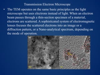
Carbon dots
Characterizations and Applications
By
P. Chandrasekaran
CONTENTS
 What is Carbon Dots?
 Properties of Carbon Dots(CD)
 Synthesis of CDs
 Characterization of CDs
 Applications of CDs
• CDs are small carbon nanoparticle(less than
10nm in size )with some form of surface
passivation.
• CDs were First discovered by XU et al., in 2004
accidently during the purification of single
walled carbon nanotubes. This Discovery
triggered extensive studies to exploit the
florescence properties of CDs.
• CDs are Biocompatible , have small size with
relatively large surface area , photostable and
having photoluminescence properties.
• CDs are currently emerging as a class of
promising fluorescent probe on account of
their low photo bleaching ,versatile surfaces
and excellent biocompatibility.
Contd.,
• Fluorescent carbon Dots have attracted
increasing attention due to their potential
application in sensing, catalysis and
biomedicine.
• Heteroatom doped carbon dots are enhance
the fluorescent properties.
Properties of CDs
• Excellent water solubility
• Biocompatibility
• Good conductivity
• Photochemical stability
• Low toxicity
• Environmental friendly
Synthesis of CDs
• Carbonization procedure
• Microwave synthesis
• Hydrothermal method
• Solvothermal method
• Laser ablation technique
• Chemical route (thermolysis)
• Electrochemical method
• Natural method of manufacturing of CDs
Carbonization method
Watermelon peel,
Hair fiber,
Citric acid,
Sweet Red Pepper,
Bovine serum of albumin….
• Hydrothermal method
• Saccharum officinarum juice
• Orange juice
• Pomelo peel
• Strawberry juice
• Actinidia deliciosa……
• Microwave
• Sucrose
• Chitosan
• Lotus root
• Alginic acid
Applications of CDs Bio sensing
Optoronics
Catalysis
Drug Delivery
Bio imaging
UV-Visible
• Principle of ultraviolet–visible absorption. Molecules
containing π-electrons or non-bonding electrons (n-electrons)
can absorb energy in the form of ultraviolet or visible light to
excite these electrons to higher anti-bonding molecular
orbitals.
• Fluorescence is the emission of light by a substance that
has absorbed light or other electromagnetic radiation. It is a
form of luminescence. In most cases, the emitted light has a
longer wavelength, and therefore lower energy, than the
absorbed radiation.
• The TEM operates on the same basic principles as the light
microscope but uses electrons instead of light. When an electron
beam passes through a thin-section specimen of a material,
electrons are scattered. A sophisticated system of electromagnetic
lenses focuses the scattered electrons into an image or a
diffraction pattern, or a Nano-analytical spectrum, depending on
the mode of operation.
Transmission Electron Microscope
X-ray Diffraction
• The periodic lattice found in crystalline structure may act as
diffraction grating for wave particles of electromagnetic
radiation with wavelength of a similar order of magnitude
(1Aº).
• The atomic planes of a crystal causes an incident beam of X-
rays to interfere with one another as they come out from the
crystal. This phenomenon is called X-ray diffraction.
Raman spectroscopy
• In Raman spectroscopy, sample is illuminated with a
monochromatic laser beam which interacts with the molecules
of sample and originates a scattered light. The scattered light
having a frequency different from that of incident light
(inelastic scattering) is used to construct a Raman spectrum.
Raman spectra arise due to inelastic collision between incident
monochromatic radiation and molecules of sample.
Infrared spectroscopy
• IR spectroscopy is concerned with the study of
absorption of infrared radiation, which causes
vibrational transition in the molecule.
• Molecules are made up of atoms linked by
chemical bonds.
• When energy in the form of infrared radiation
is applied then it causes the molecular
vibration.
• XPS spectra are obtained by irradiating a material with a beam
of X-rays while simultaneously measuring the kinetic energy and
number of electrons that escape from the top 0 to 10 nm of the
material being analyze.
Applications of CDs
• Bio imaging
• Optronics
• Catalysis
• Drug Delivery
• Bio sensing
• CDs with superior photo stability and low
cytotoxicity have been widely studied in optical
imaging applications as an alternative to QDs.
Both in vitro and in vivo evaluations indicated
that CDs are excellent candidates in bio
applications due to their visible excitation and
emission wavelengths, high brightness at the
individual dot level.
• An efficient approach for targeting and
detecting cancer cells has been developed
through the design of the assembly of
fluorescent CDs and folic acid (C-dots–FA),
which is endocytosible by the overexpressed
folate receptor (FR) molecule.
• Two-photon excited fluorescence images of MCF-
7 cancer cells after incubation with FTNP0 (A) and
FTNP40 (B) for 2 h at 37 1C ([T1] = 1 mM). The
images were recorded upon 800 nm excitation
with a 505 nm longpass barrier filter.
Carbon dots characterization and applications

Carbon dots characterization and applications

  • 1.
    Carbon dots Characterizations andApplications By P. Chandrasekaran
  • 2.
    CONTENTS  What isCarbon Dots?  Properties of Carbon Dots(CD)  Synthesis of CDs  Characterization of CDs  Applications of CDs
  • 3.
    • CDs aresmall carbon nanoparticle(less than 10nm in size )with some form of surface passivation. • CDs were First discovered by XU et al., in 2004 accidently during the purification of single walled carbon nanotubes. This Discovery triggered extensive studies to exploit the florescence properties of CDs.
  • 4.
    • CDs areBiocompatible , have small size with relatively large surface area , photostable and having photoluminescence properties. • CDs are currently emerging as a class of promising fluorescent probe on account of their low photo bleaching ,versatile surfaces and excellent biocompatibility.
  • 5.
    Contd., • Fluorescent carbonDots have attracted increasing attention due to their potential application in sensing, catalysis and biomedicine. • Heteroatom doped carbon dots are enhance the fluorescent properties.
  • 6.
    Properties of CDs •Excellent water solubility • Biocompatibility • Good conductivity • Photochemical stability • Low toxicity • Environmental friendly
  • 7.
    Synthesis of CDs •Carbonization procedure • Microwave synthesis • Hydrothermal method • Solvothermal method • Laser ablation technique • Chemical route (thermolysis) • Electrochemical method
  • 8.
    • Natural methodof manufacturing of CDs Carbonization method Watermelon peel, Hair fiber, Citric acid, Sweet Red Pepper, Bovine serum of albumin….
  • 9.
    • Hydrothermal method •Saccharum officinarum juice • Orange juice • Pomelo peel • Strawberry juice • Actinidia deliciosa……
  • 10.
    • Microwave • Sucrose •Chitosan • Lotus root • Alginic acid
  • 11.
    Applications of CDsBio sensing Optoronics Catalysis Drug Delivery Bio imaging
  • 12.
    UV-Visible • Principle ofultraviolet–visible absorption. Molecules containing π-electrons or non-bonding electrons (n-electrons) can absorb energy in the form of ultraviolet or visible light to excite these electrons to higher anti-bonding molecular orbitals.
  • 13.
    • Fluorescence isthe emission of light by a substance that has absorbed light or other electromagnetic radiation. It is a form of luminescence. In most cases, the emitted light has a longer wavelength, and therefore lower energy, than the absorbed radiation.
  • 14.
    • The TEMoperates on the same basic principles as the light microscope but uses electrons instead of light. When an electron beam passes through a thin-section specimen of a material, electrons are scattered. A sophisticated system of electromagnetic lenses focuses the scattered electrons into an image or a diffraction pattern, or a Nano-analytical spectrum, depending on the mode of operation. Transmission Electron Microscope
  • 15.
    X-ray Diffraction • Theperiodic lattice found in crystalline structure may act as diffraction grating for wave particles of electromagnetic radiation with wavelength of a similar order of magnitude (1Aº). • The atomic planes of a crystal causes an incident beam of X- rays to interfere with one another as they come out from the crystal. This phenomenon is called X-ray diffraction.
  • 16.
    Raman spectroscopy • InRaman spectroscopy, sample is illuminated with a monochromatic laser beam which interacts with the molecules of sample and originates a scattered light. The scattered light having a frequency different from that of incident light (inelastic scattering) is used to construct a Raman spectrum. Raman spectra arise due to inelastic collision between incident monochromatic radiation and molecules of sample.
  • 18.
    Infrared spectroscopy • IRspectroscopy is concerned with the study of absorption of infrared radiation, which causes vibrational transition in the molecule. • Molecules are made up of atoms linked by chemical bonds. • When energy in the form of infrared radiation is applied then it causes the molecular vibration.
  • 20.
    • XPS spectraare obtained by irradiating a material with a beam of X-rays while simultaneously measuring the kinetic energy and number of electrons that escape from the top 0 to 10 nm of the material being analyze.
  • 21.
    Applications of CDs •Bio imaging • Optronics • Catalysis • Drug Delivery • Bio sensing
  • 22.
    • CDs withsuperior photo stability and low cytotoxicity have been widely studied in optical imaging applications as an alternative to QDs. Both in vitro and in vivo evaluations indicated that CDs are excellent candidates in bio applications due to their visible excitation and emission wavelengths, high brightness at the individual dot level.
  • 23.
    • An efficientapproach for targeting and detecting cancer cells has been developed through the design of the assembly of fluorescent CDs and folic acid (C-dots–FA), which is endocytosible by the overexpressed folate receptor (FR) molecule.
  • 24.
    • Two-photon excitedfluorescence images of MCF- 7 cancer cells after incubation with FTNP0 (A) and FTNP40 (B) for 2 h at 37 1C ([T1] = 1 mM). The images were recorded upon 800 nm excitation with a 505 nm longpass barrier filter.