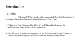
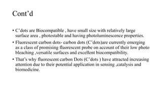
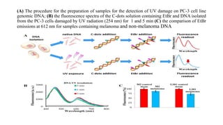
Carbon Dots are used for the detection of
UV-DNA Damage
Bio-Analytical chemistry and High performance
Ahmad Ali
University of Camerino
Outlines
1. What is C-dots:
2. Nature of C-dots
3. Size of C-dots
4. How can be c-dots modified for the detection
5. Genomics DNA isolation and damage experiment
6. Interaction of C-dots and DNA and Fluorescence energy
Transfer
7. conclusion
Introduction
C-Dots:
C’dots (or CD’s)are small carbon nanoparticle(less than10nm in size )
with same form of surface passivation.( Creation of Outer Layer)
• C’dots were First discovered by XU et al., in 2004 accidently during the
purification of single walled carbon nanotubes .
• This discovery achieved much progress in the florescence property of c-dots in
various sector of biological, analytical chemistry and their Applications.
Cont’d
• C’dots are Biocompatible , have small size with relatively large
surface area , photostable and having photoluminescence properties.
• Fluorescent carbon dots- carbon dots (C’dots)are currently emerging
as a class of promising fluorescent probe on account of their low photo
bleaching ,versatile surfaces and excellent biocompatibility.
• That’s why fluorescent carbon Dots (C’dots ) have attracted increasing
attention due to their potential application in sensing ,catalysis and
biomedicine.
Merits of C-Dots:
• C-dots, in contrast to other’s semiconductor quantum dots,
possess several merits such as;
• low cost, versatile surface modifications, excellent photo stability,
small size, chemical inertness, water solubility, etc.
• These extraordinary properties, together with biocompatibility and low
toxicity, make them an ideal candidate for biomedical, biochemical
and chemical applications.
Chemistry of C-dots and how it can be modified?
• The chemistry of c-dots consist of an amorphous to non crystalline
core with mostly SP2 carbons and highly abundant oxygen.
Cont’d
• The size and surface passivation of c-dots influence the absorption and
fluorescent characteristics. however, they harvest light in the
ultraviolet (UV) region.
Modification of carbon Dots : for the
Detection Of Damage DNA
• The c-dots were modified with their surface amine moieties, using the
microwave assisted pyrolysis of CA (Citric acid), in the presence of
DETA (diethylenetriamine).
• Amine moiety rich molecule such as 1,2-ethylenediamine,
polyethyleneimine were previously used to stabilize the C-dots
surface.
• DETA was chosen as the surface passivation agent in order to obtain
C-dots with a positive surface charge and to promote their interaction
with the negatively charged backbone of the DNA.
Genomics DNA isolation and damage
experiment:
• Actually in this experiment genomics DNA were taken, and control
condition were applied:
• Native DNA
• UV Exposed DNA
• Condition
Isolation of genomic DNA from human prostate cancer cell lines (PC-3)
and exposed to UV at 254 nm for 1 and 5 min.
• Damaged DNA was prepared using UV irradiation at 254 nm and was
monitored using UV/Vis spectrophotometry.
Cont’d
• C-dots incorporated in both Native and UV exposed DNA.
• Similarly in the next step EtBr incorporated to detect the Fluorescence
resonance energy transfer between these two molecule.
• At the very last step fluorescence readout will be noticed for samples
containing melanoma and non-melanoma DNA detection.
(A) The procedure for the preparation of samples for the detection of UV damage on PC-3 cell line
genomic DNA; (B) the fluorescence spectra of the C-dots solution containing EtBr and DNA isolated
from the PC-3 cells damaged by UV radiation (254 nm) for 1 and 5 min (C) the comparison of EtBr
emissions at 612 nm for samples containing melanoma and non-melanoma DNA
First Case:
• In the First case, when EtBr was taken alone for both DNA, and was
damaged by UV radiation for 20 min (λ=254 nm).
• which show 33% lower emission intensity, than the control sample
containing non-damaged DNA.
• The decrease is due to total amount of canonical base pairs, which are
partially replaced by UV photoproducts.
Cont’d
2nd Case:
• The FRET between the C-dots and EtBr took place (λex=360 nm).
• In this case, the EtBr emission intensity decreased by 50%.
• In the first case photoproducts are not able to accommodate EtBr or
shield the intercalated EtBr from quenching.
• Now in this case the non-canonical base pair are completely replaced
by the UV Photoproduct.
Conclusion:
• The FRET between the C-dots and EtBr was used to quantify the UV
damage to the DNA and show how we distinguish native DNA from
DNA Damaged by UV.
• C-dots with a positive surface charge were attached to DNA and
served as a donor of excitation energy.
• suggested that the FRET-based mechanism between C-dots and EtBr is
more sensitive to UV-induced damage to DNA than EtBr alone.
Thank you so much

Carbon dots

  • 1.
    Carbon Dots areused for the detection of UV-DNA Damage Bio-Analytical chemistry and High performance Ahmad Ali University of Camerino
  • 2.
    Outlines 1. What isC-dots: 2. Nature of C-dots 3. Size of C-dots 4. How can be c-dots modified for the detection 5. Genomics DNA isolation and damage experiment 6. Interaction of C-dots and DNA and Fluorescence energy Transfer 7. conclusion
  • 3.
    Introduction C-Dots: C’dots (or CD’s)aresmall carbon nanoparticle(less than10nm in size ) with same form of surface passivation.( Creation of Outer Layer) • C’dots were First discovered by XU et al., in 2004 accidently during the purification of single walled carbon nanotubes . • This discovery achieved much progress in the florescence property of c-dots in various sector of biological, analytical chemistry and their Applications.
  • 4.
    Cont’d • C’dots areBiocompatible , have small size with relatively large surface area , photostable and having photoluminescence properties. • Fluorescent carbon dots- carbon dots (C’dots)are currently emerging as a class of promising fluorescent probe on account of their low photo bleaching ,versatile surfaces and excellent biocompatibility. • That’s why fluorescent carbon Dots (C’dots ) have attracted increasing attention due to their potential application in sensing ,catalysis and biomedicine.
  • 5.
    Merits of C-Dots: •C-dots, in contrast to other’s semiconductor quantum dots, possess several merits such as; • low cost, versatile surface modifications, excellent photo stability, small size, chemical inertness, water solubility, etc. • These extraordinary properties, together with biocompatibility and low toxicity, make them an ideal candidate for biomedical, biochemical and chemical applications.
  • 6.
    Chemistry of C-dotsand how it can be modified? • The chemistry of c-dots consist of an amorphous to non crystalline core with mostly SP2 carbons and highly abundant oxygen.
  • 7.
    Cont’d • The sizeand surface passivation of c-dots influence the absorption and fluorescent characteristics. however, they harvest light in the ultraviolet (UV) region.
  • 8.
    Modification of carbonDots : for the Detection Of Damage DNA • The c-dots were modified with their surface amine moieties, using the microwave assisted pyrolysis of CA (Citric acid), in the presence of DETA (diethylenetriamine). • Amine moiety rich molecule such as 1,2-ethylenediamine, polyethyleneimine were previously used to stabilize the C-dots surface. • DETA was chosen as the surface passivation agent in order to obtain C-dots with a positive surface charge and to promote their interaction with the negatively charged backbone of the DNA.
  • 9.
    Genomics DNA isolationand damage experiment: • Actually in this experiment genomics DNA were taken, and control condition were applied: • Native DNA • UV Exposed DNA • Condition Isolation of genomic DNA from human prostate cancer cell lines (PC-3) and exposed to UV at 254 nm for 1 and 5 min. • Damaged DNA was prepared using UV irradiation at 254 nm and was monitored using UV/Vis spectrophotometry.
  • 10.
    Cont’d • C-dots incorporatedin both Native and UV exposed DNA. • Similarly in the next step EtBr incorporated to detect the Fluorescence resonance energy transfer between these two molecule. • At the very last step fluorescence readout will be noticed for samples containing melanoma and non-melanoma DNA detection.
  • 11.
    (A) The procedurefor the preparation of samples for the detection of UV damage on PC-3 cell line genomic DNA; (B) the fluorescence spectra of the C-dots solution containing EtBr and DNA isolated from the PC-3 cells damaged by UV radiation (254 nm) for 1 and 5 min (C) the comparison of EtBr emissions at 612 nm for samples containing melanoma and non-melanoma DNA
  • 12.
    First Case: • Inthe First case, when EtBr was taken alone for both DNA, and was damaged by UV radiation for 20 min (λ=254 nm). • which show 33% lower emission intensity, than the control sample containing non-damaged DNA. • The decrease is due to total amount of canonical base pairs, which are partially replaced by UV photoproducts.
  • 13.
    Cont’d 2nd Case: • TheFRET between the C-dots and EtBr took place (λex=360 nm). • In this case, the EtBr emission intensity decreased by 50%. • In the first case photoproducts are not able to accommodate EtBr or shield the intercalated EtBr from quenching. • Now in this case the non-canonical base pair are completely replaced by the UV Photoproduct.
  • 14.
    Conclusion: • The FRETbetween the C-dots and EtBr was used to quantify the UV damage to the DNA and show how we distinguish native DNA from DNA Damaged by UV. • C-dots with a positive surface charge were attached to DNA and served as a donor of excitation energy. • suggested that the FRET-based mechanism between C-dots and EtBr is more sensitive to UV-induced damage to DNA than EtBr alone.
  • 15.