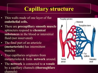
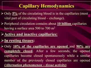
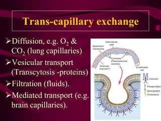
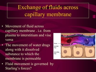
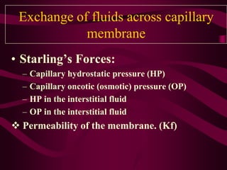
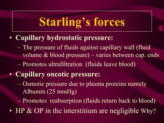
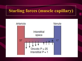
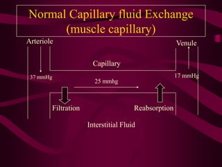
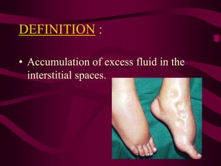
Capillary Circulation &
Trans-Capillary Exchange
Dr. Aamir Magzoub
MB BS, MSc,PhD
Objectives
• Capillary structure
• Capillary functions
• Capillary Hemodynamics
• Trans-capillary exchange
• Edema
– Definition
– Causes
– Pathophysiology
Capillary structure
• Thin walls made of one layer of flat
endothelial cells.
• There are precapillary smooth muscle
sphincters respond to chemical
substances in the blood or interstitial
fluid (not innervated)
• The final part of an arteriole
(metarteriole) has intermittent
muscles
• True capillaries originates from
metarterioles & form network around.
• The network is connected to a venule
by a capillary channels (thoroughfare
channels)
• The diameter of a true capillary is about 5 μm
(arteriolar end) and about 9 μm (venular end).
• Therefore, the red cells have to squeeze through
the capillaries and become convex discs; this
favors exchange of O2
• Also low velocity of blood flow in the
capillaries. Significance?
• More time for exchange at tissue level.
Capillary structure
• Special types of “pores” in the capillaries of
certain organs. (fenestrated capillaries - kidney)
• Tight junctions in brain capillaries.
• Exchange of water, nutrients, and other substances
between the blood and interstitial fluid.
Capillary structure
Interstitium and Interstitial
Fluid
Collagen fibers:
Tissue strength
Proteoglycans +
water Tissue gel
(holds water)
Free water:
Small amount vesicles
& rivulets – large
amounts in edema
Capillary Functions
• Sites of exchange between blood and tissues
(nutrients & Oxygen).
• Drainage of body waste products.
• Temperature regulation.
• Exchange vessels.
Capillary Hemodynamics
 Only 5% of the circulating blood is in the capillaries (most
vital part of circulating blood – exchange).
 Peripheral circulation contains about 10 billion capillaries
having a surface area 500 to 700 m2
Active and inactive capillaries:
In resting tissues:
 Only 10% of the capillaries are opened and 90% are
completely closed. After a few seconds, the opened
capillaries become closed spontaneously while a similar
number of the previously closed capillaries are opened
(Alternation phenomenon – tissue activity).
Alternation phenomenon can be explained
as follows:
- Gradual  in O2 and ↑ CO2 and H+ around the
closed capillaries  open them.
- Blood flow in this area provides O2 and removes
waste products (CO2 + H+)  abolishing their
vasodilator effect  capillaries are closed again.
In active tissues (e.g. muscular exercise):
The vasodilator metabolites formed in these tissues
dilatation of precapillary sphincters  blood
flow through the closed capillaries (recruitment).
Trans-capillary exchange
Diffusion, e.g. O2 &
CO2 (lung capillaries)
Vesicular transport
(Transcytosis -proteins)
Filtration (fluids).
Mediated transport (e.g.
brain capillaries).
Exchange of fluids across
capillary membrane
• Movement of fluid across
capillary membrane . i.e. from
plasma to interstitium and vise
versa
• The movement of water drags
along with it dissolved
substance to which the
membrane is permeable
• Fluid movement is governed by
Starling’s forces?
Exchange of fluids across capillary
membrane
• Starling’s Forces:
– Capillary hydrostatic pressure (HP)
– Capillary oncotic (osmotic) pressure (OP)
– HP in the interstitial fluid
– OP in the interstitial fluid
 Permeability of the membrane. (Kf)
Starling’s forces
• Capillary hydrostatic pressure:
– The pressure of fluids against capillary wall (fluid
volume & blood pressure) – varies between cap. ends
– Promotes ultrafiltration (fluids leave blood)
• Capillary oncotic pressure:
– Osmotic pressure due to plasma proteins namely
Albumin (25 mmHg)
– Promotes reabsorption (fluids return back to blood)
• HP & OP in the interstitium are negligible Why?
What are the important determinants of fluid
movement across capillary endothelium?
Starling forces (muscle capillary)
Normal Capillary fluid Exchange
(muscle capillary)
37 mmHg
25 mmhg
17 mmHg
Arteriole Venule
Capillary
Interstitial Fluid
Filtration Reabsorption
Arteriole
Venule
Capillary
Interstitial fluid
37 mmHg 25 mmhg
17 mmHg
100 %
90 %
~( EDEMA )~
DEFINITION :
• Accumulation of excess fluid in the
interstitial spaces.
Mechanism of edema
(pathophysiology)
1. Increased capillary hydrostatic pressure
2. Decreased capillary oncotic (colloid)
osmotic pressure
3. Lymphatic obstruction (or inadequate flow)
4. Increased capillary permeability
1. Increased hydrostatic (filtration)
pressure:
• Increased capillary hydrostatic pressure
(venous ):
– Heart failure, venous obstruction (DVT)
– Increased total ECF volume (salt & H2O retention)
– Orthostatic edema (prolonged sitting or
standing) – effect of gravity.
• Generalized or localized edema.
Mechanism:
1  Increased filtration (hydrostatic) pressure :
37 mmHg
25 mmhg
17 mmHg
Arteriole
Venule
Capillary
* *
2. Decreased oncotic osmotic
pressure
Decreased plasma albumin level due to:
Liver damage (decreased synthesis) - cirrhosis
Renal diseases (Nephrotic syndrome - Loss of
proteins in urine).
Protein malnutrition (Kwashiorkor- decreased
protein intake).
Generalized edema.
Mechanism
37 mmHg 17 mmHg
25 mmHg
Capillary
Arteriole Venule
Interstitial Fluid
*
Absorption
Filtration
Edema in nephrotic syndrome
(A) & protein malnutrition (B)
A B
3. Blockage of lymph vessels or
inadequate flow
Filariasis (Elephantiasis) – worms
Lymph nodes enlargement or
surgical removal (malignancy)
Localized edema
Mechanism
Arteriole
Venule
Capillary
Interstitial fluid
37 mmHg 25 mmhg
17 mmHg
100 %
90 %
Lymphedema
4.Increased capillary permeability:
Allergy (serious) ,why?
Due to histamine , substance P & others
Burns (leaky or damaged membrane)
Localized or generalized
Pitting & Non-pitting edema
Pitting edema
Most types
Pitting edema
Non- pitting edema
• Does not pit on pressure (fluids unite with
the tissue elements)
• Occurs in:
– Myxedema of severe hypothyroidism.
– Edema due to lymphatic obstruction.
Advanced myxedema (non-
pitting edema)
Pulmonary Edema
Pulmonary Edema
(left ventricular failure)
Ascites
Edema of the hand
Bilateral lower limb edema

Capillary circulation & fluid exchange.pdf

  • 1.
    Capillary Circulation & Trans-CapillaryExchange Dr. Aamir Magzoub MB BS, MSc,PhD
  • 2.
    Objectives • Capillary structure •Capillary functions • Capillary Hemodynamics • Trans-capillary exchange • Edema – Definition – Causes – Pathophysiology
  • 3.
    Capillary structure • Thinwalls made of one layer of flat endothelial cells. • There are precapillary smooth muscle sphincters respond to chemical substances in the blood or interstitial fluid (not innervated) • The final part of an arteriole (metarteriole) has intermittent muscles • True capillaries originates from metarterioles & form network around. • The network is connected to a venule by a capillary channels (thoroughfare channels)
  • 4.
    • The diameterof a true capillary is about 5 μm (arteriolar end) and about 9 μm (venular end). • Therefore, the red cells have to squeeze through the capillaries and become convex discs; this favors exchange of O2 • Also low velocity of blood flow in the capillaries. Significance? • More time for exchange at tissue level. Capillary structure
  • 5.
    • Special typesof “pores” in the capillaries of certain organs. (fenestrated capillaries - kidney) • Tight junctions in brain capillaries. • Exchange of water, nutrients, and other substances between the blood and interstitial fluid. Capillary structure
  • 6.
    Interstitium and Interstitial Fluid Collagenfibers: Tissue strength Proteoglycans + water Tissue gel (holds water) Free water: Small amount vesicles & rivulets – large amounts in edema
  • 7.
    Capillary Functions • Sitesof exchange between blood and tissues (nutrients & Oxygen). • Drainage of body waste products. • Temperature regulation. • Exchange vessels.
  • 8.
    Capillary Hemodynamics  Only5% of the circulating blood is in the capillaries (most vital part of circulating blood – exchange).  Peripheral circulation contains about 10 billion capillaries having a surface area 500 to 700 m2 Active and inactive capillaries: In resting tissues:  Only 10% of the capillaries are opened and 90% are completely closed. After a few seconds, the opened capillaries become closed spontaneously while a similar number of the previously closed capillaries are opened (Alternation phenomenon – tissue activity).
  • 9.
    Alternation phenomenon canbe explained as follows: - Gradual  in O2 and ↑ CO2 and H+ around the closed capillaries  open them. - Blood flow in this area provides O2 and removes waste products (CO2 + H+)  abolishing their vasodilator effect  capillaries are closed again. In active tissues (e.g. muscular exercise): The vasodilator metabolites formed in these tissues dilatation of precapillary sphincters  blood flow through the closed capillaries (recruitment).
  • 10.
    Trans-capillary exchange Diffusion, e.g.O2 & CO2 (lung capillaries) Vesicular transport (Transcytosis -proteins) Filtration (fluids). Mediated transport (e.g. brain capillaries).
  • 11.
    Exchange of fluidsacross capillary membrane • Movement of fluid across capillary membrane . i.e. from plasma to interstitium and vise versa • The movement of water drags along with it dissolved substance to which the membrane is permeable • Fluid movement is governed by Starling’s forces?
  • 12.
    Exchange of fluidsacross capillary membrane • Starling’s Forces: – Capillary hydrostatic pressure (HP) – Capillary oncotic (osmotic) pressure (OP) – HP in the interstitial fluid – OP in the interstitial fluid  Permeability of the membrane. (Kf)
  • 13.
    Starling’s forces • Capillaryhydrostatic pressure: – The pressure of fluids against capillary wall (fluid volume & blood pressure) – varies between cap. ends – Promotes ultrafiltration (fluids leave blood) • Capillary oncotic pressure: – Osmotic pressure due to plasma proteins namely Albumin (25 mmHg) – Promotes reabsorption (fluids return back to blood) • HP & OP in the interstitium are negligible Why?
  • 14.
    What are theimportant determinants of fluid movement across capillary endothelium?
  • 15.
  • 16.
    Normal Capillary fluidExchange (muscle capillary) 37 mmHg 25 mmhg 17 mmHg Arteriole Venule Capillary Interstitial Fluid Filtration Reabsorption
  • 17.
  • 18.
  • 19.
    DEFINITION : • Accumulationof excess fluid in the interstitial spaces.
  • 20.
    Mechanism of edema (pathophysiology) 1.Increased capillary hydrostatic pressure 2. Decreased capillary oncotic (colloid) osmotic pressure 3. Lymphatic obstruction (or inadequate flow) 4. Increased capillary permeability
  • 21.
    1. Increased hydrostatic(filtration) pressure: • Increased capillary hydrostatic pressure (venous ): – Heart failure, venous obstruction (DVT) – Increased total ECF volume (salt & H2O retention) – Orthostatic edema (prolonged sitting or standing) – effect of gravity. • Generalized or localized edema.
  • 22.
    Mechanism: 1 Increasedfiltration (hydrostatic) pressure : 37 mmHg 25 mmhg 17 mmHg Arteriole Venule Capillary * *
  • 23.
    2. Decreased oncoticosmotic pressure Decreased plasma albumin level due to: Liver damage (decreased synthesis) - cirrhosis Renal diseases (Nephrotic syndrome - Loss of proteins in urine). Protein malnutrition (Kwashiorkor- decreased protein intake). Generalized edema.
  • 24.
    Mechanism 37 mmHg 17mmHg 25 mmHg Capillary Arteriole Venule Interstitial Fluid * Absorption Filtration
  • 25.
    Edema in nephroticsyndrome (A) & protein malnutrition (B) A B
  • 26.
    3. Blockage oflymph vessels or inadequate flow Filariasis (Elephantiasis) – worms Lymph nodes enlargement or surgical removal (malignancy) Localized edema
  • 27.
  • 28.
  • 29.
    4.Increased capillary permeability: Allergy(serious) ,why? Due to histamine , substance P & others Burns (leaky or damaged membrane) Localized or generalized
  • 30.
  • 31.
  • 32.
  • 33.
    Non- pitting edema •Does not pit on pressure (fluids unite with the tissue elements) • Occurs in: – Myxedema of severe hypothyroidism. – Edema due to lymphatic obstruction.
  • 34.
  • 35.
  • 36.
  • 37.
  • 38.
  • 39.