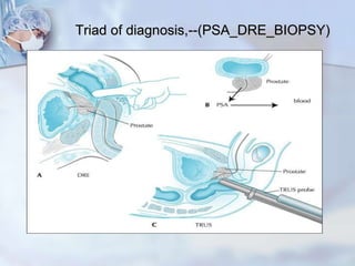
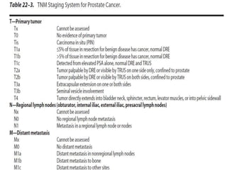
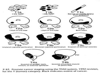
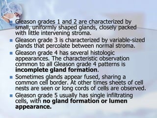
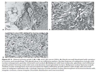
Facts About the Prostate
• At birth, prostate is about the size of a pea.
• Prostate experiences 2 growth spurts
- at puberty
- around age 50
• Prostate sits at base of the bladder with
urethral urine outflow tube tunneling through
its center.
• Its function is to produce the majority of the
seminal fluid that carries sperm forward with
ejaculation.
Prostate Anatomy
 Prostate weights ~20g
 Measures ~3 by 4 by 2 cm
 Apex = inferior portion of prostate, continuous with
striated sphincter.
 Base superior portion and continuous with bladder neck.
Prostatic urethra
 Covered by transitional epithelium.
 Hugs anterior portion of gland .
Verumontanum
 Just distal to urethral angulation's.
 Ejaculatory ducts (union of seminal vesicles and each vas
deferens) drain to each side of prostatic utricle (vestigial
Mullerian duct structure).
Gross Anatomy
 According to the Lowsley classification,
prostate consists of 5 lobes.
 Anterior
 Posterior
 Median
 Two lateral lobes.
Gross Anatomy
 According to McNeal (1972),
the prostate has
 Peripheral zone
 Central zone
 Transitional zone
Prostate zones
Central zone (CZ)
 Cone shaped region that surround the ejaculatory ducts (extends from bladder base
to the veru).
 25% of glandular tissue in young adults.
 Only 1-5% of prostate cancer from this region (likely because of Wolffian duct
embryologic origin).
 Peripheral zone (PZ)
 Posterio-lateral prostate.
 Mesodermal in origin.
 Majority of prostatic glandular tissue.
 Origin of up to 70% of prostate adenocarcinoma.
 Transitional zone (TZ)
 Surrounds the prostatic urethra proximal to the veru (preprostatic urethra).
 Endodermal in origin.
 In young men, accounts for only 5-10% of prostatic glandular tissue.
 Only ~20% of prostate cancer.
 It is only site for BPH.
 Arterial supply is derived from the inferior vesical,
internal pudendal and middle rectal arteries.
 The veins from the prostate drain into the
periprostatic plexus, which has connections with the
deep dorsal vein of the penis and the internal iliac
(hypogastric) veins.
 Prostate gland receives a rich nerve supply from the
sympathetic and parasympathetic nerve plexuses.
 Lymphatics from the prostate drain obturator nodes
and then into internal iliac (hypogastric), sacral,
vesical and external iliac lymph nodes.
CARCINOMA OF PROSTATE
Incidence & Epidemiology
 Prostate cancer is the most common cancer
detected in American men.
 It is 2nd leading cause of cancer death for men.
 Of all cancers, the prevalence of CaP increases the
most rapidly with age.
 Unlike most cancers, which have a peak age of
incidence, the incidence of CaP continues to
increase with advancing age.
 The lifetime risk of a 50year old man for latent CaP
(detected as incidental finding at autopsy, not
related to the cause of death) is 40%; for clinically
apparent CaP 9.5% and for death from CaP 2.9%.
Risk Factors
 Environmental factors
(diet, saturated fats)
 Genetic factors(family history)
 2.4 times increased risk of first degree relative)
 Chromosomal rearrangements or copy number
abnormalities at 8p, 10q, 11q, 13q, 16q, 17p, and
18q.
 Some of these such as specific loss at 8p23.2 and/or
gain at 11q13.1 are predictive of prostate cancer
progression.
 Men with a family history of breast/ovarian cancer
who carry the family specific BRCA1/2 mutations.
 Geography/race
 The age of disease onset in the family member also
affects a patient’s relative risk.
 If the age of onset is 70, the relative risk is
increased fourfold; if the age of onset is 60, the
relative risk is increased fivefold; and if the age of
onset is 50, the relative risk is increased 7 folds.
 Additionally, lycopene, selenium, omega-3 fatty
acids (fish) and vitamin E intake have been
shown to be protective.
 Whereas vitamin D and calcium increase risk. ?
 Previous vasectomy has been suggested as
factor that heightens the risk for CaP, but these
data are controversial.
Pathology
 95% of the cancers of prostate are adenocarcinoma.
 Other 5%, 90% are transitional cell carcinomas and the
remaining cancers are neuroendocrine (“small cell”)
carcinomas or sarcomas.
 Cytological characteristics of CaP include
hyperchromatic enlarged nuclei with prominent nucleoli.
Cytoplasm is often abundant; thus, nuclear to
cytoplasmic ratios are not often helpful in making a
diagnosis of CaP.
 The basal cell layer is absent in CaP, although it is
present in normal glands, BPH & precursor lesions of
CaP.
 If the diagnosis of CaP is in question, high-molecular-
weight keratin immuno-histochemical staining is useful, as
it preferentially stains basal cells.
 Absence of staining is thus consistent with CaP.
 Histology of the remaining 5% of prostate cancer is
heterogenous, arising from stromal, epithelial or ectopic
cells.
 Non-adenocarcinoma variants can be categorized into 2
groups based on the cellular origin: epithelial and non-
epithelial.
 Epithelial variants consist of endometrioid, mucinous,
signet-ring, adenoid cystic, adeno-squamous, squamous
cell, transitional cell, neuroendocrine and
comedocarcinoma.
 Non-epithelial variants include rhabdomyosarcoma,
leiomyosarcoma, osteosarcoma, angiosarcoma,
carcinosarcoma, malignant lymphoma, and metastatic
neoplasms among others.
Clinical presentation
 Asymptomatic.
 Incidental pathological finding/occult.
 BOO/abnormal DRE/raised PSA
level(47%)
 Urinary irritative and obstructive
symptoms.
 Hemato-spermia, hematuria, perineal
discomfort, renal failure.
 Metastatic symptoms bone pain,
Pathological fractures, anorexia, spinal
cord compression.
 Difficult erection and ejaculation.
Diagnosis
 General physical examination.
 DRE
 PSA
 TRUS
 BIOPSY
 Bone scan
 CT scan
 MRI
DRE
 DRE does not palpate
entire prostate gland
 Abnormal: hard nodular.
 Mucosa fixed.
 Median sulcus not
palpable.
LABORATORY FINDINGS
 Azotemia can result from bilateral
ureteral obstruction either from direct
extension into the trigone or from
retroperitoneal adenopathy.
 Anemia may be present in metastatic
disease.
 Alkaline phosphatase may be elevated in
the presence of bone metastases.
 Serum acid phosphatase may be
elevated with disease outside the
confines of the prostate.
TUMOR MARKERS
PROSTATE-SPECIFIC ANTIGEN
 PSA is a serine protease produced by benign and
malignant prostate epithelium not the stroma.
 Its function is to un clot semen.
 It circulates in serum as free or bound forms.
 Normal PSA values are those ≤4 ng/mL.
 PSA is prostate specific not CaP specific.
 Positive predictive value of a serum PSA between
4 and 10ng/mL is approximately 20–30%.
 For levels 10 & in excess of 10 ng/mL, the positive
predictive value increases from 42% to 71.4%.
 Given that most men with elevated serum PSA
levels do not have prostate cancer.
 Numerous strategies to refine PSA for cancer detection
have been explored. Their common goal is to decrease
the number of false-positive test results to increase
specificity and positive predictive value.
 Attempts at refining PSA have included PSA velocity
(change of PSA over time), PSA density (standardizing
levels in relation to the size of the prostate), age-
adjusted PSA reference ranges (accounting for age-
dependent prostate growth and occult prostatic disease)
and PSA forms (free versus protein bound molecular
forms of PSA).
PSA Velocity
 PSA velocity refers to rate of change of serum PSA.
 A study has shown that men with prostate cancer
have a more rapidly rising serum PSA in the years
before diagnosis than do men without prostate cancer.
 Patients whose serum PSA increases by 0.75 ng/mL/y
with atleast 3 determinations over atleast 18 months
appear to be at an increased risk of harboring cancer.
 According to AUA Phoenix def: PSA of nadir +2 ng/ml.
 However, rapid PSA velocity (ie, PSA doubling times
≤6 months) both before diagnosis and/or after
treatment are at an increased risk of failure of initial
treatment, the development of metastases and
prostate cancer specific mortality.
PSA DENSITY
 The ratio of PSA to gland volume is termed the
PSA density.
 PSA levels are elevated approximately 0.12 ng/mL/g
of BPH tissue.
 Some investigators advocate prostate biopsy only if
the PSA density exceeds 0.10 or 0.15, while others
have not found PSA density to be useful.
 Problems with this approach include the facts that (1)
epithelial-stromal ratios vary from gland to gland and
only the epithelium produces PSA, and (2) errors in
calculating prostatic volume may approach 25%.
 Instead of adjusting the PSA to total prostate volume,
some have advocated adjusting it to transition zone
volume (PSA transition zone density).
Age-adjusted reference
ranges for PSA
 Elevated indicates possible Carcinoma prostate
 PSA 4 – 10 indicates 25-35% risk of cancer diagnosis.
 PSA 10 – 20 indicates 65% risk of cancer diagnosis.
 PSA > 20 indicates possible metastatic disease.
Molecular forms of PSA
 Approximately 90% of the serum PSA is
bound to alpha 1 anti-chymotrypsin and
lesser amount is free or bound to alpha 2
macro-globulins.
 Early studies suggest that prostate
cancer patients demonstrate a lower
percentage of free PSA than do patients
with benign disease.
Traditional indication for Prostate
Biopsy:
Usually with Life Expectancy >10yrs
 Abnormal DRE regardless of PSA.
 Abnormal PSA velocity (.75 ng/dL/yr)
 PSA > 4.0 or age appropriate range.
 Elevated PSA does not mean prostate
cancer.
Prostate biopsy
 Traditionally, 6 (sextant) biopsies were taken along a
parasagittal line between lateral edge and midline of
prostate at the apex, midgland and base bilaterally.
 Several investigators shown that increasing the number
≥10 and performing more laterally directed biopsies of
peripheral zone will increase detection rates 14–20%
over the more traditional sextant technique.
 There is ongoing interest in use of even more extended
biopsy schemes (“saturation biopsy”) to improve cancer
detection, who have had a negative biopsy, but are
thought to be at an increased risk of prostate cancer
based on a persistently abnormal serum PSA.
 Prostate biopsy is usually performed using local
anesthesia and pre procedure antibiotic prophylaxis.
kidney.niddk.nih.gov/kudiseases
TRUS Guided Biopsy
 Hypoechoic
shows
abnormal
area
needing
biopsy
Transrectal
sonogram of the
prostate. Looking
up from the feet of
a patient toward his
head.
Flow chart
1-PSA + DRE-----results-------if both results
normal-consider regular DRE and PSA
2-PSA + DRE ---one or both result abnormal
consider Free PSA, if low ,--Ca suspected--
biopsy----Gleason's scoring---- treatment
options.
3- If biopsy negative then go for regular DRE and PSA.
Triad of diagnosis,--(PSA_DRE_BIOPSY)
Endorectal magnetic resonance
imaging (MRI)
 Use of an endorectal coil improves cancer detection and staging
compared to the use of a standard body coil. While rendering
high image quality.
 Use of an endorectal coil appears to be operator dependent,
requiring education and expertise.
 Use of magnetic resonance spectroscopy (MRS) in conjunction
with MRI may improve the accuracy of imaging.
 Prostate cancer is associated with proportionately lower levels of
citrate and higher levels of choline and creatine compared to BPH
or normal prostate tissue.
 The combined metabolic and anatomic information provided by
MRI and MRS may allow for a more accurate assessment of
cancer location and stage.
 The reported staging accuracy of endorectal MRI varies from
51% to 92%.
Axial imaging (CT, MRI)
 Cross-sectional imaging of the pelvis with CaP is
selectively performed to exclude lymph node
metastases in high-risk patients who are candidates
for definitive local therapy, whether it be surgery or
radiation.
 Patients identified as having lymphadenopathy on
imaging may undergo CT-guided fine-needle
aspiration.
 Incidence of lymph node metastases in contemporary
radical prostatectomy series is low (<10%).
 Imaging is costly and its sensitivity is limited 30–40%.
 Various criteria can be used to identify patients for
axial imaging, including negative bone scans and
either T3 cancers or a PSA >20 ng/mL and primary
Gleason grade 4 or 5 cancers.
Bone scan
 Prostate cancer metastasizes most commonly to
bone.
 Soft tissue metastases (e.g lung and liver) are rare at
the time of initial presentation.
 Good evidence has been accumulated that it can be
excluded in most of these men on the basis of serum
PSA.
 Several investigators have shown that bone scans
can be omitted in patients with newly diagnosed,
untreated prostate cancer who are asymptomatic,
have T1 and T2 disease, and have serum PSA
concentrations <15 ng/mL.
 However, patients with PSA 15 ng/mL or greater,
locally advanced disease (T3B, T4) are at higher risk
for bone metastases and should be considered for
bone scan.
CaP, TNM STAGING
 The TNM staging system for CaP is presented in
American Joint Committee on Cancer 1997, with
respect to the primary tumor categorization (T stage),
the clinical staging system uses results of the DRE and
TRUS, but not the results of the biopsy.
 Some examples to illustrate this staging system follow.
If a patient has a palpable abnormality on one side of
the prostate, even though biopsies demonstrate
bilateral disease, his clinical stage remains T2a.
 If a patient has a normal DRE, with TRUS
demonstrating a lesion on one side and a biopsy
confirming cancer, his clinical stage is also T2a (using
results of DRE and TRUS).
 A T1c cancer must have both a normal DRE and a
normal TRUS.
Gleason's grading score system
 The Gleason grading system relies upon the low-power appearance
of the glandular architecture under the microscope.
 Grades range from 1 to 5.
 If the entire specimen has only one pattern present, then both the
primary and secondary grade are reported as the same grade.
 The Gleason score or Gleason sum is obtained by adding
the primary and secondary grades together.
 As Gleason grades range from 1 to 5, Gleason scores or sums thus
range from 2 to 10.
 Well-differentiated tumors have a Gleason sum of 2–4, moderately
differentiated tumors have 5–6, and poorly differentiated tumors
have 8–10.
 Patients with a Gleason sum of 7 who have a primary Gleason
grade of 4 (4 + 3) tend to have a worse prognosis than those who
have a primary Gleason grade of 3 (3 + 4).
 Gleason grades 1 and 2 are characterized by
small, uniformly shaped glands, closely packed
with little intervening stroma.
 Gleason grade 3 is characterized by variable-sized
glands that percolate between normal stroma.
 Gleason grade 4 has several histologic
appearances. The characteristic observation
common to all Gleason grade 4 patterns is
incomplete gland formation.
 Sometimes glands appear fused, sharing a
common cell border. At other times sheets of cell
nests are seen or long cords of cells are observed.
 Gleason grade 5 usually has single infiltrating
cells, with no gland formation or lumen
appearance.
Differential Diagnosis
 Not all patients with an elevated PSA
concentration have CaP.
 Induration of the prostate is associated not
only with CaP, but also with chronic
granulomatous prostatitis, previous TURP or
needle biopsy or prostatic calculi.
 Sclerotic lesions on plain x-ray films and
elevated levels of alkaline phosphatase can
be seen in Paget’s disease and can often be
difficult to distinguish from metastatic CaP.
 In Paget’s disease PSA levels are usually
normal and x-ray findings demonstrate
subperiosteal cortical thickening.
Pretreatment Risk
Assessment
Treatment LOCALIZED DISEASE
 General considerations Treatment dilemma persist
in management of localized disease (T1 & T2) b/c of
uncertainty in efficacy of various modalities including
radical prostatectomy, radiotherapy & surveillance.
 Current treatment decisions are based on the grade
and stage of the tumor,
 The life expectancy of the patient,
 The ability of each therapy to ensure disease-free
survival, its associated morbidity,
 Patient and physician preferences.
 Advantage of surgery is most apparent in younger
patients.
 Watchful waiting; is based on the premise that
some patients will NOT benefit from definitive
treatment of CaP.
 Decision is made @ time of diagnosis to forgo definitive
treatment or to provide palliative treatment for local &
metastatic progression if & when it occurs.
 Active surveillance; is based on concept that
some, but not all, patients may drive benefits
from treatment of CaP.
 Goals of active surveillance are to provide definitive
treatment for man with localized tumor that are likely to
progress & to reduce the risk of treatment related
complications for man with cancers that are not likely to
progress hence maintaining quality of life.
 Disadvantages are increased anxiety, physician visits &
tests while causing subsequent treatment to be more
aggressive.
Retropubic Redical Prostectomy
Advantages
 Whole prostate and thus the entire tumor can
be examined histologically.
 Surgeon has access to regional lymph nodes
to test if prostate cancer cells have left the
tumor.
 Surgical margin can be examined.
 complications are Incontinence Impotence
 Bleeding, Thrombo-embolism
TT
Negative
Surgical
Margin
Positive
Surgical
Margin
Not all
of tumor
removed
OR
Radical prostectomy
Surgical Approach
Bladder
Prostate
Urethra
Rectum
Excision of entire
prostate along with
prostatic urethra &
seminal vesicles.
Usually epidural
anesthesia.
Incision: Lower
midline staying
extraperitoneal, the
retropubic space is
opened.
Remaining Urethra
is sewn to bladder.
neck over a catheter.
Surgical
Approach
Pelvic
Bone
(Pubis)
Radical Perineal Prostatectomy
Bladder
Prostate
Urethra
Rectum
Surgical
Approach
Very similar to
Retropubic protocol
(nerve sparing, sewing
of urethra, etc.)
Incision:
Between Anus and base
of Scrotum.
Comparison with RRP: Comparable
cure rates as well as similar urinary
and potency complications.
Disadvantages:
 Cannot access regional
lymph nodes
 Slight increase in risk of
rectal injury and associated
complications.
Pelvic
Bone
Emerging Therapy: Laparoscopic
Radical Prostatectomy
 Advantages:
 Less blood loss.
 No large incision.
 Shorter hospital stay
and earlier return to
activities.
 Disadvantages:
 Longer procedure
 Variable surgical
margins rates.
 Slower return of
urinary continence.
 Variable potency rates.
Robotic prostatectomy
 Surgeon operates from a
console with a 3-D screen.
 Grasp controls to manipulate
surgical tools within the
patient.
 Robotic arms translate finger,
hand and wrist movements.
 Shortens learning curve of
surgeons.
 Very High-Precision
 Cost, Benefit unclear.
Radiation therapy
 High-Powered X-Rays that damage DNA
and kill prostate cancer cells.
1. External Beam Radiation Therapy (EBRT):
X-rays aimed at prostate.
2. Brachytherapy: Radioactive seed implants
into prostate.
EBRT and Brachytherapy
 EBRT:
1. Map precise area that will receive radiation.
2. Multiple treatments ~5 days/week for 6~8
weeks. Each treatment takes about 10 minutes
and no anesthesia is required.
 Brachytherapy
1. 40-100 rice-sized radioactive seeds are
implanted into the prostate via ultrasound-guided
needles. Anesthesia is required.
2. All radiation inside the pellets is generally
exhausted within a year.
External Beam Radiation
Goal: Maximize damage to the prostate
and minimize damage to surrounding
tissues (i.e. bladder and rectum)
Prostate
Seminal
Vesicles
Image of Prostate With
Radioactive Bead Implants
Cryotherapy & HIFU
 Two minimal invasive treatment for organ
confined recurrent disease following
radiotherapy.
 CRYOTHERAPY
 Destroys tissue by freezing tissue @ -20 to -40c
through trans-perineal u/s cryo-probe deliver
Argon or liquid nitrogen.
 High intensity focused ultrasound (HIFU);
 It is the selective destruction of tissues @ 85c by
using trans-rectal device that causes coagulative
necrosis.
Management of locally advanced
Disease; Hormone Therapy
 Prostate cells and prostate cancer cells are dependent upon
androgens (male sex hormones) for survival and growth.
 Removal of androgens kills a majority of prostate cancer cells.
Testes
Prostate
Growth and
Function
Testosterone
95%
Adrenal
Androgen
5
%
Management of locally
advanced Disease
Mechanisms of androgen deprivation
1. Surgical castration: Bilateral Orchiectomy.
2. Medical castration: LHRH agonists, Antagonists,
estrogen.
3. Anti-androgens: which block the effects of
testosterone. (Blocks binding of DHT to androgen
receptors.)
4. 5-AR inhibitor; enhances intracellular androgen
blockade.
5. Maximum androgen blockade (MAB): Medical or
Surgical castration plus anti-androgen.
 Both form of castration have equal efficacy, so
patients should be given choice.
 Estrogens are no longer 1st line because of
cardiovascular morbidity.
 Anti-androgens alone are less effective in treating
metastatic disease, but equivalent for non-
metastatic disease.
 MAB has a theoretical advantage over castration in
blocking the effect of adrenal androgens.
 5AR inhibitors are not used in treatment of prostate
cancer but appear to have a role in prevention.
LHRH Analogs
 Goserelin, Leuprolide, hisrelin & triptorelin are
04 FDA approved LHRH analogs.
 LHRH agonists are alternative to bilateral
orchiectomy with clinically equal efficacy.
Anti-androgens.
 Flutamide, Bicalutamide, Nilutamide
 Combined androgen blockade not superior to
LHRH therapy alone.
 Higher cost and more side effects than LHRH
therapy alone.
 Primary value when starting LHRH to limit the
flare reaction that occurs with LHRH antagonists.
Results of Androgen Removal
 Impotence
 Loss of sexual desire (libido)
 Hot flashes
 Weight gain
 Fatigue
 Reduced brain function
 Loss of muscle and bone mass
 Some cardiovascular risks
Hormone-Refractory Prostate
Cancer (HRPC)
 Despite initial response rates of 80-90%, nearly
all men with advanced prostate cancer develop
hormone-resistant prostate cancer after 18-36
months.
 These “hormone-refractory” (HR) prostate
cancer cells can grow in the absence of
androgens.
 The behavior of HR prostate cancers differ
widely between patients.
Treatment of Symptomatic, Hormone
Refractory Metastatic Disease
1. Cytotoxic chemotherapy
 Docetaxel (every three weeks) and prednisone
improves pain and reduces need for analgesic
agents.
 Mitoxantrone
2. Bisphosphonates - decreases skeletal
complications.
Complications of Systemic Prostate
Cancer Therapy
 All hormonal therapies can cause sexual
dysfunction and decreased libido; less with
finasteride and anti-androgen.
 Orchiectomy - rarely local infection or hematoma
 Anti-androgen - diarrhea, hepatic dysfunction
 Estrogen - thromboembolic disease, fluid
retention, cardiac disease.
 Chemotherapy - nausea, vomiting, mucositis,
marrow suppression and alopecia.
Management of Prostate Cancer
Bone Metastases
 Goal: prevent pain, improve mobility,
prevent complications such as fractures or
compression & maintain acceptable quality
of life.
 Methods: bis-phosphonates, radiation of
detected metastatic lesions, Calcium &
vitamin D supplementation & surgery.
Ca prostate

Ca prostate

  • 1.
    Facts About theProstate • At birth, prostate is about the size of a pea. • Prostate experiences 2 growth spurts - at puberty - around age 50 • Prostate sits at base of the bladder with urethral urine outflow tube tunneling through its center. • Its function is to produce the majority of the seminal fluid that carries sperm forward with ejaculation.
  • 2.
    Prostate Anatomy  Prostateweights ~20g  Measures ~3 by 4 by 2 cm  Apex = inferior portion of prostate, continuous with striated sphincter.  Base superior portion and continuous with bladder neck. Prostatic urethra  Covered by transitional epithelium.  Hugs anterior portion of gland . Verumontanum  Just distal to urethral angulation's.  Ejaculatory ducts (union of seminal vesicles and each vas deferens) drain to each side of prostatic utricle (vestigial Mullerian duct structure).
  • 3.
    Gross Anatomy  Accordingto the Lowsley classification, prostate consists of 5 lobes.  Anterior  Posterior  Median  Two lateral lobes.
  • 4.
    Gross Anatomy  Accordingto McNeal (1972), the prostate has  Peripheral zone  Central zone  Transitional zone
  • 5.
    Prostate zones Central zone(CZ)  Cone shaped region that surround the ejaculatory ducts (extends from bladder base to the veru).  25% of glandular tissue in young adults.  Only 1-5% of prostate cancer from this region (likely because of Wolffian duct embryologic origin).  Peripheral zone (PZ)  Posterio-lateral prostate.  Mesodermal in origin.  Majority of prostatic glandular tissue.  Origin of up to 70% of prostate adenocarcinoma.  Transitional zone (TZ)  Surrounds the prostatic urethra proximal to the veru (preprostatic urethra).  Endodermal in origin.  In young men, accounts for only 5-10% of prostatic glandular tissue.  Only ~20% of prostate cancer.  It is only site for BPH.
  • 6.
     Arterial supplyis derived from the inferior vesical, internal pudendal and middle rectal arteries.  The veins from the prostate drain into the periprostatic plexus, which has connections with the deep dorsal vein of the penis and the internal iliac (hypogastric) veins.  Prostate gland receives a rich nerve supply from the sympathetic and parasympathetic nerve plexuses.  Lymphatics from the prostate drain obturator nodes and then into internal iliac (hypogastric), sacral, vesical and external iliac lymph nodes.
  • 7.
    CARCINOMA OF PROSTATE Incidence& Epidemiology  Prostate cancer is the most common cancer detected in American men.  It is 2nd leading cause of cancer death for men.  Of all cancers, the prevalence of CaP increases the most rapidly with age.  Unlike most cancers, which have a peak age of incidence, the incidence of CaP continues to increase with advancing age.  The lifetime risk of a 50year old man for latent CaP (detected as incidental finding at autopsy, not related to the cause of death) is 40%; for clinically apparent CaP 9.5% and for death from CaP 2.9%.
  • 8.
    Risk Factors  Environmentalfactors (diet, saturated fats)  Genetic factors(family history)  2.4 times increased risk of first degree relative)  Chromosomal rearrangements or copy number abnormalities at 8p, 10q, 11q, 13q, 16q, 17p, and 18q.  Some of these such as specific loss at 8p23.2 and/or gain at 11q13.1 are predictive of prostate cancer progression.  Men with a family history of breast/ovarian cancer who carry the family specific BRCA1/2 mutations.  Geography/race
  • 9.
     The ageof disease onset in the family member also affects a patient’s relative risk.  If the age of onset is 70, the relative risk is increased fourfold; if the age of onset is 60, the relative risk is increased fivefold; and if the age of onset is 50, the relative risk is increased 7 folds.  Additionally, lycopene, selenium, omega-3 fatty acids (fish) and vitamin E intake have been shown to be protective.  Whereas vitamin D and calcium increase risk. ?  Previous vasectomy has been suggested as factor that heightens the risk for CaP, but these data are controversial.
  • 10.
    Pathology  95% ofthe cancers of prostate are adenocarcinoma.  Other 5%, 90% are transitional cell carcinomas and the remaining cancers are neuroendocrine (“small cell”) carcinomas or sarcomas.  Cytological characteristics of CaP include hyperchromatic enlarged nuclei with prominent nucleoli. Cytoplasm is often abundant; thus, nuclear to cytoplasmic ratios are not often helpful in making a diagnosis of CaP.  The basal cell layer is absent in CaP, although it is present in normal glands, BPH & precursor lesions of CaP.
  • 11.
     If thediagnosis of CaP is in question, high-molecular- weight keratin immuno-histochemical staining is useful, as it preferentially stains basal cells.  Absence of staining is thus consistent with CaP.  Histology of the remaining 5% of prostate cancer is heterogenous, arising from stromal, epithelial or ectopic cells.  Non-adenocarcinoma variants can be categorized into 2 groups based on the cellular origin: epithelial and non- epithelial.  Epithelial variants consist of endometrioid, mucinous, signet-ring, adenoid cystic, adeno-squamous, squamous cell, transitional cell, neuroendocrine and comedocarcinoma.  Non-epithelial variants include rhabdomyosarcoma, leiomyosarcoma, osteosarcoma, angiosarcoma, carcinosarcoma, malignant lymphoma, and metastatic neoplasms among others.
  • 12.
    Clinical presentation  Asymptomatic. Incidental pathological finding/occult.  BOO/abnormal DRE/raised PSA level(47%)  Urinary irritative and obstructive symptoms.  Hemato-spermia, hematuria, perineal discomfort, renal failure.  Metastatic symptoms bone pain, Pathological fractures, anorexia, spinal cord compression.  Difficult erection and ejaculation.
  • 13.
    Diagnosis  General physicalexamination.  DRE  PSA  TRUS  BIOPSY  Bone scan  CT scan  MRI
  • 14.
    DRE  DRE doesnot palpate entire prostate gland  Abnormal: hard nodular.  Mucosa fixed.  Median sulcus not palpable.
  • 15.
    LABORATORY FINDINGS  Azotemiacan result from bilateral ureteral obstruction either from direct extension into the trigone or from retroperitoneal adenopathy.  Anemia may be present in metastatic disease.  Alkaline phosphatase may be elevated in the presence of bone metastases.  Serum acid phosphatase may be elevated with disease outside the confines of the prostate.
  • 16.
    TUMOR MARKERS PROSTATE-SPECIFIC ANTIGEN PSA is a serine protease produced by benign and malignant prostate epithelium not the stroma.  Its function is to un clot semen.  It circulates in serum as free or bound forms.  Normal PSA values are those ≤4 ng/mL.  PSA is prostate specific not CaP specific.  Positive predictive value of a serum PSA between 4 and 10ng/mL is approximately 20–30%.  For levels 10 & in excess of 10 ng/mL, the positive predictive value increases from 42% to 71.4%.  Given that most men with elevated serum PSA levels do not have prostate cancer.
  • 17.
     Numerous strategiesto refine PSA for cancer detection have been explored. Their common goal is to decrease the number of false-positive test results to increase specificity and positive predictive value.  Attempts at refining PSA have included PSA velocity (change of PSA over time), PSA density (standardizing levels in relation to the size of the prostate), age- adjusted PSA reference ranges (accounting for age- dependent prostate growth and occult prostatic disease) and PSA forms (free versus protein bound molecular forms of PSA).
  • 18.
    PSA Velocity  PSAvelocity refers to rate of change of serum PSA.  A study has shown that men with prostate cancer have a more rapidly rising serum PSA in the years before diagnosis than do men without prostate cancer.  Patients whose serum PSA increases by 0.75 ng/mL/y with atleast 3 determinations over atleast 18 months appear to be at an increased risk of harboring cancer.  According to AUA Phoenix def: PSA of nadir +2 ng/ml.  However, rapid PSA velocity (ie, PSA doubling times ≤6 months) both before diagnosis and/or after treatment are at an increased risk of failure of initial treatment, the development of metastases and prostate cancer specific mortality.
  • 19.
    PSA DENSITY  Theratio of PSA to gland volume is termed the PSA density.  PSA levels are elevated approximately 0.12 ng/mL/g of BPH tissue.  Some investigators advocate prostate biopsy only if the PSA density exceeds 0.10 or 0.15, while others have not found PSA density to be useful.  Problems with this approach include the facts that (1) epithelial-stromal ratios vary from gland to gland and only the epithelium produces PSA, and (2) errors in calculating prostatic volume may approach 25%.  Instead of adjusting the PSA to total prostate volume, some have advocated adjusting it to transition zone volume (PSA transition zone density).
  • 20.
    Age-adjusted reference ranges forPSA  Elevated indicates possible Carcinoma prostate  PSA 4 – 10 indicates 25-35% risk of cancer diagnosis.  PSA 10 – 20 indicates 65% risk of cancer diagnosis.  PSA > 20 indicates possible metastatic disease.
  • 21.
    Molecular forms ofPSA  Approximately 90% of the serum PSA is bound to alpha 1 anti-chymotrypsin and lesser amount is free or bound to alpha 2 macro-globulins.  Early studies suggest that prostate cancer patients demonstrate a lower percentage of free PSA than do patients with benign disease.
  • 22.
    Traditional indication forProstate Biopsy: Usually with Life Expectancy >10yrs  Abnormal DRE regardless of PSA.  Abnormal PSA velocity (.75 ng/dL/yr)  PSA > 4.0 or age appropriate range.  Elevated PSA does not mean prostate cancer.
  • 23.
    Prostate biopsy  Traditionally,6 (sextant) biopsies were taken along a parasagittal line between lateral edge and midline of prostate at the apex, midgland and base bilaterally.  Several investigators shown that increasing the number ≥10 and performing more laterally directed biopsies of peripheral zone will increase detection rates 14–20% over the more traditional sextant technique.  There is ongoing interest in use of even more extended biopsy schemes (“saturation biopsy”) to improve cancer detection, who have had a negative biopsy, but are thought to be at an increased risk of prostate cancer based on a persistently abnormal serum PSA.  Prostate biopsy is usually performed using local anesthesia and pre procedure antibiotic prophylaxis.
  • 24.
    kidney.niddk.nih.gov/kudiseases TRUS Guided Biopsy Hypoechoic shows abnormal area needing biopsy
  • 25.
    Transrectal sonogram of the prostate.Looking up from the feet of a patient toward his head.
  • 26.
    Flow chart 1-PSA +DRE-----results-------if both results normal-consider regular DRE and PSA 2-PSA + DRE ---one or both result abnormal consider Free PSA, if low ,--Ca suspected-- biopsy----Gleason's scoring---- treatment options. 3- If biopsy negative then go for regular DRE and PSA.
  • 27.
  • 28.
    Endorectal magnetic resonance imaging(MRI)  Use of an endorectal coil improves cancer detection and staging compared to the use of a standard body coil. While rendering high image quality.  Use of an endorectal coil appears to be operator dependent, requiring education and expertise.  Use of magnetic resonance spectroscopy (MRS) in conjunction with MRI may improve the accuracy of imaging.  Prostate cancer is associated with proportionately lower levels of citrate and higher levels of choline and creatine compared to BPH or normal prostate tissue.  The combined metabolic and anatomic information provided by MRI and MRS may allow for a more accurate assessment of cancer location and stage.  The reported staging accuracy of endorectal MRI varies from 51% to 92%.
  • 29.
    Axial imaging (CT,MRI)  Cross-sectional imaging of the pelvis with CaP is selectively performed to exclude lymph node metastases in high-risk patients who are candidates for definitive local therapy, whether it be surgery or radiation.  Patients identified as having lymphadenopathy on imaging may undergo CT-guided fine-needle aspiration.  Incidence of lymph node metastases in contemporary radical prostatectomy series is low (<10%).  Imaging is costly and its sensitivity is limited 30–40%.  Various criteria can be used to identify patients for axial imaging, including negative bone scans and either T3 cancers or a PSA >20 ng/mL and primary Gleason grade 4 or 5 cancers.
  • 30.
    Bone scan  Prostatecancer metastasizes most commonly to bone.  Soft tissue metastases (e.g lung and liver) are rare at the time of initial presentation.  Good evidence has been accumulated that it can be excluded in most of these men on the basis of serum PSA.  Several investigators have shown that bone scans can be omitted in patients with newly diagnosed, untreated prostate cancer who are asymptomatic, have T1 and T2 disease, and have serum PSA concentrations <15 ng/mL.  However, patients with PSA 15 ng/mL or greater, locally advanced disease (T3B, T4) are at higher risk for bone metastases and should be considered for bone scan.
  • 32.
    CaP, TNM STAGING The TNM staging system for CaP is presented in American Joint Committee on Cancer 1997, with respect to the primary tumor categorization (T stage), the clinical staging system uses results of the DRE and TRUS, but not the results of the biopsy.  Some examples to illustrate this staging system follow. If a patient has a palpable abnormality on one side of the prostate, even though biopsies demonstrate bilateral disease, his clinical stage remains T2a.  If a patient has a normal DRE, with TRUS demonstrating a lesion on one side and a biopsy confirming cancer, his clinical stage is also T2a (using results of DRE and TRUS).  A T1c cancer must have both a normal DRE and a normal TRUS.
  • 35.
    Gleason's grading scoresystem  The Gleason grading system relies upon the low-power appearance of the glandular architecture under the microscope.  Grades range from 1 to 5.  If the entire specimen has only one pattern present, then both the primary and secondary grade are reported as the same grade.  The Gleason score or Gleason sum is obtained by adding the primary and secondary grades together.  As Gleason grades range from 1 to 5, Gleason scores or sums thus range from 2 to 10.  Well-differentiated tumors have a Gleason sum of 2–4, moderately differentiated tumors have 5–6, and poorly differentiated tumors have 8–10.  Patients with a Gleason sum of 7 who have a primary Gleason grade of 4 (4 + 3) tend to have a worse prognosis than those who have a primary Gleason grade of 3 (3 + 4).
  • 36.
     Gleason grades1 and 2 are characterized by small, uniformly shaped glands, closely packed with little intervening stroma.  Gleason grade 3 is characterized by variable-sized glands that percolate between normal stroma.  Gleason grade 4 has several histologic appearances. The characteristic observation common to all Gleason grade 4 patterns is incomplete gland formation.  Sometimes glands appear fused, sharing a common cell border. At other times sheets of cell nests are seen or long cords of cells are observed.  Gleason grade 5 usually has single infiltrating cells, with no gland formation or lumen appearance.
  • 38.
    Differential Diagnosis  Notall patients with an elevated PSA concentration have CaP.  Induration of the prostate is associated not only with CaP, but also with chronic granulomatous prostatitis, previous TURP or needle biopsy or prostatic calculi.  Sclerotic lesions on plain x-ray films and elevated levels of alkaline phosphatase can be seen in Paget’s disease and can often be difficult to distinguish from metastatic CaP.  In Paget’s disease PSA levels are usually normal and x-ray findings demonstrate subperiosteal cortical thickening.
  • 39.
  • 40.
    Treatment LOCALIZED DISEASE General considerations Treatment dilemma persist in management of localized disease (T1 & T2) b/c of uncertainty in efficacy of various modalities including radical prostatectomy, radiotherapy & surveillance.  Current treatment decisions are based on the grade and stage of the tumor,  The life expectancy of the patient,  The ability of each therapy to ensure disease-free survival, its associated morbidity,  Patient and physician preferences.  Advantage of surgery is most apparent in younger patients.
  • 41.
     Watchful waiting;is based on the premise that some patients will NOT benefit from definitive treatment of CaP.  Decision is made @ time of diagnosis to forgo definitive treatment or to provide palliative treatment for local & metastatic progression if & when it occurs.  Active surveillance; is based on concept that some, but not all, patients may drive benefits from treatment of CaP.  Goals of active surveillance are to provide definitive treatment for man with localized tumor that are likely to progress & to reduce the risk of treatment related complications for man with cancers that are not likely to progress hence maintaining quality of life.  Disadvantages are increased anxiety, physician visits & tests while causing subsequent treatment to be more aggressive.
  • 42.
    Retropubic Redical Prostectomy Advantages Whole prostate and thus the entire tumor can be examined histologically.  Surgeon has access to regional lymph nodes to test if prostate cancer cells have left the tumor.  Surgical margin can be examined.  complications are Incontinence Impotence  Bleeding, Thrombo-embolism TT Negative Surgical Margin Positive Surgical Margin Not all of tumor removed OR
  • 43.
    Radical prostectomy Surgical Approach Bladder Prostate Urethra Rectum Excisionof entire prostate along with prostatic urethra & seminal vesicles. Usually epidural anesthesia. Incision: Lower midline staying extraperitoneal, the retropubic space is opened. Remaining Urethra is sewn to bladder. neck over a catheter. Surgical Approach Pelvic Bone (Pubis)
  • 44.
    Radical Perineal Prostatectomy Bladder Prostate Urethra Rectum Surgical Approach Verysimilar to Retropubic protocol (nerve sparing, sewing of urethra, etc.) Incision: Between Anus and base of Scrotum. Comparison with RRP: Comparable cure rates as well as similar urinary and potency complications. Disadvantages:  Cannot access regional lymph nodes  Slight increase in risk of rectal injury and associated complications. Pelvic Bone
  • 45.
    Emerging Therapy: Laparoscopic RadicalProstatectomy  Advantages:  Less blood loss.  No large incision.  Shorter hospital stay and earlier return to activities.  Disadvantages:  Longer procedure  Variable surgical margins rates.  Slower return of urinary continence.  Variable potency rates.
  • 46.
    Robotic prostatectomy  Surgeonoperates from a console with a 3-D screen.  Grasp controls to manipulate surgical tools within the patient.  Robotic arms translate finger, hand and wrist movements.  Shortens learning curve of surgeons.  Very High-Precision  Cost, Benefit unclear.
  • 47.
    Radiation therapy  High-PoweredX-Rays that damage DNA and kill prostate cancer cells. 1. External Beam Radiation Therapy (EBRT): X-rays aimed at prostate. 2. Brachytherapy: Radioactive seed implants into prostate.
  • 48.
    EBRT and Brachytherapy EBRT: 1. Map precise area that will receive radiation. 2. Multiple treatments ~5 days/week for 6~8 weeks. Each treatment takes about 10 minutes and no anesthesia is required.  Brachytherapy 1. 40-100 rice-sized radioactive seeds are implanted into the prostate via ultrasound-guided needles. Anesthesia is required. 2. All radiation inside the pellets is generally exhausted within a year.
  • 49.
    External Beam Radiation Goal:Maximize damage to the prostate and minimize damage to surrounding tissues (i.e. bladder and rectum) Prostate Seminal Vesicles
  • 50.
    Image of ProstateWith Radioactive Bead Implants
  • 51.
    Cryotherapy & HIFU Two minimal invasive treatment for organ confined recurrent disease following radiotherapy.  CRYOTHERAPY  Destroys tissue by freezing tissue @ -20 to -40c through trans-perineal u/s cryo-probe deliver Argon or liquid nitrogen.  High intensity focused ultrasound (HIFU);  It is the selective destruction of tissues @ 85c by using trans-rectal device that causes coagulative necrosis.
  • 52.
    Management of locallyadvanced Disease; Hormone Therapy  Prostate cells and prostate cancer cells are dependent upon androgens (male sex hormones) for survival and growth.  Removal of androgens kills a majority of prostate cancer cells. Testes Prostate Growth and Function Testosterone 95% Adrenal Androgen 5 %
  • 53.
    Management of locally advancedDisease Mechanisms of androgen deprivation 1. Surgical castration: Bilateral Orchiectomy. 2. Medical castration: LHRH agonists, Antagonists, estrogen. 3. Anti-androgens: which block the effects of testosterone. (Blocks binding of DHT to androgen receptors.) 4. 5-AR inhibitor; enhances intracellular androgen blockade. 5. Maximum androgen blockade (MAB): Medical or Surgical castration plus anti-androgen.
  • 54.
     Both formof castration have equal efficacy, so patients should be given choice.  Estrogens are no longer 1st line because of cardiovascular morbidity.  Anti-androgens alone are less effective in treating metastatic disease, but equivalent for non- metastatic disease.  MAB has a theoretical advantage over castration in blocking the effect of adrenal androgens.  5AR inhibitors are not used in treatment of prostate cancer but appear to have a role in prevention.
  • 55.
    LHRH Analogs  Goserelin,Leuprolide, hisrelin & triptorelin are 04 FDA approved LHRH analogs.  LHRH agonists are alternative to bilateral orchiectomy with clinically equal efficacy. Anti-androgens.  Flutamide, Bicalutamide, Nilutamide  Combined androgen blockade not superior to LHRH therapy alone.  Higher cost and more side effects than LHRH therapy alone.  Primary value when starting LHRH to limit the flare reaction that occurs with LHRH antagonists.
  • 57.
    Results of AndrogenRemoval  Impotence  Loss of sexual desire (libido)  Hot flashes  Weight gain  Fatigue  Reduced brain function  Loss of muscle and bone mass  Some cardiovascular risks
  • 58.
    Hormone-Refractory Prostate Cancer (HRPC) Despite initial response rates of 80-90%, nearly all men with advanced prostate cancer develop hormone-resistant prostate cancer after 18-36 months.  These “hormone-refractory” (HR) prostate cancer cells can grow in the absence of androgens.  The behavior of HR prostate cancers differ widely between patients.
  • 59.
    Treatment of Symptomatic,Hormone Refractory Metastatic Disease 1. Cytotoxic chemotherapy  Docetaxel (every three weeks) and prednisone improves pain and reduces need for analgesic agents.  Mitoxantrone 2. Bisphosphonates - decreases skeletal complications.
  • 60.
    Complications of SystemicProstate Cancer Therapy  All hormonal therapies can cause sexual dysfunction and decreased libido; less with finasteride and anti-androgen.  Orchiectomy - rarely local infection or hematoma  Anti-androgen - diarrhea, hepatic dysfunction  Estrogen - thromboembolic disease, fluid retention, cardiac disease.  Chemotherapy - nausea, vomiting, mucositis, marrow suppression and alopecia.
  • 61.
    Management of ProstateCancer Bone Metastases  Goal: prevent pain, improve mobility, prevent complications such as fractures or compression & maintain acceptable quality of life.  Methods: bis-phosphonates, radiation of detected metastatic lesions, Calcium & vitamin D supplementation & surgery.