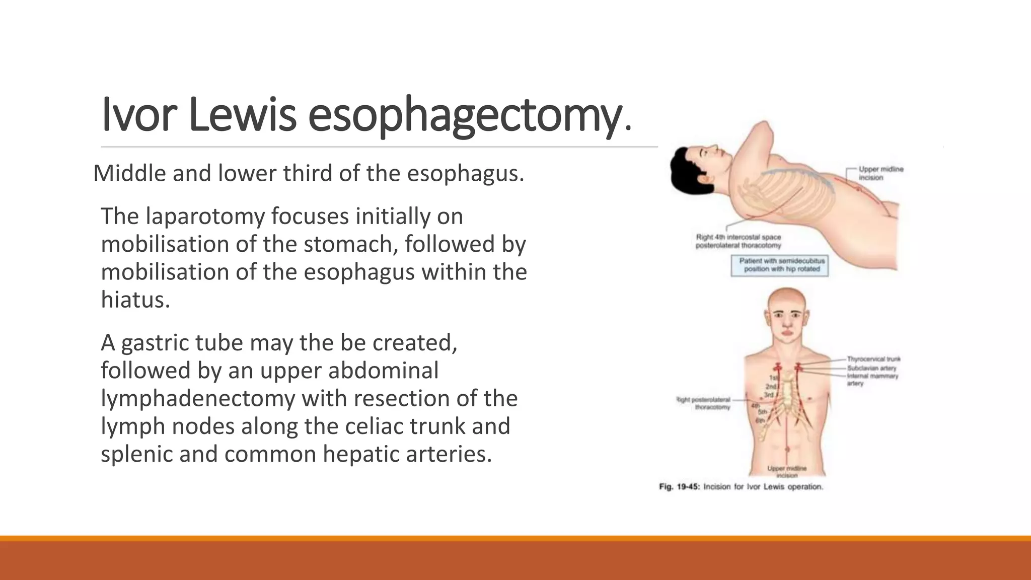
Carcinoma Esophagus
PRESENTER : DR ADITHI S RAO , PG
MODERATOR : DR MURALI MOHAN R, ASSO PROF
DEPARTMENT OF SURGERY,
MIMS, MANDYA
Contents
1. Anatomy
2. Epidemiology
3. Risk factors
4. Staging
5.Lymphatic drainage
6.Types
7.Management
8.Complications
Anatomy
• 25 cm in length
• Lined by Stratified squamous epithelium
• Extent: Cricopharyngeus to GEJ
• Has 3 layers : Mucosa , submucosa ,
Muscularis propria
• Mucosa has M1 – Epithelium
• M2 – Lamina propria
• M3 – Muscularia mucosae
• No Serosa is present , facilitating
Extraesophageal spread of disease.
AJCC 8th ed : 4 parts Cervical, upper thoracic , middle thoracic , lower
thoracic
Cervical – Cricoid cartilage – thoracic inlet (15 -18cm from incisors)
Upper thoracic- thoracic inlet to tracheal bifurcation
Mid thoracic – Tracheal bifurcation to just above GEJ (24-32cm)
Lower thoracic – GEJ to 2cm (32-42cm)
Epidemiology and Incidence
Esophageal cancer is the 8th mc cancer worldwide .
Incidence is 160/1lakh
In India 8-20/1lakh , 6th mc in males
Squamous cell ca accounts for most oesophageal cancers diagnosed.
M:F 3:1 … Adenocacinoma 15:1
Risk factors –SCC EAC
Smoking
Alcohol
Aerodigestive malignancy
Hot beverages
N-nitroso containing foods
Betel nut
Corrosive strictures
Mediastinal radiation
Risk factors
Tobacco and Alcohol use (Independent Risk factors)- Adenoca >1pack/day for
10yrs
Diet and Nutrition : PROTECTIVE – RAW VEG AND FRUITS
Obesity Increased ADENOCA with more BMI
Increased SCC with Low BMI
Abdominal Obesity
GERD – Barret’s Esophagus - AdenoCa
Syndromes : Plummer Vinson Syndrome , Fanconi’s anemia , ZE syndrome
Achalasia – 10-50 fold increase in SCC , AdenoCa (not well established )
usually 2-4 yrs after onset of Achalasia.
Vit C , Selenium , Zinc are protective .
HPV : HPV 16 and 18
Barrett’s Esophagus : Annual Risk per year
No Dysplasia -0.25%
High grade – 6%
Long Segment >3cm -0.22%
PPI – Are Double edge swords
Reduces Acid reflux
In patients with reflux , Increased Bile reflux
Metaplasia , Barrett’s
LES , relaxing drugs : NTG and Anticholinergic (long term )
H.Pylori : CagA production and reflux
Pattern of Spread
SCC is characterised by extensive local growth and proclivity to lymph node mets
As esophagus has no covering Serosa , direct invasion of Contiguous structures
may occur early .
Lesions in the upper esophagus can impinge on or invade the RLN , Carotid A
and Trachea.
If esophageal extension occurs in the mediastinum , tracheo-esophageal
extension occurs in the mediastinum , TEF or BEF may occur .
Tumors in lower 1/3rd can Invade Aorta or pericardium , resulting in
mediastinits, massive haemorrhage or empyema.
Distribution of spread
Commonly occurs by lymphatics :
70%
Hematogenous spread : 25-30%
Mc sites of spread are Lung, liver ,
pleura,bone, kideny
Staging
Rice TW, Patil DT, Blackstone EH. 8th edition AJCC/UICC staging of cancers of the esophagus and esophagogastric junction:
application to clinical practice. Ann Cardiothorac Surg. 2017 Mar;6(2):119-130. doi: 10.21037/acs.2017.03.14. PMID: 28447000;
PMCID: PMC5387145.
cSCC cEAC
Siewert’s Classification
Type 1 : from >1cm to 5cm above the
GEJ
Type 2 : within 1cm cephalad to 2 cm
Caudal to GEJ
Type 3: tumor located 5cm below GEJ
AJCC 8th : Cancers within the proximal 2cm of stomach are staged as
Esophageal CA
If Epicenter is >2cm distal to GEJ is classified as Stomach Cancer
Lymphatics
Lymphatic arrangement in oesophagus is longitudinal
and so spread of carcinoma to distant lymph nodes
occurs early.
Longitudinal lymphatics are 6 times more than
transverse vessels.
More lymph vessels in submucosa than blood vessels.
Lymph nodes are:
-paraoesophageal groups located immediately
adjacent to oesophageal wall. They are deep cervical,
scalene, paratracheal, mediastinal, diaphragmatic,
gastric and coeliac lymph nodes.
-lateral oesophageal groups receive lymph
from para and perioesophageal lymph nodes.
Lymph can travel the entire length of the
Esophagus before draining into the lymph
nodes, the entire esophagus is at potential risk
for lymphatic involvement .
Upto 8cm or more of “Normal “tissue can exist
b/w gross tumor and micrometastais , skip
areas .
Depth of tumor penetration (T stage )
affects the Lymph node Involvement
(LNI)
Intramucosal T1a – lesions (18%LNI)
Submucosal T1b lesions –(55%LNI)
T2 lesions (60%LNI)
T3 lesions (80%LNI)
Management *
LN<50% - Conservative resection f/b
Neoadjuvant therapy
LN>50% - Neoadjuvant f/b resection
Histology
Squamous and Adeno – 95% of all ca
Pseudosarcoma – Spindle cell ca (SCC variant )
Adeno Variants – Adenocystic and Mucoepiermoid (poor prognosis)
Small cell Ca- Arise from Argyrophillic cells – Paraneoplastic
Syndromes like, ADH , Hypercalcemia , similar to SCLC
Non epithelial origin – rare
Malignant Melanomas
Lymphoma – mostly extension , as primary very rare
Barrett’s Esophagus
The presence of Columnar mucosa extending minimum 3cm
unto the Esophagus .
Diagnosis is presently made by presence of any length of
endoscopically identifiable columnar mucosa that is biopsy
proven .
10% GERD develop – Barrett’s
1 in 100 patients on yearly follow-up develop
Adenoca
Clinical manifestations
Early – Asymptomatic
Dysphagia >90% mc , 2/3rd lumen should be obstructed , initially for solids later
progresses to liquids - Odynophagia
Weight loss (40-70%) – chemokines release
Vomiting , blood tinged/ frank bleed
Pain – later stages of disease (bone mets )
Regurgitation – due to total/near total obstruction
Aspiration Pneumonia(TEF)
Cachexia , malnutrition , anaemia , dehydration
Distant metastasis
Invasion of nearby structures
oRLN – Hoarseness of voice
oTrachea – stridor and TEF – cough , choking and cyanosis
oPerforation into pleural cavity – Empyema
oBack pain in celiac axis node involvement
Work-up for Esophageal Ca
Detailed history and Physical examination
Endoscopy
CT
EUS
PET – evaluate mets if any
MRI- not done routinely, inv of vessels and Nerves, detects mets
Bronchoscopy – upper 1/3rd growth
Layngoscopy – vocal cord palsy
Endoscopy
Best , first line –dysphagia
Can differentiate – intra luminal from intra mural
intrinsic from extrinsic
Critical Points:
Location of lesion
Nature of lesion
Extent and Relation to Cricopharynx, GEJ
Chromoendoscopy
>6 BIOPSIES from suspicious lesions are taken .
For tight malignant strictures : Brush cytology can be done .
Criteria for node Positive on EUS:
Hypoechoeic node
Smooth, round border
Width >5mm
>5 LN
Celiac nodes
T3/T4 tumor
EUS: Endoscopic Ultrasound
More accuracy in assessing
- periesophageal and celiac LN involvement
-Transmural extent of disease
Limitation
-less significant accuracy in neoadjuvant f/u cases cannot differentiate tumor
from fibrosis
Accuracy is 85-90%
EUS
Normal
I layer :interface b/w balloon and mucosa
II layer lamina propria and muscularis mucosa
III layer submucosa
IV layer Muscularis propria
V layer interface b/w adventitia and surrounding tissue
Barium swallow
Apple core irregular filling defect
Gives the location and length of the tumor
Sticks to the tumor making biopsy difficult
TEF can also be detected .
CT scan
Important for staging
Normal wall thickness is 3-4mm
Chest and Abdomen – Length, thickness,
LN Liver and Lung mets
Accuracy – 57% T-24% n-74%, M—83%
Many unresectable tumors by CT scan are
deemed resectable at the time of surgery.
Picus angle
Angle between the centre of Aorta
to border of the tumor.
>90 – involvement of Aorta
PET
FDG –PET
Evaluates the Primary mass LN mets
Sn and Sp slightly greater than CT
Value only in evaluating patients on
chemo and Radiotherapy
N staging – limitations : cannot accurately
distinguish infections and ca
Detects distant Mets
Molecular consideration
MANAGEMENT OF
CARCINOMA
ESOPHAGUS
Treatment options
Surgery
Radiation therapy
EBRT
Intraluminal Brachytherapy
Chemotherapy
Multimodality treatment combining above methods
Resectable Tumors Stage I-III
I. Resectable Esophageal and GE tumors:
II. T 1a tumors – inv mucosa but not submucosa – EMR + ablation or
Esophagectomy
III. T1b tumors – inv submucosa – esophagectomy
IV. T1-3 tumors –are resectable even with regional nodal mets
V. T4a tumors involvement of Peridcardium , pleura or diaphragm are
resectable
Unresectable tumors
I. cT4b tumors with inv of heart, great vessels , trachea or adjacent organs
including liver, pancreas and spleen are unresectable.
II. Most pts with multi-station, bulky lymphadenopathy should be considered
unresectable.
III. Pts with EGJ and Supraclavicular lymph node inv should be considered
unresctable.
IV. Pts with distant (non regional LN ) mets are unresectable .
Early Stages
• High grade Dysplasia : EMR
• Early CA Tis, T1a: EMR f/b Ablation
Endoscopic Mucosal Resection
Aim is to excise specimen in one piece
If done in piecemeal study of margins is
difficult .
Assessment : Depth,Lymphovascular
invasion , degree of infiltration .
Advantages : Lower rate of complications ,
good ling term disease control
Disadvantages : High recurrence rate
Cervical Esophagus
Proximal Esophageal tumors are treated by DEFINITIVE
CHEMORADIOTHERAPY
Total- laryngo-pharyngo-esopagectomy – causes losss of function
and is a morbid procedure .
The most common surgical techniques are:
1. Transthoracic esophagectomies, such as;
-Ivor Lewis,
-McKeown techniques.
2.Transhiatal esophagectomy.
Variations of these techniques include different choices of conduit( ie; stomach,
colon, or jejunum) to serve in lieu of the resected esophagus.
Thoracic esophagus
Ivor Lewis esophagectomy.
Middle and lower third of the esophagus.
The laparotomy focuses initially on
mobilisation of the stomach, followed by
mobilisation of the esophagus within the
hiatus.
A gastric tube may the be created,
followed by an upper abdominal
lymphadenectomy with resection of the
lymph nodes along the celiac trunk and
splenic and common hepatic arteries.
McKeown procedure
This technique involves a right thoracotomy, laparotomy, and left
neck incision for creation of a cervical anastomosis.
The key difference between Ivor Lewis and McKeown procedures is
the addition of a left neck incision with a cervical anastomosis in
the McKeown method, which allows resection of more proximal
esophageal tumors and makes management of potential
anastomotic leak easier.
The Ivor Lewis approach allows exploration of the peritoneum early
in the operation, thus avoiding a thoracotomy and the potential
morbidity of thoracic esophagus devascularisation in patients with
metastatic peritoneal disease.
POST OPERATIVE CARE:
Intercoastal tube is removed once lung has expanded fully in 5-7 days.
Respiratory physiotherapy, antibiotics, observation are done regularly.
Nasogastric tube is removed in 7 days.
Oral sips started later.
Often contrast study is done to confirm the adequacy of the stoma.
Transhiatal esophagectomy
It is used for the treatment of tumors involving the lower third of the esophagus and gastric
cardia.
The major advantage of transhiatal technique is the potential to diminish respiratory
complications by avoiding a thoracotomy and an intrathoracic anastomosis with a possible
intrathoracic anastomotic leak.
The disadvantage are an increased rate of anastomotic leak for cervical anastomosis, increased
risk for subsequent stricture formation, and higher risk for recurrent laryngeal injury.
The procedure is performed in three phases.
The first step involves a supraumbilical incision, which allows distal esophageal
dissection.
The second step, the cervical phase, involves an incision parallel to the left
sternocleidomastoid muscle for dissection of the proximal esophagus.
The third phase consists of dissection of the esophagus in the mediastinum
transhiatally.
Once the entire esophagus is mobilised, the cervical esophagus is transected.
Next, a partial gastrectomy is performed, and the esophagus is removed via the
abdominal incision.
The gastric conduit is then brought up to the neck through the posterior
mediastinum to create a cervical esophagogastric anastomosis.
Gastric conduit
Based on the right Gastric and Right Gastro-
epiploic arteries .
Adv : Needs only one anastomosis
Quick alimentation
Early mobilisation and quick operative time
Colonic conduit
Based on Left colic Artery.
Requires 3 anastomosis
Disadvantages
• Technically demanding procedure
• Higher morbidity and mortality compared
with a gastric conduit
• Longer operating times
• Creation of additional anastomose (each
with their own rates of leak and stricture
formation), postoperative pulmonary
complications.
Jejunal interposition
The jejunum as an esophageal replacement is
more resistant to bile and acid.
Use of the jejunum has also been shown to
reduce the occurence of intrinsic disease,
increase peristalsis, contribute to superior
postoperative body weight maintenance, and
decrease the incidence of gastroparesis.
The major disadvantage is that at least three
anastomoses are created, each with its own risk
factors.
Minimally invasive esophagectomy.(video
assisted approaches)
Minimally invasive esophagectomy is indicated in the same patients as the open
techniques, with a few exceptions. Stage T4 cancers are a relative
contraindication.
Laparoscopic mobilistaion of the stomach and abdominal esophagus is done .
Gastric conduit is created using endostapler.
Advantages - better visualisation and dissection, avoiding thoracotomy, faster
recovery.
Disadvantages - technical expertise, learning curve, availability, cost, prolonged
anesthesia.
EXTENT OF LYMPHADENECTOMY
CERVICAL: Carotid cartilage to upper
margin of clavicle
MEDIASTINAL:
STANDARD: Infracarinal
EXTENDED: Standard + Superior
mediastinal + Right paratracheal
COMPLETE: Extended + Subaortic +
Left paratracheal
ABDOMINAL: Superior gastric, Celiac,
Common hepatic nodes
3 field lymphadenectomy: (FOR
SCC)
Bilateral Cervical
Complete Mediastinal
Abdominal
2 field lymphadectomy: (FOR
ADENOCARCINOMA)
Complete mediastinal
Abdominal
Post –op Complications
Pulmonary complications are the most common postoperative
complications, accounting for almost two thirds of postoperative deaths.
Complications include pneumonia, aspiration, acute respiratory distress
syndrome, prolonged ventilator dependence, reintubation, pulmonary edema,
pleural effusion, pneumothorax, tracheobronchial injury, and pulmonary
embolism.
Anastomotic leaks:
Anastomotic leaks occur in 10%-44%.
First 10 days after surgery due to-
Inappropriate tension(excessive or insufficient) at the anastomosis.
Ischemia, if severe, can lead to most feared complication, which is conduit
necrosis.
Anastomotic leaks are mc seen with gastric conduits
than with colon interposition .
Anastomotic leaks also lead to increased risk for
subsequent development of anastomotic strictures.
A leak at the cervical anastomosis is less serious and
easier to treat(by reopening the neck incision) than
an intrathoracic leak, which could lead to
mediastinitis.
Technical complications
I. Recurrent laryngeal nerve injury usually occurs during cervical dissection and
has an incident of 10%-20% when a cervical anastomosis is created.
II. Hemorrhage often occurs because of injury to the spleen, azygos vein,
intercostal vessels, right gastric artery, and lung parenchyma during
retraction or dissection.
III. Loosening of the diaphragmatic hiatus during surgery predisposes to
herniation of the abdominal contents into chest.
Delayed complications.
Delayed complications are generally anastomotic strictures or
disease recurrence, with the most common symptom of both
being dysphagia.
In the early postop period, dysphagia is commonly due to
anastomotic strictures; however, in the late postop period
disease recurrence becomes an increasing concern.
Other complications discussed earlier, including functional
complications, postoperative leaks, fistulas, and diaphragmatic
hernias, may occur in the delayed postoperative period as
well.
Postoperative anastomotic leaks predispose to development
of anastomotic strictures and therefore occur more commonly
when a cervical anastomosis is created.
Palliative therapy
Luminal obstruction and bleeding
Medical c/I to surgery
Inoperable disease
SEMS
DILATATION
ABLATION
EBRT
CHEMOTHERAPY
SEMS : SELF EXPANDABLE METTALIC
STENTS
Chemotherapy
The advantages of adding chemotherapy to esophageal cancer treatment
include potential tumor downstaging prior to surgery as well as targeting
micrometastases, thus decreasing the risk of distant metastasis.
Postoperative taxane-based adjuvant chemotherapy improved the OS of
patients with lymphnode positive thoracic esophageal squamous cell carcinoma
as compared with surgery alone.
NeoAdjuvant CT ( pre-op )
Pre-op Radiation did not show significant
improvements.*
Perioperative chemo – pre+ post OP
Neoadjuvant CT+RT
Definitive Chemoradiotherapy when
surgery is not feasible
*Burmeister B. H. (2015). Role of radiotherapy in the pre-operative management of carcinoma of the esophagus. World
journal of gastrointestinal oncology, 7(1), 1–5. https://doi.org/10.4251/wjgo.v7.i1.1
Pre-op Chemotherapy
Downstaging the disease
Increases rate of R0 resection.
Eradicates Occult mets.
Improves local control
Absolute survival benefit 2yr-13%
5yr – 6.5%
Regimens:
Paclitaxel + Carboplatin
Cisplatin + 5FU
5FU+Oxaliplatin , Irinotecan
Definitive Chemoradiation
Indications :
SCC:
cT1b, T4 N1
Cervical esophagus
Lack of consent for surgery
cT4b
Adenoca :
cT1b, T4 N1
Lack of consent for
surgery
cT4b
Radiotherapy techniques
Jaw tracing technique
T –shaped field RT for
Upper Esophageal tumors.
Targeted therapy
Transtuzumab:
Used as first line in recurrent or advanced metastatic adenoca over expressing
HER2neu.
Response rate is 34%-50%
Ramicirumab –newest congener .
Pembrolizumab
Ipilimumab
Follow-up and Surveillance
Conclusion
Esophagectomy and restoration of gastrointestinal continuity are complex and challenging
procedures.
Understanding the different surgical techniques and recognising their postoperative
appearances is imperative to evaluate postoperative patients.
Radiologists should review the operative report with specific attention the type of conduit used
and the location of anastomosis.
Pulmonary complications and anastomosis are the leading causes of postoperative mortality
after esophagectomy.
Knowledge of the potential comlpications is critical for radiologists to provide effective
postoperative patient care.
Carcinoma Esophagus new.pptx

Carcinoma Esophagus new.pptx

  • 1.
    Carcinoma Esophagus PRESENTER :DR ADITHI S RAO , PG MODERATOR : DR MURALI MOHAN R, ASSO PROF DEPARTMENT OF SURGERY, MIMS, MANDYA
  • 2.
    Contents 1. Anatomy 2. Epidemiology 3.Risk factors 4. Staging 5.Lymphatic drainage 6.Types 7.Management 8.Complications
  • 3.
    Anatomy • 25 cmin length • Lined by Stratified squamous epithelium • Extent: Cricopharyngeus to GEJ • Has 3 layers : Mucosa , submucosa , Muscularis propria • Mucosa has M1 – Epithelium • M2 – Lamina propria • M3 – Muscularia mucosae • No Serosa is present , facilitating Extraesophageal spread of disease.
  • 4.
    AJCC 8th ed: 4 parts Cervical, upper thoracic , middle thoracic , lower thoracic Cervical – Cricoid cartilage – thoracic inlet (15 -18cm from incisors) Upper thoracic- thoracic inlet to tracheal bifurcation Mid thoracic – Tracheal bifurcation to just above GEJ (24-32cm) Lower thoracic – GEJ to 2cm (32-42cm)
  • 6.
    Epidemiology and Incidence Esophagealcancer is the 8th mc cancer worldwide . Incidence is 160/1lakh In India 8-20/1lakh , 6th mc in males Squamous cell ca accounts for most oesophageal cancers diagnosed. M:F 3:1 … Adenocacinoma 15:1
  • 7.
    Risk factors –SCCEAC Smoking Alcohol Aerodigestive malignancy Hot beverages N-nitroso containing foods Betel nut Corrosive strictures Mediastinal radiation
  • 8.
    Risk factors Tobacco andAlcohol use (Independent Risk factors)- Adenoca >1pack/day for 10yrs Diet and Nutrition : PROTECTIVE – RAW VEG AND FRUITS Obesity Increased ADENOCA with more BMI Increased SCC with Low BMI Abdominal Obesity GERD – Barret’s Esophagus - AdenoCa Syndromes : Plummer Vinson Syndrome , Fanconi’s anemia , ZE syndrome
  • 9.
    Achalasia – 10-50fold increase in SCC , AdenoCa (not well established ) usually 2-4 yrs after onset of Achalasia. Vit C , Selenium , Zinc are protective . HPV : HPV 16 and 18 Barrett’s Esophagus : Annual Risk per year No Dysplasia -0.25% High grade – 6% Long Segment >3cm -0.22%
  • 10.
    PPI – AreDouble edge swords Reduces Acid reflux In patients with reflux , Increased Bile reflux Metaplasia , Barrett’s LES , relaxing drugs : NTG and Anticholinergic (long term ) H.Pylori : CagA production and reflux
  • 11.
    Pattern of Spread SCCis characterised by extensive local growth and proclivity to lymph node mets As esophagus has no covering Serosa , direct invasion of Contiguous structures may occur early . Lesions in the upper esophagus can impinge on or invade the RLN , Carotid A and Trachea. If esophageal extension occurs in the mediastinum , tracheo-esophageal extension occurs in the mediastinum , TEF or BEF may occur . Tumors in lower 1/3rd can Invade Aorta or pericardium , resulting in mediastinits, massive haemorrhage or empyema.
  • 12.
    Distribution of spread Commonlyoccurs by lymphatics : 70% Hematogenous spread : 25-30% Mc sites of spread are Lung, liver , pleura,bone, kideny
  • 13.
    Staging Rice TW, PatilDT, Blackstone EH. 8th edition AJCC/UICC staging of cancers of the esophagus and esophagogastric junction: application to clinical practice. Ann Cardiothorac Surg. 2017 Mar;6(2):119-130. doi: 10.21037/acs.2017.03.14. PMID: 28447000; PMCID: PMC5387145.
  • 15.
  • 16.
    Siewert’s Classification Type 1: from >1cm to 5cm above the GEJ Type 2 : within 1cm cephalad to 2 cm Caudal to GEJ Type 3: tumor located 5cm below GEJ AJCC 8th : Cancers within the proximal 2cm of stomach are staged as Esophageal CA If Epicenter is >2cm distal to GEJ is classified as Stomach Cancer
  • 17.
    Lymphatics Lymphatic arrangement inoesophagus is longitudinal and so spread of carcinoma to distant lymph nodes occurs early. Longitudinal lymphatics are 6 times more than transverse vessels. More lymph vessels in submucosa than blood vessels. Lymph nodes are: -paraoesophageal groups located immediately adjacent to oesophageal wall. They are deep cervical, scalene, paratracheal, mediastinal, diaphragmatic, gastric and coeliac lymph nodes.
  • 18.
    -lateral oesophageal groupsreceive lymph from para and perioesophageal lymph nodes. Lymph can travel the entire length of the Esophagus before draining into the lymph nodes, the entire esophagus is at potential risk for lymphatic involvement . Upto 8cm or more of “Normal “tissue can exist b/w gross tumor and micrometastais , skip areas . Depth of tumor penetration (T stage ) affects the Lymph node Involvement (LNI) Intramucosal T1a – lesions (18%LNI) Submucosal T1b lesions –(55%LNI) T2 lesions (60%LNI) T3 lesions (80%LNI) Management * LN<50% - Conservative resection f/b Neoadjuvant therapy LN>50% - Neoadjuvant f/b resection
  • 19.
    Histology Squamous and Adeno– 95% of all ca Pseudosarcoma – Spindle cell ca (SCC variant ) Adeno Variants – Adenocystic and Mucoepiermoid (poor prognosis) Small cell Ca- Arise from Argyrophillic cells – Paraneoplastic Syndromes like, ADH , Hypercalcemia , similar to SCLC Non epithelial origin – rare Malignant Melanomas Lymphoma – mostly extension , as primary very rare
  • 20.
    Barrett’s Esophagus The presenceof Columnar mucosa extending minimum 3cm unto the Esophagus . Diagnosis is presently made by presence of any length of endoscopically identifiable columnar mucosa that is biopsy proven . 10% GERD develop – Barrett’s 1 in 100 patients on yearly follow-up develop Adenoca
  • 23.
    Clinical manifestations Early –Asymptomatic Dysphagia >90% mc , 2/3rd lumen should be obstructed , initially for solids later progresses to liquids - Odynophagia Weight loss (40-70%) – chemokines release Vomiting , blood tinged/ frank bleed Pain – later stages of disease (bone mets ) Regurgitation – due to total/near total obstruction Aspiration Pneumonia(TEF)
  • 24.
    Cachexia , malnutrition, anaemia , dehydration Distant metastasis Invasion of nearby structures oRLN – Hoarseness of voice oTrachea – stridor and TEF – cough , choking and cyanosis oPerforation into pleural cavity – Empyema oBack pain in celiac axis node involvement
  • 25.
    Work-up for EsophagealCa Detailed history and Physical examination Endoscopy CT EUS PET – evaluate mets if any MRI- not done routinely, inv of vessels and Nerves, detects mets Bronchoscopy – upper 1/3rd growth Layngoscopy – vocal cord palsy
  • 26.
    Endoscopy Best , firstline –dysphagia Can differentiate – intra luminal from intra mural intrinsic from extrinsic Critical Points: Location of lesion Nature of lesion Extent and Relation to Cricopharynx, GEJ
  • 27.
  • 28.
    >6 BIOPSIES fromsuspicious lesions are taken . For tight malignant strictures : Brush cytology can be done . Criteria for node Positive on EUS: Hypoechoeic node Smooth, round border Width >5mm >5 LN Celiac nodes T3/T4 tumor
  • 29.
    EUS: Endoscopic Ultrasound Moreaccuracy in assessing - periesophageal and celiac LN involvement -Transmural extent of disease Limitation -less significant accuracy in neoadjuvant f/u cases cannot differentiate tumor from fibrosis Accuracy is 85-90%
  • 30.
    EUS Normal I layer :interfaceb/w balloon and mucosa II layer lamina propria and muscularis mucosa III layer submucosa IV layer Muscularis propria V layer interface b/w adventitia and surrounding tissue
  • 32.
    Barium swallow Apple coreirregular filling defect Gives the location and length of the tumor Sticks to the tumor making biopsy difficult TEF can also be detected .
  • 33.
    CT scan Important forstaging Normal wall thickness is 3-4mm Chest and Abdomen – Length, thickness, LN Liver and Lung mets Accuracy – 57% T-24% n-74%, M—83% Many unresectable tumors by CT scan are deemed resectable at the time of surgery.
  • 34.
    Picus angle Angle betweenthe centre of Aorta to border of the tumor. >90 – involvement of Aorta
  • 36.
    PET FDG –PET Evaluates thePrimary mass LN mets Sn and Sp slightly greater than CT Value only in evaluating patients on chemo and Radiotherapy N staging – limitations : cannot accurately distinguish infections and ca Detects distant Mets
  • 37.
  • 38.
  • 39.
    Treatment options Surgery Radiation therapy EBRT IntraluminalBrachytherapy Chemotherapy Multimodality treatment combining above methods
  • 40.
    Resectable Tumors StageI-III I. Resectable Esophageal and GE tumors: II. T 1a tumors – inv mucosa but not submucosa – EMR + ablation or Esophagectomy III. T1b tumors – inv submucosa – esophagectomy IV. T1-3 tumors –are resectable even with regional nodal mets V. T4a tumors involvement of Peridcardium , pleura or diaphragm are resectable
  • 41.
    Unresectable tumors I. cT4btumors with inv of heart, great vessels , trachea or adjacent organs including liver, pancreas and spleen are unresectable. II. Most pts with multi-station, bulky lymphadenopathy should be considered unresectable. III. Pts with EGJ and Supraclavicular lymph node inv should be considered unresctable. IV. Pts with distant (non regional LN ) mets are unresectable .
  • 43.
    Early Stages • Highgrade Dysplasia : EMR • Early CA Tis, T1a: EMR f/b Ablation
  • 44.
    Endoscopic Mucosal Resection Aimis to excise specimen in one piece If done in piecemeal study of margins is difficult . Assessment : Depth,Lymphovascular invasion , degree of infiltration . Advantages : Lower rate of complications , good ling term disease control Disadvantages : High recurrence rate
  • 45.
    Cervical Esophagus Proximal Esophagealtumors are treated by DEFINITIVE CHEMORADIOTHERAPY Total- laryngo-pharyngo-esopagectomy – causes losss of function and is a morbid procedure .
  • 46.
    The most commonsurgical techniques are: 1. Transthoracic esophagectomies, such as; -Ivor Lewis, -McKeown techniques. 2.Transhiatal esophagectomy. Variations of these techniques include different choices of conduit( ie; stomach, colon, or jejunum) to serve in lieu of the resected esophagus. Thoracic esophagus
  • 47.
    Ivor Lewis esophagectomy. Middleand lower third of the esophagus. The laparotomy focuses initially on mobilisation of the stomach, followed by mobilisation of the esophagus within the hiatus. A gastric tube may the be created, followed by an upper abdominal lymphadenectomy with resection of the lymph nodes along the celiac trunk and splenic and common hepatic arteries.
  • 48.
    McKeown procedure This techniqueinvolves a right thoracotomy, laparotomy, and left neck incision for creation of a cervical anastomosis. The key difference between Ivor Lewis and McKeown procedures is the addition of a left neck incision with a cervical anastomosis in the McKeown method, which allows resection of more proximal esophageal tumors and makes management of potential anastomotic leak easier. The Ivor Lewis approach allows exploration of the peritoneum early in the operation, thus avoiding a thoracotomy and the potential morbidity of thoracic esophagus devascularisation in patients with metastatic peritoneal disease.
  • 49.
    POST OPERATIVE CARE: Intercoastaltube is removed once lung has expanded fully in 5-7 days. Respiratory physiotherapy, antibiotics, observation are done regularly. Nasogastric tube is removed in 7 days. Oral sips started later. Often contrast study is done to confirm the adequacy of the stoma.
  • 50.
    Transhiatal esophagectomy It isused for the treatment of tumors involving the lower third of the esophagus and gastric cardia. The major advantage of transhiatal technique is the potential to diminish respiratory complications by avoiding a thoracotomy and an intrathoracic anastomosis with a possible intrathoracic anastomotic leak. The disadvantage are an increased rate of anastomotic leak for cervical anastomosis, increased risk for subsequent stricture formation, and higher risk for recurrent laryngeal injury.
  • 52.
    The procedure isperformed in three phases. The first step involves a supraumbilical incision, which allows distal esophageal dissection. The second step, the cervical phase, involves an incision parallel to the left sternocleidomastoid muscle for dissection of the proximal esophagus. The third phase consists of dissection of the esophagus in the mediastinum transhiatally. Once the entire esophagus is mobilised, the cervical esophagus is transected. Next, a partial gastrectomy is performed, and the esophagus is removed via the abdominal incision. The gastric conduit is then brought up to the neck through the posterior mediastinum to create a cervical esophagogastric anastomosis.
  • 53.
    Gastric conduit Based onthe right Gastric and Right Gastro- epiploic arteries . Adv : Needs only one anastomosis Quick alimentation Early mobilisation and quick operative time
  • 54.
    Colonic conduit Based onLeft colic Artery. Requires 3 anastomosis Disadvantages • Technically demanding procedure • Higher morbidity and mortality compared with a gastric conduit • Longer operating times • Creation of additional anastomose (each with their own rates of leak and stricture formation), postoperative pulmonary complications.
  • 55.
    Jejunal interposition The jejunumas an esophageal replacement is more resistant to bile and acid. Use of the jejunum has also been shown to reduce the occurence of intrinsic disease, increase peristalsis, contribute to superior postoperative body weight maintenance, and decrease the incidence of gastroparesis. The major disadvantage is that at least three anastomoses are created, each with its own risk factors.
  • 56.
    Minimally invasive esophagectomy.(video assistedapproaches) Minimally invasive esophagectomy is indicated in the same patients as the open techniques, with a few exceptions. Stage T4 cancers are a relative contraindication. Laparoscopic mobilistaion of the stomach and abdominal esophagus is done . Gastric conduit is created using endostapler. Advantages - better visualisation and dissection, avoiding thoracotomy, faster recovery. Disadvantages - technical expertise, learning curve, availability, cost, prolonged anesthesia.
  • 57.
    EXTENT OF LYMPHADENECTOMY CERVICAL:Carotid cartilage to upper margin of clavicle MEDIASTINAL: STANDARD: Infracarinal EXTENDED: Standard + Superior mediastinal + Right paratracheal COMPLETE: Extended + Subaortic + Left paratracheal ABDOMINAL: Superior gastric, Celiac, Common hepatic nodes 3 field lymphadenectomy: (FOR SCC) Bilateral Cervical Complete Mediastinal Abdominal 2 field lymphadectomy: (FOR ADENOCARCINOMA) Complete mediastinal Abdominal
  • 58.
    Post –op Complications Pulmonarycomplications are the most common postoperative complications, accounting for almost two thirds of postoperative deaths. Complications include pneumonia, aspiration, acute respiratory distress syndrome, prolonged ventilator dependence, reintubation, pulmonary edema, pleural effusion, pneumothorax, tracheobronchial injury, and pulmonary embolism.
  • 59.
    Anastomotic leaks: Anastomotic leaksoccur in 10%-44%. First 10 days after surgery due to- Inappropriate tension(excessive or insufficient) at the anastomosis. Ischemia, if severe, can lead to most feared complication, which is conduit necrosis.
  • 60.
    Anastomotic leaks aremc seen with gastric conduits than with colon interposition . Anastomotic leaks also lead to increased risk for subsequent development of anastomotic strictures. A leak at the cervical anastomosis is less serious and easier to treat(by reopening the neck incision) than an intrathoracic leak, which could lead to mediastinitis.
  • 62.
    Technical complications I. Recurrentlaryngeal nerve injury usually occurs during cervical dissection and has an incident of 10%-20% when a cervical anastomosis is created. II. Hemorrhage often occurs because of injury to the spleen, azygos vein, intercostal vessels, right gastric artery, and lung parenchyma during retraction or dissection. III. Loosening of the diaphragmatic hiatus during surgery predisposes to herniation of the abdominal contents into chest.
  • 63.
    Delayed complications. Delayed complicationsare generally anastomotic strictures or disease recurrence, with the most common symptom of both being dysphagia. In the early postop period, dysphagia is commonly due to anastomotic strictures; however, in the late postop period disease recurrence becomes an increasing concern. Other complications discussed earlier, including functional complications, postoperative leaks, fistulas, and diaphragmatic hernias, may occur in the delayed postoperative period as well. Postoperative anastomotic leaks predispose to development of anastomotic strictures and therefore occur more commonly when a cervical anastomosis is created.
  • 64.
    Palliative therapy Luminal obstructionand bleeding Medical c/I to surgery Inoperable disease SEMS DILATATION ABLATION EBRT CHEMOTHERAPY
  • 65.
    SEMS : SELFEXPANDABLE METTALIC STENTS
  • 67.
    Chemotherapy The advantages ofadding chemotherapy to esophageal cancer treatment include potential tumor downstaging prior to surgery as well as targeting micrometastases, thus decreasing the risk of distant metastasis. Postoperative taxane-based adjuvant chemotherapy improved the OS of patients with lymphnode positive thoracic esophageal squamous cell carcinoma as compared with surgery alone.
  • 68.
    NeoAdjuvant CT (pre-op ) Pre-op Radiation did not show significant improvements.* Perioperative chemo – pre+ post OP Neoadjuvant CT+RT Definitive Chemoradiotherapy when surgery is not feasible *Burmeister B. H. (2015). Role of radiotherapy in the pre-operative management of carcinoma of the esophagus. World journal of gastrointestinal oncology, 7(1), 1–5. https://doi.org/10.4251/wjgo.v7.i1.1
  • 69.
    Pre-op Chemotherapy Downstaging thedisease Increases rate of R0 resection. Eradicates Occult mets. Improves local control Absolute survival benefit 2yr-13% 5yr – 6.5% Regimens: Paclitaxel + Carboplatin Cisplatin + 5FU 5FU+Oxaliplatin , Irinotecan
  • 70.
    Definitive Chemoradiation Indications : SCC: cT1b,T4 N1 Cervical esophagus Lack of consent for surgery cT4b Adenoca : cT1b, T4 N1 Lack of consent for surgery cT4b
  • 71.
    Radiotherapy techniques Jaw tracingtechnique T –shaped field RT for Upper Esophageal tumors.
  • 72.
    Targeted therapy Transtuzumab: Used asfirst line in recurrent or advanced metastatic adenoca over expressing HER2neu. Response rate is 34%-50% Ramicirumab –newest congener . Pembrolizumab Ipilimumab
  • 73.
  • 75.
    Conclusion Esophagectomy and restorationof gastrointestinal continuity are complex and challenging procedures. Understanding the different surgical techniques and recognising their postoperative appearances is imperative to evaluate postoperative patients. Radiologists should review the operative report with specific attention the type of conduit used and the location of anastomosis. Pulmonary complications and anastomosis are the leading causes of postoperative mortality after esophagectomy. Knowledge of the potential comlpications is critical for radiologists to provide effective postoperative patient care.