The cardiac conduction system is a network of specialized cardiac muscle cells that initiate and transmit the electrical impulses responsible for the coordinated contractions of each cardiac cycle. These special cells are able to generate an action potential on their own (self-excitation) and pass it on to other nearby cells (conduction), including cardiomyocytes.
The cardiac conduction system is a network of specialized cardiac muscle cells that initiate and transmit the electrical impulses responsible for the coordinated contractions of each cardiac cycle. These special cells are able to generate an action potential on their own (self-excitation) and pass it on to other nearby cells (conduction), including cardiomyocytes.
Cardiac cycle refers to a complete heartbeat from its generation to the beginning of the next beat.
Cardiac events that occur from –
beginning of one heart beat to the beginning of the next are called the cardiac cycle.
Cardiac cycle refers to a complete heartbeat from its generation to the beginning of the next beat.
Cardiac events that occur from –
beginning of one heart beat to the beginning of the next are called the cardiac cycle.
Cardiac muscle has three types of membrane ion channels that play important roles in causing the voltage changes of the action potential. They are (1) fast sodium channels, (2) slow sodium-calcium channels, and (3) potassium channels
Depolarization: First, the action potential of cardiac muscle is caused almost entirely by sudden opening of large numbers of so-called fast sodium channels that allow tremendous numbers of sodium ions to enter the cardiac muscle fiber from the extracellular fluid. These channels are called “fast” channels because they remain open for only a few thousandths of a second and then abruptly close. After depolarization, there's a brief repolarization that takes place with the efflux of potassium through fast acting potassium channels.
Plateau: Secondly, another entirely different population of slow calcium channels, which are also called calcium-sodium channels. This second population of channels differs from the fast sodium channels in that they are slower to open and, even more important, remain open for several tenths of a second. During this time, a large quantity of both calcium and sodium ions flows through these channels to the interior of the cardiac muscle fiber, and this maintains a prolonged period of depolarization, causing the plateau in the action potential.
Repolarization: When the slow calcium-sodium channels do close at the end of 0.2 to 0.3 second and the influx of calcium and sodium ions ceases, the membrane permeability for potassium ions also increases rapidly; this rapid loss of potassium from the fiber immediately returns the membrane potential to its resting level, thus ending the action potential.
Electrocardiography: is the recording of the electrical impulses that are generated in the heart. These impulses initiate the contraction of cardiac muscles.
CARDIAC CYCLE-The cardiac cycle is the performance of the human heart from th...zaaprotta
The cardiac cycle refers to all of the events that occur from the beginning of one heartbeat to the beginning of the next and can be divided into two parts: a period of relaxation known as diastole and a period of contraction known as systole.
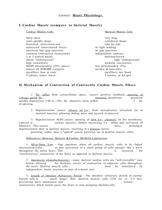
1. Lecture: Heart Phys iol o g y
I. Cardiac Muscl e (comp ar e to Skel e t al Muscl e )
Cardiac Muscle Cells Skelet al Muscle Cells
fairly short very long
semi- spindle shap e cylindrical shap e
branch e d, interconn e c t e d side- by- side
conne c t e d (intercal at e d discs) no tight binding
electrical link (gap junction) no gap junctions
commo n contraction (syncytium) indepe n d e n t contrac t
1 or 2 central nuclei multinucle a t e d
dens e "endomy sium" light "endomy sium"
high vasculatur e medium vasculatur e
MANY mitochon dri a (25% spac e) less mitochon dri a (2%)
almos t all AEROBIC (oxygen) aerobic & ana e robic
myofiber s fuse at ends myofiber s not fused
T tubules wider, fewer T tubules at A/I spot
II. Mechani sm of Contrac t i on of Contrac t i l e Cardiac Muscl e Fiber s
1. Na + influx from extrac ellular spac e, caus e s positive feedb a ck opening of
voltage- gate d Na + chann el s ;membr a n e potential
quickly depolarize s (-90 to +30); Na + chann el s close within 3 ms
of opening.
2. Depolarization caus e s relea s e of Ca ++ from sarcopl a smic reticulum (as in
skelet al muscle), allowing sliding actin and myosin to proce e d.
3. Depolarization ALSO caus e s opening of slow Ca ++ chann el s on the membr a n e
(special to cardiac muscl e), further increa sing Ca ++ influx and activation of
filame nt s . This caus e s more prolonge d
depolarization than in skelet al muscl e, resulting in a plate a u action
potential, rather than a "spiked" action potenti al (as in skelet al muscl e cells).
Differenc e s Betwe e n Skelet al & Cardiac MUSCLE Contraction
1. All-or-None Law - Gap junctions allow all cardiac muscle cells to be linked
electroch emi c ally, so that activation of a small group of cells spre a d s like a wave
througho u t the entire hear t. This is esse nti al for
"synchroni s tic" contraction of the hear t as oppos e d to skelet al muscle.
2. Automicity (Autorhythmicity) - some skelet al cardiac cells are "self- excitabl e" (see
below, allowing for rhythmic waves of contraction to adjac e nt cells throughout
the hear t. Skelet al muscle cells mus t be stimulat e d by
indepe n d e n t motor neurons as part of a motor unit.
3. Length of Absolute Refractory Period - The absolut e refractory period of cardiac
muscle cells is much longer than skelet al muscle cells (250 ms vs. 2- 3 ms),
preventing wave summa t ion and tet anic
contractions which would caus e the hear t to stop pumping rhythmic ally.
2. III. Internal Conduc t i on (Stimul a t i o n) Sys t em of the Heart
A. Gener al Proper tie s of Conduction
1. hear t can beat rhythmic ally without nervous input
2. nodal system (cardiac conduction syst em) - special aut orhy thmi c cell s of
hear t that initiate impulse s for wave- like
contraction of entire hear t (no nervous stimulation need e d for thes e)
3. gap junctions - electrically couple all cardiac muscle cells so that depol arization
sweep s acros s hear t in seque nt i al fashion from atria to ventricles
B. "Pacema k e r " Featur e s of Autorhythmic Cells
1. pacema k e r potenti al s - "autorhythmi c cells" of hear t muscl e creat e action
potential s in rhythmic fashion; this is due to
unst a bl e resting potenti al s which slowly drift back toward
threshold voltage after repolarization from a previous cycle.
Theoretic al Mechanism of Pacemak e r Potential :
a. K + leak chann el s allow K+ OUT of the cell more slowly than in skelet al muscl e
b. Na + slowly leaks into cell, causing membr a n e potenti al to slowly drift up to
the threshold to trigger Ca ++ influx from outside (-40 mv)
c. when threshold for voltage- gat e d Ca ++ chann el s is reache d (-40 mv), fast
3. calcium chann el s open, permit ting explosive entry of Ca ++ from of the
cell, causing sharp rise in level of
depolarization
d. when peak depol arization is achieve d, voltage- gat ed K+ chann el s open,
causing repolarization to the "unst a bl e resting pot enti al"
e. cycle begins again at step a.
C. Anatomical Seque n c e of Excitation of the Hear t
1. Autorhythmi c Cell Location & Order of Impulse s
(right atrium) sinoatrial node (SA) ->
(right AV valve) atriovent ricular node (AV)
->
atriovent ricular bundle (bundle of His) ->
right & left bundle of His branch e s ->
Purkinje fibers of ventricular walls
(from SA through compl et e hear t contraction = 220 ms = 0.22 s)
a. sinoatrial node (SA node) "the pacema k e r " - has the faste s t autorhythmic rate
(70- 80 per minut e), and set s the pac e for the entire hear t; this rhythm is
called the sinus rhythm ; locat ed in
right atrial wall, just inferior to the superior vena cava
b. atriovent ricular node (AV node) - impulse s pas s from SA via gap junctions in
about 40 ms.; impulse s are delaye d about 100 ms to allow comple tion of the
contraction of both atria; locat ed just
above tricuspid valve (betwe e n right atrium & ventricle)
c. atriovent ricular bundle (bundle of His) - in the interATRIAL septum (conne c t s L
and R atria)
4. d. L and R bundl e of His branch e s - within the interVENTRICULAR septum (betwe e n
L and R ventricles)
e. Purkinje fibers - within the lateral walls of both the L and R ventricles; since left
ventricle much larger, Purkinjes more elabor a t e here; Purkinje fibers
innervat e "papillary muscles" before
ventricle walls so AV can valves preve nt backflow
D. Special Consider a tions of Wave of Excitation
1. initial SA node excitation caus e s contraction of both the R and L atria
2. contraction of R and L ventricles begins at APEX of hear t (inferior point), ejecting
blood superiorly to aorta and pulmon a ry artery
3. the bundl e of His is the ONLY link betwe e n atrial contr action and ventricular
contraction; AV node and bundl e mus t work for ventricular
contractions
4. since cells in the SA node has the faste s t autorhythmic rate (70- 80 per minut e) ,
it drives all other autorhythmic cent er s in a normal hear t
5. arrhythmi a s - uncoordina t e d hear t contractions
6. fibrillation - rapid and irregular contr actions of the hear t chamb e r s ; reduc e s
efficiency of hear t
7. defibrillation - application of electric shock to hear t in att emp t to retain normal
SA node rate
5. 8. ectopic focus - autorhythmic cells other than SA node take over hear t rhythm
9. nodal rhythm - when AV node takes over pacema k e r function (40- 60 per
minut e)
10. extrasys tole - when outside influenc e (such as drugs) leads to prema tur e
contraction
11. hear t block - when AV node or bundle of His is not transmi t ting sinus rhythm
to ventricles
E. External Innerva tion Regulating Heart Function
1. hear t can beat without external innervation
2. ext ernal innervation is from AUTONOMIC SYSTEM
parasymp a t h e t i c - (acetylcholine) DECREASES rate of contrac tions
cardioinhibitory cent er (medulla) ->
vagus nerve (cranial X) ->
hear t
sympa th e t ic - (norepine p hrine) INCREASES rate of contractions
cardioa cc el er a tory cent er (medulla) ->
lateral horn of spinal cord to prega n glionics T1-T5 ->
postg a nlionics cervical/thor a cic ganglia ->
hear t
IV. Electroc ardi o g r a p h y : Electrical Activity of the Heart
6. A. Deflection Waves of ECG
1. P wave - initial wave, demon s t r a t e s the depolarization from SA Node through
both ATRIA; the ATRIA contract about 0.1 s after start of P Wave
2. QRS compl ex - next series of deflections , demo n s t r a t e s the depolarization of AV
node through both ventricles; the ventricles contract througho ut the period of
the QRS complex, with a short delay after the end of atrial contraction;
repolarization of atria also obscur e d
3. T Wave - repolarization of the ventricles (0.16 s)
4. PR (PQ) Interval - time period from beginning of atrial contrac tion to beginning
of ventricular contraction (0.16 s)
5. QT Interval - the time of ventricular contrac tion (about 0.36 s); from beginning
of ventricular depolarization to end of repolarization
V. The Normal Cardiac Cycle
A. Gener al Concept s
1. systole - period of chamb e r contra ction
2. diastole - period of chamb e r relaxation
3. cardiac cycle - all event s of systole and diastol e during one hear t flow cycle
B. Event s of Cardiac Cycle
7. 1. mid- to- late diastole: ventricles filled
* pres sur e: LOW in chamb e r s ; HIGH in aorta/pulmon a ry trunk
* aortic/pulmo n a ry semilun ar valves CLOSED
* blood flows from vena cavas /pulmo n a ry vein INTO atria
* blood flows through AV valves INTO ventricles (70%)
* atrial systole propels more blood > ventricles (30%)
* atrial dias tole returns through end of cycle
2. ventricular systole: blood eject ed from hear t
* filled ventricles begin to contract, AV valves CLOSE
* isovolume t ric contraction phas e - ventricles CLOSED
* contr action of closed ventricles incre a s e s pres sur e
* ventricular ejection phas e - blood forced out
* semiluna r valves open, blood -> aorta & pulmon a ry trunk
3. isovolume t ric relaxation: early diastole
* ventricles relax, ventricular pres sur e become s LOW
* semiluna r valves close, aorta & pulmon a ry trunk backflow
* dicrotic notch - brief incre a s e in aortic pres sur e
TOTAL CARDIAC CYCLE TIME = 0.8 second
(normal 70 beat s /minut e )
atrial systole (contra ction) = 0.1 second
ventricular systole (contra ction) = 0.3 second
quiesc e nt period (relaxation) = 0.4 second
8. VI. Heart Sound s : St e th o s c o p e List enin g
A. Overview of Heart Sounds
1. lub- dub, - , lub,dub, -
2. lub - closur e of AV valves, onse t of ventricular systole
3. dub - closur e of semiluna r valves, onset of diastole
4. paus e - quiesc e n t period of cardiac cycle
5. tricuspid valve (lub) - RT 5th intercos t al, medial
6. mitral valve (lub) - LT 5th intercos t al, lateral
7. aortic semilunar valve (dub) - RT 2nd intercos t al
8. pulmon a ry semilunar valve (dub) - LT 2nd intercos t al
B. Heart Murmurs
1. murmur - sounds other than the typical "lub- dub"; typically cause d by
disruptions in flow
2. incomp e t e n t valve - swishing sound just AFTER the normal "lub" or "dub"; valve
does not compl et ely close, some regurgit ation of blood
3. stenotic valve - high pitched swishing sound when blood should be flowing
through valve; narrowing of outlet in the open stat e
9. VII. Cardiac Output - Blood Pumpin g of the Heart
A. Gener al Variable s of Cardiac Output
1. Cardiac Output (CO) - blood amount pump e d per minut e
2. Stroke Volume (SV) - ventricle blood pump e d per min.
3. Heart Rate (HR) - cardiac cycles per minut e
CO (ml/min) = HR (beat s /min) X SV (ml/be a t )
normal CO = 75 beat s /min X 70 ml/be a t = 5.25 L/min
B. Regulation of Stroke Volume (SV)
1. end diastolic volume (EDV) - total blood collect ed in ventricle at end of dias tole;
det ermin e d by length of diastol e and venous pres sur e (~120 ml)
2. end systolic volume (ESV) - blood left over in ventricle at end of contrac tion (not
pump e d out); det ermin e d by force of ventricle contraction and arterial
blood pres sur e (~50 ml)
SV (ml/be a t ) = EDV (ml/be a t ) - ESV (ml/be a t )
normal SV = 120 ml/be at - 50 ml/be a t = 70 ml/be a t
3. Frank- Starling Law of the Heart - critical factor for stroke volume is "degre e of
stretch of cardiac muscle cells"; more stretch = more contraction force
a. increa s e d EDV = more contraction force
i. slow hear t rate = more time to fill
ii. exercise = more venous blood return
C. Regulation of Heart Rate (Autonomic, Chemic al, Other)
10. 1. Autonomic Regulation of Heart Rate (HR)
a. symp a t h e t ic - NOREPINEPHRINE (NE) increa s e s hear t rate (maint ains Stroke
Volume)
b. para symp a t h e t i c - ACETYLCHOLINE (ACh) decre a s e s hear t rate
c. vagal tone - parasymp a t h e t ic inhibition of inher ent rate of SA node, allowing
normal HR
d. baror ec e pt or s , pres sor e c e p tor s - monitor chang e s in blood pres sur e and
allow reflex activity with the autonomic nervous syst em
2. Hormon al and Chemic al Regulation of Hear t Rate (HR)
a. epinep hrine - hormon e relea s e d by adren al medulla during stres s ; incre a s e s
hear t rate
b. thyroxine - hormon e relea s e d by thryroid; increa s e s hear t rate in large
quantitie s; amplifies effect of epinephr ine
c. Ca ++ , K + , and Na + levels very import ant ;
* hyperkal emi a - incre a s e d K+ level; KCl used to stop hear t on lethal injection
* hypokal emi a - lower K+ levels; leads to abnormal hear t rate rhythms
* hypoc alc emi a - depr e s s e s hear t function
* hyperc alc emi a - incre a s e s contrac tion phas e
* hypern a t r emi a - HIGH Na + conc ent r a t ion; can block Na + transpor t & muscl e
contraction
3. Other Factors Effecting Heart Rate (HR)
a. normal hear t rate - fetus 140- 160 beat s /minut e
femal e 72- 80 beat s /minut e
male 64- 72 beat s /minut e
b. exercise - lowers resting hear t rate (40- 60)
c. heat - incre a s e s hear t rate significantly
d. cold - decre a s e s hear t rate significantly
e. tachyc ar di a - HIGHER than normal resting hear t rate (over 100); may lead to
fibrillation
f. bradyc ardi a - LOWER than normal resting hear t rate (below 60);
para symp a t h e t i c drug side effect s ;
physical conditioning; sign of pathology in non- healthy patient
VIII. Imbalanc e of Cardiac Output & Heart Pathol o gi e s
A. Imbalanc e of Cardiac Output
1. conge s tive hear t failure - hear t cannot pump sufficiently to me e t needs of the
body
a. coronary atherosclerosi s - leads to gradu al occlusion of hear t ves s el s ,
reducing oxyge n nutrient supply to
cardiac muscle cells; (fat & salt diet, smoking, stres s )
b. high blood pres sur e - when aortic pres sur e get s to large, left ventricle
cannot pump properly, incre a sing ESV, and lowering SV
11. c. myoc ardi al infarct (MI) - "hear t cell death" due to numero u s factors,
including coronary artery occlusion
d. pulmon a ry conge s tion - failure of LEFT hear t; leads to buildup of blood in
the lungs
e. peripher al conge s tion - failure of RIGHT hear t; pools in body, leading to
edema (fluid buildup in are a s such as feet, ankles, finger s)
B. Heart Pathologie s (Disea s e s of the Heart)
1. conge ni t al hear t defect s - hear t problems that are pres e nt at the time of birth
a. pat ent ductus arteriosus - bypa s s hole betwe e n pulmon a ry trunk and aort a
does not close
2. sclerosis of AV valves - fatty deposit s on valves; particularly the mitral valve of
LEFT side; leads to hear t murmur
3. decline in cardiac reserve - hear t efficiency decre a s e s with age
4. fibrosis and conduction problems - node s and conduction fibers become scarred
over time; may lead to arrhythmi a s