SlideShare a Scribd company logo
Introduction
Materials and Methods
Results
Spectroscopic Characterization of Protein Structure Dynamics in Aqueous States
Yemin Xu1, Chris Carpenter2, Korben Knudson3, Rina Dukor4, John Carpenter5, Theodore Randolph3
1Biochemistry Department, University of Colorado Boulder
2Dept. of Physics, University of Colorado Boulder
3Dept. of Chemical and Biological Engineering, University of Colorado Boulder
4BioTools, Inc
5Dept. of Pharmaceutical Sciences, University of Colorado, Denver
Maintaining protein high order structure integrity is critical for
developing protein pharmaceuticals. Mis-folded or unfolded protein
pharmaceuticals will compromise their efficacy, change the
pharmacokinetics profile and/or cause severe immune-responses.
Thus, it is important to develop fast and accurate methods to
routinely monitor the proteins pharmaceutical’s structures.
In this study, lysozyme was used as model protein, formulated at
50mg/ml and incubated at elevated temperatures (40 and 65 °C) for
one week. Raman spectroscopy and Circular Dichroism (CD) were
employed to measure the lysozyme structure in aqueous solution.
Particle formation was monitored by Flowcam, and soluble
aggregates were examined and quantified by size exclusion
chromatography (SEC).
The extent of structural perturbation observed during incubation
exhibited good correlation with the rate of aggregation and sub-
visible particle formation under accelerated conditions. The results
demonstrate that Raman spectroscopy could be a quick and reliable
way to predict protein stability and high order structure integrity
during incubation.
50mg/mL
Lysozyme (H2O)
pH 4.0
Samples divided
into two groups
(40oCand 65oC)
40oC 65oC
Samples were placed in the incubators for measuring at various
time points (0-Point, Day 1, Day 4 , and Day 7, respectively).
At each time point, triplicate samples were removed from the
incubator and measured by different methods.
Lysozyme from chicken egg white (Sigma-L6876):
50mg/ml (H2O, pH 4.0)
Incubation temperature:
40±2 and 65±2oC
Incubation time point:
0, 1, 4 and 7 days
Protein particle measurement:
Flowcam (0.2mL sample size)
Soluble protein aggregation:
SEC (mobile phase: 0.1 M Phosphate buffer 0.1 M Na2SO4, pH 6.7 )
Non-Reducing SDS Gel
Proteins structure measurement:
CD (Far-UV 0.3mg/ml, Near-UV 50mg/ml)
Raman (50mg/ml, laser power 100mW at 780nm)
2013 Colorado Protein Stability Conference
Discussion
Fig 4: Protein secondary and tertiary structure measured by circular dichroism (CD)
A) Far-UV: measuring secondary structure; B) near-UV: measuring tertiary structure.
Each color curve is the average of triplicate result. Different colors show different samples incubated at different temperatures.
CD data show similar secondary structures for all samples, however, tertiary structures measured by near-UV indicate protein
tertiary strcutures of samples incubated at 65oC differ from others after 4 days. Each spectra represents average of triplicate
measurements.
A B
A
Disulfide bond S-S
C
Amide III
B
Tyrosine
D
Amide I
Fig 1: Protein structure measured by Raman
Center panel is the entire spectra of 50mg/ml lysozyme in aqueous solution (control) and 80oC for 20 hours.
Close look (A, B, C, D)of each representative region of disulfide bond, tyrosine, amide III and amide I indicates that protein
structures were significantly perturbed after storing at high temperature (80oC).
A
Disulfide bond S-S
C
Amide III
B
Tyrosine
D
Amide I
Fig 2: Raman spectra of proteins incubated for various times at 40oC
Center panel is the entire spectra of 50mg/ml lysozyme at 40oC for different time points
Close look (A, B, C, D)of each representative region of disulfide bond, tyrosine, amide III and amide I indicates that protein
structures were similar after storing at 40oC for 1 week. Each spectra represents average of triplicate measurements.
A
Disulfide bond S-S
C
Amide III
B
Tyrosine
D
Amide I
Fig 3: Raman spectra of proteins incubated for various times at 60oC
Center panel is the entire spectra of 50mg/ml lysozyme at 60oC for different time points
Close look (A, B, C, D)of each representative region of disulfide bond, tyrosine, amide III and amide I indicates that protein
structures were disrupted after 4 days storing at 60oC. Each spectra represents average of triplicate measurements.
Fig 5: percentage of remaining monomer measured by SEC
Results of triplicate measurements show that no
noticeable protein loss was seen for samples incubated at
40oC for one week, in contrast, there was around 10%
monomer lysozyme lost after one week at 65oC.
Fig 6: non-reducing SDS gel (coomassie blue stain)
Non-reducing SDS gel shows that there are noticeable
aggregates from samples incubated at 65oC after 4 days.
MW kD
50
37
25
20
15
10
Fig 7: Sub-visible particle concentration measured by flowcam
Within the flowcam measurable size range, protein particle size and
concentration increase for both 40oC and 60oC samples. Representative
images from each size range are shown
Conclusions References
 Structure information from Raman spectroscopy and CD measurement show that there are structure perturbations in
samples incubated at 65oC after 4 days.
 Particle concentration decreased after 4 days, it may due to the size of protein aggregates is beyond the capable range of
flowcam
 Far-UV measurement did not pick up noticeable differences between samples, but near-UV did, which indicates that
under experiment condition, lysozyme likely undergoes tertiary structural perturbation.
 Raman spectroscopy is a useful tool to examine the protein high order structure, and it is capable to identify the
structural differences occurring at various position (eg. Disulfide bond, tyrosine, amide III and amide I region).)
 It is critical and helpful to use orthogonal methodologies to evaluate protein stability.
1. R. S. Porubcan, K. L. Watters and J. T. McFarland, Arch. Biochem. Biophys., 1978, 186, 255-264.
2.J. L. Lippert, D. Tyminski and P. J. Desmeules, Journal of the American Chemical Society, 1976, 98, 7075-7080.
3.A. T. Tu, Raman spectroscopy in biology : principles and applications, Wiley, New York, 1982.
4.Z.-Q. Wen, J. Pharm. Sci., 2007, 96, 2861-2878.

More Related Content

What's hot

Characterization of protein
Characterization of proteinCharacterization of protein
Characterization of protein
KAUSHAL SAHU
 
In-vitro antioxidant and GC-MS analysis ethanolic extract of poly herbal drug
In-vitro antioxidant and GC-MS analysis ethanolic extract of poly herbal drugIn-vitro antioxidant and GC-MS analysis ethanolic extract of poly herbal drug
In-vitro antioxidant and GC-MS analysis ethanolic extract of poly herbal drug
Skyfox Publishing Group
 
Aquasome
AquasomeAquasome
Chapter 3(part2) - Protein purification and analysis
Chapter 3(part2) - Protein purification and analysisChapter 3(part2) - Protein purification and analysis
Chapter 3(part2) - Protein purification and analysis
Ammedicine Medicine
 
Various techniques for study of Crystal Properties
Various techniques for study of Crystal PropertiesVarious techniques for study of Crystal Properties
Various techniques for study of Crystal Properties
Cooling Crystal
 
Crystal
CrystalCrystal
MABs Stability – Analytical Techniques
MABs Stability – Analytical TechniquesMABs Stability – Analytical Techniques
MABs Stability – Analytical Techniques
Pharmaxo
 
RSC advances
RSC advancesRSC advances
Analytical techniques for separation or purification of proteins
Analytical techniques for separation or purification of proteinsAnalytical techniques for separation or purification of proteins
Analytical techniques for separation or purification of proteins
rohini sane
 
SELF-ASSEMBLY AMPHIPHILIC CHITOSAN NANOCARRIERS FOR ORAL CO-DELIVERY OF HYDRO...
SELF-ASSEMBLY AMPHIPHILIC CHITOSAN NANOCARRIERS FOR ORAL CO-DELIVERY OF HYDRO...SELF-ASSEMBLY AMPHIPHILIC CHITOSAN NANOCARRIERS FOR ORAL CO-DELIVERY OF HYDRO...
SELF-ASSEMBLY AMPHIPHILIC CHITOSAN NANOCARRIERS FOR ORAL CO-DELIVERY OF HYDRO...
Tomsk Polytechnic University
 
Electrosomes preparation and application
Electrosomes preparation and applicationElectrosomes preparation and application
Electrosomes preparation and application
mahesh745
 
Protein separation
Protein separationProtein separation
Protein separation
Sourav Sharma
 
PRINCIPLE AND APPLICATIONS OF CELL VIABILITY, GLUCOSE UPTAKE AND CALCIUM UPT...
PRINCIPLE AND APPLICATIONS OF CELL VIABILITY, GLUCOSE UPTAKE  AND CALCIUM UPT...PRINCIPLE AND APPLICATIONS OF CELL VIABILITY, GLUCOSE UPTAKE  AND CALCIUM UPT...
PRINCIPLE AND APPLICATIONS OF CELL VIABILITY, GLUCOSE UPTAKE AND CALCIUM UPT...
Raghavendra institute of pharmaceutical education and research .
 
Protein Purification Lecture
Protein Purification LectureProtein Purification Lecture
Protein Purification Lecture
ouopened
 
Aquasomes
AquasomesAquasomes
Aquasomes
Zahid1392
 
AQUASOMES: A NOVEL CARRIER FOR DRUG DELIVERY SYSTEM
AQUASOMES: A NOVEL CARRIER FOR DRUG DELIVERY SYSTEMAQUASOMES: A NOVEL CARRIER FOR DRUG DELIVERY SYSTEM
AQUASOMES: A NOVEL CARRIER FOR DRUG DELIVERY SYSTEM
MUSTAFIZUR RAHMAN
 
Aquasomes
AquasomesAquasomes
10.analytical biochemistry
10.analytical biochemistry10.analytical biochemistry
10.analytical biochemistry
Happy Learning
 
Poster Spring 2016
Poster Spring 2016Poster Spring 2016
Poster Spring 2016
Adam Hildebrandt
 
A review on antioxidant methods
A review on antioxidant methodsA review on antioxidant methods
A review on antioxidant methods
neeluyadav
 

What's hot (20)

Characterization of protein
Characterization of proteinCharacterization of protein
Characterization of protein
 
In-vitro antioxidant and GC-MS analysis ethanolic extract of poly herbal drug
In-vitro antioxidant and GC-MS analysis ethanolic extract of poly herbal drugIn-vitro antioxidant and GC-MS analysis ethanolic extract of poly herbal drug
In-vitro antioxidant and GC-MS analysis ethanolic extract of poly herbal drug
 
Aquasome
AquasomeAquasome
Aquasome
 
Chapter 3(part2) - Protein purification and analysis
Chapter 3(part2) - Protein purification and analysisChapter 3(part2) - Protein purification and analysis
Chapter 3(part2) - Protein purification and analysis
 
Various techniques for study of Crystal Properties
Various techniques for study of Crystal PropertiesVarious techniques for study of Crystal Properties
Various techniques for study of Crystal Properties
 
Crystal
CrystalCrystal
Crystal
 
MABs Stability – Analytical Techniques
MABs Stability – Analytical TechniquesMABs Stability – Analytical Techniques
MABs Stability – Analytical Techniques
 
RSC advances
RSC advancesRSC advances
RSC advances
 
Analytical techniques for separation or purification of proteins
Analytical techniques for separation or purification of proteinsAnalytical techniques for separation or purification of proteins
Analytical techniques for separation or purification of proteins
 
SELF-ASSEMBLY AMPHIPHILIC CHITOSAN NANOCARRIERS FOR ORAL CO-DELIVERY OF HYDRO...
SELF-ASSEMBLY AMPHIPHILIC CHITOSAN NANOCARRIERS FOR ORAL CO-DELIVERY OF HYDRO...SELF-ASSEMBLY AMPHIPHILIC CHITOSAN NANOCARRIERS FOR ORAL CO-DELIVERY OF HYDRO...
SELF-ASSEMBLY AMPHIPHILIC CHITOSAN NANOCARRIERS FOR ORAL CO-DELIVERY OF HYDRO...
 
Electrosomes preparation and application
Electrosomes preparation and applicationElectrosomes preparation and application
Electrosomes preparation and application
 
Protein separation
Protein separationProtein separation
Protein separation
 
PRINCIPLE AND APPLICATIONS OF CELL VIABILITY, GLUCOSE UPTAKE AND CALCIUM UPT...
PRINCIPLE AND APPLICATIONS OF CELL VIABILITY, GLUCOSE UPTAKE  AND CALCIUM UPT...PRINCIPLE AND APPLICATIONS OF CELL VIABILITY, GLUCOSE UPTAKE  AND CALCIUM UPT...
PRINCIPLE AND APPLICATIONS OF CELL VIABILITY, GLUCOSE UPTAKE AND CALCIUM UPT...
 
Protein Purification Lecture
Protein Purification LectureProtein Purification Lecture
Protein Purification Lecture
 
Aquasomes
AquasomesAquasomes
Aquasomes
 
AQUASOMES: A NOVEL CARRIER FOR DRUG DELIVERY SYSTEM
AQUASOMES: A NOVEL CARRIER FOR DRUG DELIVERY SYSTEMAQUASOMES: A NOVEL CARRIER FOR DRUG DELIVERY SYSTEM
AQUASOMES: A NOVEL CARRIER FOR DRUG DELIVERY SYSTEM
 
Aquasomes
AquasomesAquasomes
Aquasomes
 
10.analytical biochemistry
10.analytical biochemistry10.analytical biochemistry
10.analytical biochemistry
 
Poster Spring 2016
Poster Spring 2016Poster Spring 2016
Poster Spring 2016
 
A review on antioxidant methods
A review on antioxidant methodsA review on antioxidant methods
A review on antioxidant methods
 

Similar to Breckenridge Protein Stability Conference - 2013

Bioanalytical method development and validation .
Bioanalytical method development and validation .Bioanalytical method development and validation .
Bioanalytical method development and validation .
Shubham Bora
 
POSTER- 1091210015, 1091210038 & 1091210040
POSTER- 1091210015, 1091210038 & 1091210040POSTER- 1091210015, 1091210038 & 1091210040
POSTER- 1091210015, 1091210038 & 1091210040
Aditya Rishi
 
Protein Chromatography
Protein ChromatographyProtein Chromatography
Protein Chromatography
Nicole Gomez
 
Effectiveness Of The Treatment Before And After The...
Effectiveness Of The Treatment Before And After The...Effectiveness Of The Treatment Before And After The...
Effectiveness Of The Treatment Before And After The...
Olga Bautista
 
-Unprotected Docs-Student Posters 2013-Marchelle Meza (2)
-Unprotected Docs-Student Posters 2013-Marchelle Meza (2)-Unprotected Docs-Student Posters 2013-Marchelle Meza (2)
-Unprotected Docs-Student Posters 2013-Marchelle Meza (2)
Marchelle Meza
 
In-vitro evaluation techniques of anticancer, anti oxidant, anti microbial
 In-vitro evaluation techniques of anticancer, anti oxidant, anti microbial  In-vitro evaluation techniques of anticancer, anti oxidant, anti microbial
In-vitro evaluation techniques of anticancer, anti oxidant, anti microbial
ZakiyaUsmani
 
Analysis of bioreactor parameters online and offline
Analysis of bioreactor parameters online and offlineAnalysis of bioreactor parameters online and offline
Analysis of bioreactor parameters online and offline
vikash_94
 
Biophysics 2016_Kayla Washenberger
Biophysics 2016_Kayla WashenbergerBiophysics 2016_Kayla Washenberger
Biophysics 2016_Kayla Washenberger
Kayla Washenberger
 
Analytical tech in pre formulation 112070804009
Analytical tech in pre formulation 112070804009Analytical tech in pre formulation 112070804009
Analytical tech in pre formulation 112070804009
Patel Parth
 
207 progress with the calibration of a 3 fr near infrared catheter
207 progress with the calibration of a 3 fr near infrared catheter207 progress with the calibration of a 3 fr near infrared catheter
207 progress with the calibration of a 3 fr near infrared catheter
SHAPE Society
 
Progress with the a 3 fr nir fiber optic catheter
Progress with the a 3 fr nir fiber optic catheterProgress with the a 3 fr nir fiber optic catheter
Progress with the a 3 fr nir fiber optic catheter
Society for Heart Attack Prevention and Eradication
 
Nir catheter final
Nir catheter finalNir catheter final
207 progress with the calibration of a 3 fr near infrared catheter
207 progress with the calibration of a 3 fr near infrared catheter207 progress with the calibration of a 3 fr near infrared catheter
207 progress with the calibration of a 3 fr near infrared catheter
Society for Heart Attack Prevention and Eradication
 
Conventional and modern methods for detection of spoilage
Conventional and modern methods for detection of spoilageConventional and modern methods for detection of spoilage
Conventional and modern methods for detection of spoilage
AnuKiruthika
 
poster presentattion on algae-final rkb-final
poster presentattion on algae-final rkb-finalposter presentattion on algae-final rkb-final
poster presentattion on algae-final rkb-final
chaitanyakumar palakonda
 
Characteristics used in classification.pptx
Characteristics used in classification.pptxCharacteristics used in classification.pptx
Characteristics used in classification.pptx
Micro biology
 
Voss et al. - 2006 - Identification and characterization of riproximin,
Voss et al. - 2006 - Identification and characterization of riproximin,Voss et al. - 2006 - Identification and characterization of riproximin,
Voss et al. - 2006 - Identification and characterization of riproximin,
Cristina Voss
 
Identification of bacteria for micro.pptx
Identification of bacteria for micro.pptxIdentification of bacteria for micro.pptx
Identification of bacteria for micro.pptx
RamDayalVishwakarma
 
biophysical &biomedical analysis.pptx
biophysical  &biomedical analysis.pptxbiophysical  &biomedical analysis.pptx
biophysical &biomedical analysis.pptx
breenaawan
 
Proteomics in VSC for crop improvement programme
Proteomics in VSC for crop improvement programmeProteomics in VSC for crop improvement programme
Proteomics in VSC for crop improvement programme
SumanthBT1
 

Similar to Breckenridge Protein Stability Conference - 2013 (20)

Bioanalytical method development and validation .
Bioanalytical method development and validation .Bioanalytical method development and validation .
Bioanalytical method development and validation .
 
POSTER- 1091210015, 1091210038 & 1091210040
POSTER- 1091210015, 1091210038 & 1091210040POSTER- 1091210015, 1091210038 & 1091210040
POSTER- 1091210015, 1091210038 & 1091210040
 
Protein Chromatography
Protein ChromatographyProtein Chromatography
Protein Chromatography
 
Effectiveness Of The Treatment Before And After The...
Effectiveness Of The Treatment Before And After The...Effectiveness Of The Treatment Before And After The...
Effectiveness Of The Treatment Before And After The...
 
-Unprotected Docs-Student Posters 2013-Marchelle Meza (2)
-Unprotected Docs-Student Posters 2013-Marchelle Meza (2)-Unprotected Docs-Student Posters 2013-Marchelle Meza (2)
-Unprotected Docs-Student Posters 2013-Marchelle Meza (2)
 
In-vitro evaluation techniques of anticancer, anti oxidant, anti microbial
 In-vitro evaluation techniques of anticancer, anti oxidant, anti microbial  In-vitro evaluation techniques of anticancer, anti oxidant, anti microbial
In-vitro evaluation techniques of anticancer, anti oxidant, anti microbial
 
Analysis of bioreactor parameters online and offline
Analysis of bioreactor parameters online and offlineAnalysis of bioreactor parameters online and offline
Analysis of bioreactor parameters online and offline
 
Biophysics 2016_Kayla Washenberger
Biophysics 2016_Kayla WashenbergerBiophysics 2016_Kayla Washenberger
Biophysics 2016_Kayla Washenberger
 
Analytical tech in pre formulation 112070804009
Analytical tech in pre formulation 112070804009Analytical tech in pre formulation 112070804009
Analytical tech in pre formulation 112070804009
 
207 progress with the calibration of a 3 fr near infrared catheter
207 progress with the calibration of a 3 fr near infrared catheter207 progress with the calibration of a 3 fr near infrared catheter
207 progress with the calibration of a 3 fr near infrared catheter
 
Progress with the a 3 fr nir fiber optic catheter
Progress with the a 3 fr nir fiber optic catheterProgress with the a 3 fr nir fiber optic catheter
Progress with the a 3 fr nir fiber optic catheter
 
Nir catheter final
Nir catheter finalNir catheter final
Nir catheter final
 
207 progress with the calibration of a 3 fr near infrared catheter
207 progress with the calibration of a 3 fr near infrared catheter207 progress with the calibration of a 3 fr near infrared catheter
207 progress with the calibration of a 3 fr near infrared catheter
 
Conventional and modern methods for detection of spoilage
Conventional and modern methods for detection of spoilageConventional and modern methods for detection of spoilage
Conventional and modern methods for detection of spoilage
 
poster presentattion on algae-final rkb-final
poster presentattion on algae-final rkb-finalposter presentattion on algae-final rkb-final
poster presentattion on algae-final rkb-final
 
Characteristics used in classification.pptx
Characteristics used in classification.pptxCharacteristics used in classification.pptx
Characteristics used in classification.pptx
 
Voss et al. - 2006 - Identification and characterization of riproximin,
Voss et al. - 2006 - Identification and characterization of riproximin,Voss et al. - 2006 - Identification and characterization of riproximin,
Voss et al. - 2006 - Identification and characterization of riproximin,
 
Identification of bacteria for micro.pptx
Identification of bacteria for micro.pptxIdentification of bacteria for micro.pptx
Identification of bacteria for micro.pptx
 
biophysical &biomedical analysis.pptx
biophysical  &biomedical analysis.pptxbiophysical  &biomedical analysis.pptx
biophysical &biomedical analysis.pptx
 
Proteomics in VSC for crop improvement programme
Proteomics in VSC for crop improvement programmeProteomics in VSC for crop improvement programme
Proteomics in VSC for crop improvement programme
 

Breckenridge Protein Stability Conference - 2013

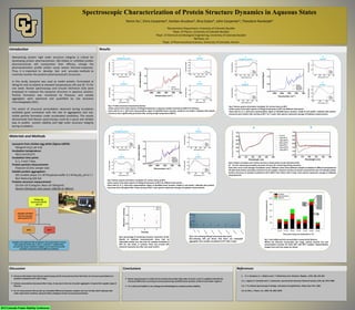
  • 1. Introduction Materials and Methods Results Spectroscopic Characterization of Protein Structure Dynamics in Aqueous States Yemin Xu1, Chris Carpenter2, Korben Knudson3, Rina Dukor4, John Carpenter5, Theodore Randolph3 1Biochemistry Department, University of Colorado Boulder 2Dept. of Physics, University of Colorado Boulder 3Dept. of Chemical and Biological Engineering, University of Colorado Boulder 4BioTools, Inc 5Dept. of Pharmaceutical Sciences, University of Colorado, Denver Maintaining protein high order structure integrity is critical for developing protein pharmaceuticals. Mis-folded or unfolded protein pharmaceuticals will compromise their efficacy, change the pharmacokinetics profile and/or cause severe immune-responses. Thus, it is important to develop fast and accurate methods to routinely monitor the proteins pharmaceutical’s structures. In this study, lysozyme was used as model protein, formulated at 50mg/ml and incubated at elevated temperatures (40 and 65 °C) for one week. Raman spectroscopy and Circular Dichroism (CD) were employed to measure the lysozyme structure in aqueous solution. Particle formation was monitored by Flowcam, and soluble aggregates were examined and quantified by size exclusion chromatography (SEC). The extent of structural perturbation observed during incubation exhibited good correlation with the rate of aggregation and sub- visible particle formation under accelerated conditions. The results demonstrate that Raman spectroscopy could be a quick and reliable way to predict protein stability and high order structure integrity during incubation. 50mg/mL Lysozyme (H2O) pH 4.0 Samples divided into two groups (40oCand 65oC) 40oC 65oC Samples were placed in the incubators for measuring at various time points (0-Point, Day 1, Day 4 , and Day 7, respectively). At each time point, triplicate samples were removed from the incubator and measured by different methods. Lysozyme from chicken egg white (Sigma-L6876): 50mg/ml (H2O, pH 4.0) Incubation temperature: 40±2 and 65±2oC Incubation time point: 0, 1, 4 and 7 days Protein particle measurement: Flowcam (0.2mL sample size) Soluble protein aggregation: SEC (mobile phase: 0.1 M Phosphate buffer 0.1 M Na2SO4, pH 6.7 ) Non-Reducing SDS Gel Proteins structure measurement: CD (Far-UV 0.3mg/ml, Near-UV 50mg/ml) Raman (50mg/ml, laser power 100mW at 780nm) 2013 Colorado Protein Stability Conference Discussion Fig 4: Protein secondary and tertiary structure measured by circular dichroism (CD) A) Far-UV: measuring secondary structure; B) near-UV: measuring tertiary structure. Each color curve is the average of triplicate result. Different colors show different samples incubated at different temperatures. CD data show similar secondary structures for all samples, however, tertiary structures measured by near-UV indicate protein tertiary strcutures of samples incubated at 65oC differ from others after 4 days. Each spectra represents average of triplicate measurements. A B A Disulfide bond S-S C Amide III B Tyrosine D Amide I Fig 1: Protein structure measured by Raman Center panel is the entire spectra of 50mg/ml lysozyme in aqueous solution (control) and 80oC for 20 hours. Close look (A, B, C, D)of each representative region of disulfide bond, tyrosine, amide III and amide I indicates that protein structures were significantly perturbed after storing at high temperature (80oC). A Disulfide bond S-S C Amide III B Tyrosine D Amide I Fig 2: Raman spectra of proteins incubated for various times at 40oC Center panel is the entire spectra of 50mg/ml lysozyme at 40oC for different time points Close look (A, B, C, D)of each representative region of disulfide bond, tyrosine, amide III and amide I indicates that protein structures were similar after storing at 40oC for 1 week. Each spectra represents average of triplicate measurements. A Disulfide bond S-S C Amide III B Tyrosine D Amide I Fig 3: Raman spectra of proteins incubated for various times at 60oC Center panel is the entire spectra of 50mg/ml lysozyme at 60oC for different time points Close look (A, B, C, D)of each representative region of disulfide bond, tyrosine, amide III and amide I indicates that protein structures were disrupted after 4 days storing at 60oC. Each spectra represents average of triplicate measurements. Fig 5: percentage of remaining monomer measured by SEC Results of triplicate measurements show that no noticeable protein loss was seen for samples incubated at 40oC for one week, in contrast, there was around 10% monomer lysozyme lost after one week at 65oC. Fig 6: non-reducing SDS gel (coomassie blue stain) Non-reducing SDS gel shows that there are noticeable aggregates from samples incubated at 65oC after 4 days. MW kD 50 37 25 20 15 10 Fig 7: Sub-visible particle concentration measured by flowcam Within the flowcam measurable size range, protein particle size and concentration increase for both 40oC and 60oC samples. Representative images from each size range are shown Conclusions References  Structure information from Raman spectroscopy and CD measurement show that there are structure perturbations in samples incubated at 65oC after 4 days.  Particle concentration decreased after 4 days, it may due to the size of protein aggregates is beyond the capable range of flowcam  Far-UV measurement did not pick up noticeable differences between samples, but near-UV did, which indicates that under experiment condition, lysozyme likely undergoes tertiary structural perturbation.  Raman spectroscopy is a useful tool to examine the protein high order structure, and it is capable to identify the structural differences occurring at various position (eg. Disulfide bond, tyrosine, amide III and amide I region).)  It is critical and helpful to use orthogonal methodologies to evaluate protein stability. 1. R. S. Porubcan, K. L. Watters and J. T. McFarland, Arch. Biochem. Biophys., 1978, 186, 255-264. 2.J. L. Lippert, D. Tyminski and P. J. Desmeules, Journal of the American Chemical Society, 1976, 98, 7075-7080. 3.A. T. Tu, Raman spectroscopy in biology : principles and applications, Wiley, New York, 1982. 4.Z.-Q. Wen, J. Pharm. Sci., 2007, 96, 2861-2878.