Brain stem.pptx
•
0 likes•38 views

For medical, nursing, and physiotherapy undergraduates, and postgraduates

Recommended
More Related Content
Similar to Brain stem.pptx
Similar to Brain stem.pptx (20)
More from Sundip Charmode
More from Sundip Charmode (20)
Recently uploaded
Recently uploaded (20)
Brain stem.pptx
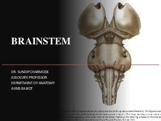
- 1. BRAINSTEM DR. SUNDIP CHARMODE ASSOCIATE PROFESSOR DEPARTMENT OF ANATOMY AIIMS RAJKOT
- 2. INTRODUCTION The brainstem is the stalk-like part of the brain which connects the spinal cord with the forebrain. From below upwards it consists of three parts: medulla oblongata, pons, and midbrain. The midbrain is continuous above with the cerebral hemispheres and the medulla oblongata is continuous below with spinal cord.
- 3. Brainstem is located in the posterior cranial fossa. Its ventral surface lies on the clivus. Posteriorly, the pons and medulla are separated from the cerebellum by the cavity of the fourth ventricle. The brainstem, like spinal cord consists of nerve fibres and nerve cells.
- 4. INTRODUCTION • Most of the nerve fibres in the brainstem are arranged longitudinally in the form of tracts as in the spinal cord. • But the nerve cells of the brainstem do not form a single central mass of grey matter as in the spinal cord; rather most of them are aggregated to form the well-defined nuclei.
- 5. INTRODUCTION • In addition to the well-defined tracts and nuclei, the brainstem consists of diffuse system of nerve cells and nerve fibres called reticular formation. • The central canal of spinal cord continues upwards in the lower part of the brainstem and then it widens and moves dorsally to form the fourth ventricle.
- 6. BRAINSTEM: FUNCTIONS 1. Provides passage to various ascending and descending tracts that connect the spinal cord to the different parts of the forebrain. 2. It contains important autonomic reflex centres (vital centres) associated with the control of respiration heart rate and blood pressure. 3. It contains Reticular Activating System which controls consciousness. 4. It contains important nuclei of the last ten cranial nerves (i.e., 3rd to 12th).
- 9. INTRODUCTION • It is the direct upward continuation of the spinal cord, extending from foramen magnum to the lower border of the pons.
- 11. LOCATION • It forms the lowest part of the brainstem. • It lies almost vertically in the anterior part of the posterior cranial fossa between the clivus (superior surface of the basi-occiput) in front and the vallecula of the cerebellum behind.
- 13. DIMENSIONS • The medulla is shaped like a truncated cone (bulb- like) hence its alternative name ‘bulb’ (cf. bulbar paralysis). • Medulla oblongata measures about 3 cm in length, 2 cm in breadth (at the widest part) and 1.25 cm in thickness).
- 14. CLINICAL SIGNIFICANCE • The medulla contains vital centres which are essential for life. These are: (a) cardiac centre, (b) vasomotor centre, and (c) respiratory centre. • Medulla provides attachment to last four cranial nerves. • The lower part of the medulla like the spinal cord contains the central canal. • In the upper part: the canal widens and moves dorsally to form the lower part of the fourth ventricle. • Thus, the medulla is divided into a lower closed part and an upper open part.
- 15. EXTERNAL FEATURES • The medulla is divided into right and left symmetrical halves by anterior median fissure and posterior median sulcus.
- 17. EXTERNAL FEATURES • The Anterior Median Fissure is continuous below with the corresponding fissure on the spinal cord, and above it ends into a small triangular depression called Foramen Caecum, at the lower border of the pons. • It is interrupted in its lower part by the bundles of fibres crossing obliquely from one side to the other, the decussation of pyramids.
- 18. EXTERNAL FEATURES • The Posterior Median Sulcus continues below with the corresponding sulcus of the cord and is present only in the lower half of the medulla. • Above its lips diverge to form the boundaries of a triangular area, the lower part of the floor of fourth ventricle. • Each half of the medulla is marked by two sulci: Antero- lateral and Postero-lateral, which are direct upward continuations of the corresponding sulci of the cord.
- 19. EXTERNAL FEATURES • The anterolateral sulcus extends along the lateral border of the pyramid and along it emerges the rootlets of the hypoglossal (XII cranial) nerve. • The posterolateral sulcus lies between the olive and the inferior cerebellar peduncle and along it emerges the rootlets of glossopharyngeal (IX), vagus (X) and accessory (XI) cranial nerves.
- 22. EXTERNAL FEATURES • The anterolateral and posterolateral sulci with nerve roots divide the surface of each half of the medulla oblongata into Anterior, Posterior and Lateral regions like that in the spinal cord.
- 24. Anterior (Ventral) Aspect of Medulla • Pyramids: – Two elongated elevations, one on either side of anterior median fissure – Produced by the Corticospinal (Pyramidal) Fibres. • Olives: – These are oval elevations, posterolateral to the pyramids and are produced by an underlying mass of grey matter called Inferior Olivary Nucleus.
- 25. Anterior (Ventral) Aspect of Medulla • Rootlets of the hypoglossal nerve: – These emerge from the anterolateral sulcus between the pyramid and the olive. • Inferior cerebellar peduncles: – These are thick bundles of fibres lying posterolateral to the olive and attach the medulla with the cerebellum.
- 27. Anterior (Ventral) Aspect of Medulla • Rootlets of the 9th, 10th, and 11th (cranial part) cranial nerves: – These emerge through the posterolateral sulcus separating the olive from the inferior cerebellar peduncle.
- 28. Posterior (Dorsal) Aspect of Medulla • The dorsal aspect the medulla is well demarcated into lower closed part and upper open part.
- 31. Posterior (Dorsal) Aspect of Medulla: Closed part • The closed part, on either side of posterior median sulcus, presents three longitudinal elevations. • From medial to lateral, these are: fasciculus gracilis, fasciculus cuneatus, and inferior cerebellar peduncle. • The upper ends of the fasciculus gracilis and fasciculus cuneatus expand to form the gracile and cuneate tubercles respectively due to underlying nuclei of the same name. • Another elevation present lateral to cuneate tubercle, the tuber cinereum is produced by the spinal nucleus of trigeminal nerve.
- 33. Posterior (Dorsal) Aspect of Medulla – Open part • The open part of the medulla forms the lower part of the floor of fourth ventricle, which presents number of features like, Median Sulcus, Hypoglossal and Vagal Triangles, Vestibular Areas, Area Postrema, Stria Medullaris, etc. (Fig. 8.3).
- 35. INTERNAL STRUCTURE • As in the spinal cord, the white matter surrounds the central mass of grey matter but in the latter, the grey matter is broken up into nuclei that are separated by nerve fibres. • The internal structure of medulla is studied by examining its transverse sections at the following four levels: 1. At the level of decussation of pyramids. 2. At the level of decussation of medial lemnisci. 3. At the level of the olives. 4. At the level just inferior to the pons.
- 37. Transverse section of medulla at the level of pyramidal decussation
- 39. Transverse section of Medulla at the level of Pyramidal Decussation • The section at this level passes through the inferior half of the medulla, and closely resembles to that of spinal cord. • The Nucleus Gracilis and Nucleus Cuneatus appear as the narrow strip like projections from the posterior aspect of the central grey matter. • The Apex of Posterior Horn gets swollen up to form the Nucleus of Spinal Tract of Trigeminal Nerve. It is an upward continuation of the substantia gelatinosa in the posterior grey column of the spinal cord. • The Spinal Tract of Trigeminal Nerve is a bundle of fibres which caps over the nucleus of spinal tract of trigeminal nerve.
- 40. Transverse section of Medulla at the level of Pyramidal Decussation • Decussation of Pyramidal Tracts forms the most important feature of medulla at this level. • About 75% fibres of pyramidal tract run backwards and laterally across the midline to reach the lateral white column of the opposite side of the spinal cord, where they run downwards as the lateral corticospinal tract. • In doing so the anterior horns are detached from the central grey matter.
- 41. Transverse section of Medulla at the level of Pyramidal Decussation • Each detached Anterior Horn divides to form the Spinal Nucleus of Accessory Nerve and Supraspinal Nucleus of First Cervical Nerve. • The Nucleus of Accessory Nerve extends downwards up to fifth cervical spinal segment. • The Supraspinal Nucleus gives off the efferent fibres of the first cervical nerve and is continuous above with the nucleus of the hypoglossal nerve. • Appearance of diffuse zone containing a network of fibres and scattered nerve cells within it the lateral white column adjacent to nucleus of spinal tract of trigeminal nerve is called Reticular Formation.
- 42. Transverse section of Medulla at the level of Sensory Decussation
- 44. Transverse section of Medulla at the level of Sensory Decussation • The Nucleus Gracilis and Nucleus Cuneatus become more pronounced and are separated from the central grey matter. • The fibres of fasciculus gracilis and fasciculus cuneatus occupy the broad posterior white column and terminate in these nuclei.
- 45. Transverse section of Medulla at the level of Sensory Decussation • The Internal Arcuate Fibres arising from the cells of gracile and cuneate nuclei (second order sensory neurons conducting sensations of discriminative touch, position and vibration) course forwards and medially around the central grey matter and decussate with corresponding fibres of opposite side in the median plane (sensory decussation) and then turn upwards to ascend as the medial lemniscus on the opposite side close to the median plane. • In this decussation the gracile fibres are medial to that of cuneate fibres.
- 46. Transverse section of Medulla at the level of Sensory Decussation
- 48. Transverse section of Medulla at the level of Sensory Decussation • The Internal Arcuate Fibres cut off the spinal nucleus and tract of trigeminal nerve from the central grey matter. • As the fibres from nucleus gracilis and nucleus cuneatus pass forwards and medially they intercross so that the most medial fibres (from the feet and leg) come to lie anteriorly in the medial lemniscus. • In the Medial Lemniscus the body is represented with the head posteriorly and the feet anteriorly. • The fibres of lemniscus relay into the corresponding thalamus.
- 49. Transverse section of Medulla at the level of Sensory Decussation
- 50. Transverse section of Medulla at the level of Sensory Decussation • Immediately dorsolateral to the cuneate nucleus lies the Accessory Cuneate Nucleus which receives the more lateral fibres (derived from the cervical segments of the cord) of the fasciculus cuneatus. • It gives rise to Posterior External Arcuate Fibres conveying proprioceptive impulses to the cerebellum of the same side through inferior cerebellar peduncle. • The separated spinal nucleus and tract of trigeminal nerve lies ventrolateral to the cuneate nucleus. • The lower part of inferior olivary nucleus is seen. • The pyramids lie on either side of the anterior median fissure.
- 52. Transverse section of Medulla at the level of Sensory Decussation • The central grey matter contains: (a) hypoglossal nucleus, (b) dorsal nucleus of vagus, and (c) nucleus of tractus solitarius. • The Hypoglossal Nucleus occupies the ventro-medial position close to the midline in the central grey matter. • The Dorsal Nucleus of Vagus lies dorsolateral to the hypoglossal nucleus and Nucleus of Tractus Solitarius lies just dorsolateral to the dorsal nucleus of vagus.
- 53. Transverse section of Medulla at the level of Sensory Decussation
- 54. Transverse section of Medulla at the level of Sensory Decussation • Medial longitudinal bundle lies posterior to the medial lemniscus. • It is small compact tract of nerve fibres which interconnect the 3rd , 4th , 6th , 8th and spinal nucleus of 11th cranial nerve nuclei. • Spinocerebellar and Lateral Spinothalamic tracts lie in the anterolateral area of lateral white column. • Lateral and Anterior Spinothalamic tracts are very close to each other and collectively form Spinal Lemniscus.
- 55. Transverse section of Medulla at the level of Sensory Decussation
- 56. Transverse section of Medulla at the level of Olive
- 58. Transverse section of Medulla at the level of Olive • The central grey matter is spread over the floor of the fourth ventricle and contains the nuclei of several cranial nerves. • From medial to lateral these are: Hypoglossal Nucleus, Nucleus Intercalatus, Dorsal Nucleus of Vagus and Vestibular Nuclei (inferior and medial). • The Nucleus of Tractus Solitarius lies ventral to vestibular nuclei.
- 59. Transverse section of Medulla at the level of Olive • The Nucleus Ambiguus lies deep within the reticular formation and gives origin to the motor fibres of 9th, 10th and 11th cranial nerves. • On either side of midline (paramedian region), from dorsal to ventral lie: Medial Longitudinal Fasciculus, Tectospinal Tract, Medial Lemniscus, and Pyramidal (Corticospinal) Tract.
- 61. Transverse section of Medulla at the level of Olive • The Arcuate Nuclei, thought to be inferiorly displaced pontine nuclei are situated on the anteromedial aspect of the pyramids. • They receive fibres from the cerebral cortex and send efferent fibres to the cerebellum of the opposite side through the Anterior External Arcuate Fibres. • Laterally, from dorsal to ventral lie two prominent structures: (a) Inferior Cerebellar Peduncle, and (b) Inferior Olivary Nucleus.
- 62. Transverse section of Medulla at the level of Olive • A. The Inferior Cerebellar Peduncle occupies posterolateral part. • B. The Inferior Olivary Nucleus is the largest mass of grey matter and forms the most prominent feature in the section through upper part of medulla. It presents a crumbled bag like appearance. • Close to the main nucleus lies Medial and Dorsal Accessory Olivary Nuclei.
- 64. BLOOD SUPPLY OF MEDULLA • The medulla is supplied by the following arteries: 1. Two vertebral arteries. 2. Anterior and Posterior Spinal arteries. 3. Anterior and Posterior Inferior Cerebellar arteries. 4. Basilar artery.
- 65. Vascular Disorders of Medulla Oblongata
- 66. Vascular Disorders of Medulla Oblongata • The common vascular lesions involving the medulla are the thrombosis of posterior inferior cerebellar and vertebral arteries leading to lateral and medial medullary syndromes, respectively.
- 67. Lateral medullary (posterior inferior cerebellar artery) syndrome of Wallenberg • The dorsolateral part of the medulla is supplied by the posterior inferior cerebellar artery, which is usually the branch of the vertebral artery. • This artery also supplies the inferior surface of the cerebellum. • Thrombosis of posterior inferior cerebellar artery, therefore, affects a wedge-shaped area on the dorsolateral aspect of medulla (Fig. 8.7) and the inferior surface of the cerebellum and produces following signs and symptoms.
- 68. Vascular Disorders of Medulla Oblongata
- 69. Lateral medullary (posterior inferior cerebellar artery) syndrome of Wallenberg • Contralateral loss of pain and temperature sensation, in the trunk and limbs, due to involvement of spinothalamic tract. • Ipsilateral loss of pain and temperature sensation over the face, due to involvement of the spinal nucleus and tract of trigeminal nerve. • Ipsilateral paralysis of muscles of palate, pharynx and larynx due to involvement of nucleus ambiguous. • Ipsilateral ataxia, due to involvement of inferior cerebellar peduncle and cerebellum. – Giddiness, due to involvement of vestibular nuclei. – Horner's syndrome, due to involvement of descending sympathetic pathway in the reticular formation of medulla.
- 70. Vascular Disorders of Medulla Oblongata
- 71. Medial Medullary (Dejerine’s Anterior Bulbar) syndrome • The paramedian region of the medulla is supplied by the branches of vertebral artery. • The vascular involvement (ischaemia) of this region produces following signs and symptoms: • Contralateral hemiplegia/paralysis of arm and leg, due to damage of pyramid. • Ipsilateral paralysis and atrophy of the half of the tongue, due to damage of hypoglossal nerve. • Contralateral loss of position and vibration sense due to damage of medial lemniscus.
- 72. PONS DR. SUNDIP CHARMODE ASSOCIATE PROFESSOR DEPARTMENT OF ANATOMY AIIMS RAJKOT
- 73. INTRODUCTION It is the middle part of Brainstem. It lies in Posterior cranial fossa on the Clivus. On either side, it is continuous with Middle cerebellar peduncle. 2.5 cm long Forms the Upper half of floor of Fourth ventricle
- 77. EXTERNAL FEATURES Surfaces – 2 (Ventral and Dorsal) Borders -2 (Superior and Inferior)
- 78. VENTRAL SURFACE Convex in both the directions Transversely striated d/t Ponto-cerebellar fibres Vertical groove in median plane Attachment of Trigeminal nerve
- 80. DORSAL SURFACE Covered by cerebellum separated by cavity of fourth ventricle.
- 83. SUPERIOR BORDER Cerebral peduncles are attached to this border Superior cerebellar arteries wind round this border.
- 84. INFERIOR BORDER The upper end of medulla is continuous with this border. Anterior Inferior cerebellar arteries curve around this border.
- 85. INTERNAL FEATURES • Large Ventral Basilar part - is continuous inferiorly with the pyramids of the medulla and on each side with the middle cerebellar peduncle. • Small Dorsal Tegmental part - is a direct upward continuation of the medulla excluding the pyramids. • The structure of basilar part is basically the same at all levels of pons, but the structure of tegmental part vary considerably in the upper and lower parts of the pons.
- 87. BASILAR PART
- 89. BASILAR PART • This part is composed of the longitudinal bundles of fibres, the transverse fibres, and the pontine nuclei. • Longitudinal bundles of fibres include corticopontine, cortico-nuclear, and corticospinal fibres. – The corticopontine fibres relay in the ipsilateral pontine nuclei. – The cortico-nuclear fibres terminate in the contralateral (and to some extent ipsilateral) motor nuclei of the cranial nerves. – The corticospinal fibres converge towards the lower part of the pons and form the pyramids of the medulla. • Transverse fibres, arise in the pontine nuclei and cross to the opposite side to form the middle cerebellar peduncle. These are pontocerebellar fibres.
- 91. Basilar part • Pontine nuclei, are scattered among the longitudinal and transverse fibres. • The pontine nuclei form an important part of the cortico-ponto-cerebellar pathway which connects the cerebral cortex of one side to the cerebellar hemisphere of the opposite side.
- 93. VENTRAL BASILAR PART Longitudinal fibres – 1. Cortico-pontine - 2. Cortico-spinal 3. Cortico-nuclear Transverse fibres – • Ponto-cerebellar fibres
- 94. VENTRAL BASILAR PART Pontine nuclei – • They establish a direct connection between I/L cerebral cortex and C/L Cerebellum. • It forms an important part of Cortico-ponto- cerebellar pathway. • They help in precision and coordination of voluntary movements.
- 95. TEGMENTAL PART
- 96. TEGMENTAL PART • The tegmental part is traversed by a number of ascending and descending tracts and contained a decussation of transversely running fibres, the trapezoid body. • It also contains the nuclei of trigeminal (V), abducent (VI), facial (VII), and vestibulocochlear (VIII) nerves. • Since, the structures of tegmentum differs in the upper (cranial) and lower (caudal) parts of the pons, it is studied by examining transverse sections at these two levels.
- 99. AT LOWER LEVEL / FACIAL COLLICULUS Grey matter : 1. Abducent nerve nucleus 2. Motor nucleus of Facial nerve 3. Neurobiotaxis 4. Superior salivatory nucleus 5. Inferior salivatory nucleus 6. Lacrimatory nucleus
- 101. AT THE LEVEL OF FACIAL COLLICULUS • The abducent nerve nucleus lies beneath the facial colliculus in the floor of fourth ventricle, just lateral to the medial longitudinal bundle (MLB). • The motor nucleus of facial nerve lie ventrolateral to the abducent nucleus. • The fibres of facial nerve first wind round the abducent nucleus, producing the facial colliculus, then pass anteriorly between the facial nucleus and the nucleus of the spinal tract of trigeminal nerve.
- 103. NEUROBIOTAXIS • In the early embryogenesis the motor nucleus of facial nerve is located dorsolateral to the cranial end of abducent nucleus. • Thereafter, it migrates caudally along the dorsal surface of that nucleus and then ventromedial to the latter until the facial nerve nucleus reaches its permanent position in close proximity to the spinal nucleus of the trigeminal nerve (a site of maximum sensory stimulus) which establishes quick reflex responses.
- 104. NEUROBIOTAXIS • Such migration of motor nucleus towards the sensory nucleus is known as the “Neurobiotaxis”. • Eventually the motor fibres of the facial nerve undergo a loop course (internal genu of facial nerve) around the dorsomedial aspect of the abducent nerve nucleus.
- 105. AT THE LEVEL OF FACIAL COLLICULUS • The Superior Salivatory, Inferior Salivatory and Lacrimatory Nuclei lie medial to the motor nucleus of facial nerve.
- 107. AT FACIAL COLLICULUS Grey matter: 7. Nucleus of Tractus Solitarius – upper end – dorso-lateral to Facial nucleus : SVA fibres 8. Vestibular nuclei 9. Dorsal and Ventral Cochlear nuclei 10. Spinal nucleus and tract of Trigeminal nerve
- 109. AT THE LEVEL OF FACIAL COLLICULUS • The nucleus of tractus solitarius lies lateral to the superior salivatory nucleus. • The vestibular nuclei lie beneath the vestibular area in the floor of the fourth ventricle. • The dorsal and ventral cochlear nuclei are situated dorsal and ventral to inferior cerebellar peduncle respectively. • The spinal nucleus of trigeminal nerve and its tract are located on the anteromedial aspect of the inferior cerebellar peduncle.
- 111. AT FACIAL COLLICULUS White matter : 1. Trapezoid body 2. Medial Lemniscus 3. Spinal Lemniscus 4. Medial Longitudinal bundle 5. Tecto-spinal tract 6. Spinal tract of Trigeminal nerve
- 112. AT FACIAL COLLICULUS • The white matter at this level comprises trapezoid body, large long (medial and spinal lemnisci) and small short (spinal tract of trigeminal nerve) tracts, medial longitudinal bundle (MLB), and small extrapyramidal and autonomic tracts. • The trapezoid body is a trapezium-shaped mass of white fibres lying in the anterior part of the tegmentum, just posterior to the basilar part of the pons. It is formed by the decussation of transversely running fibres arising from the cochlear nuclei of both the sides. • The medial lemniscus rotates as it passes from the medulla into the pons and is situated in the most anterior part of the tegmentum with its long axis running transversely (Fig. 8.8). The medial lemniscus rotates in such a way that the fibres from nucleus cuneatus are medial to those from nucleus gracilis.
- 113. AT FACIAL COLLICULUS • The somatotopic representation is therefore neck, arm, trunk, and leg, in a medial to lateral sequence. • The spinal lemniscus lies lateral to the medial lemniscus and occupies the same peripheral position as in the medulla. • The medial longitudinal bundle occupies a paramedian position in the most posterior part. • The tectospinal tracts are located ventral to the medial longitudinal bundles. • The spinal tract of trigeminal nerve lies lateral and dorsal to the motor nucleus of facial nerve.
- 116. AT UPPER LEVEL / TRIGEMINAL NUCLEUS Grey matter : • Motor nucleus of Trigeminal nerve • Principal Sensory nucleus of Trigeminal nerve • Caudal part of Nucleus Ceruleus
- 117. AT TRIGEMINAL NUCLEUS White matter : 1. Medial Lemniscus 2. Trigeminal Lemniscus 3. Spinal Lemniscus 4. Lateral Lemniscus • Trapezoid body and nuclei absent.
- 119. APPLIED ANATOMY Millard Gubler syndrome – • Lesion in lower part of Pons includes Pyramidal tract and • Emerging fibres of Abducent and Facial nerves. I/L medial squint I/L Facial palsy C/L Hemiplegia
- 121. APPLIED ANATOMY Ponto-cerebellar angle syndrome – Compression of Ponto-cerebellar angle by tumour called Acoustic neuroma of : 1. Facial nerve 2. Vestibulo-cochlear nerve 3. Flocculus of Cerebellum 4. Choroid plexus of Fourth ventricle
- 122. APPLIED ANATOMY Ponto-cerebellar angle syndrome – • Tinnitus, progressive deafness, vertigo – VIII nerve • I/L Ataxia, staggering gate – cerebellar peduncle compression • I/L LMN type of Facial palsy – VII nerve • I/L loss of pain and temperature, loss of corneal reflex – Spinal tract and nucleus of Trigeminal nerve involvement.
- 123. APPLIED ANATOMY Pontine Hemorrhage • Pinpoint pupil • Hyperpyrexia • Deep coma • B/L paralysis of face and limbs. Tumors of Pons - Astrocytoma
- 124. MIDBRAIN
- 125. INTRODUCTION • The midbrain is the upper and shortest part of the brain-stem. • It is about 2.5 cm long and 2.5 cm wide. • It connects the hindbrain with the forebrain. • Its cavity, the cerebral aqueduct (aqueduct of Sylvius) connects the third ventricle with the fourth ventricle. • The midbrain passes through the tentorial notch.
- 127. RELATIONS • On each side: Optic tract, para-hippocampal gyrus, posterior cerebral artery, and basal vein. • Anteriorly: Interpeduncular structures, viz. mammillary bodies, tuber cinereum, etc. • Posteriorly: Splenium of corpus callosum, great cerebral vein, pineal body and posterior ends of right and left thalami. • The midbrain like the other parts of the brainstem consists of grey and white matter.
- 128. EXTERNAL FEATURES - VENTRAL SURFACE • It presents two crura cerebri which emerges from the cerebral hemispheres and converge downwards to enter the pons forming the posterolateral boundaries of the interpeduncular fossa. • The superficial surface of the crus cerebri is finely corrugated by the underlying longitudinal fibres. • It is crossed transversely from above downwards by optic tract, posterior cerebral artery, superior cerebellar artery and taenia pontis.
- 129. EXTERNAL FEATURES – VENTRAL SURFACE • The Oculomotor Nerve emerges from a groove on the medial side of the crus cerebri. • The Trochlear Nerve emerges on the dorsal aspect of the midbrain and curls around the lateral aspect of the cerebral peduncle to appear on the ventral aspect of the midbrain, lateral to the oculomotor nerve. • These two nerves run forward between the posterior cerebral and the superior cerebellar arteries.
- 130. EXTERNAL FEATURES – DORSAL SURFACE • The dorsal surface of the midbrain presents four rounded elevations: two superior and two inferior colliculi (or corpora quadrigemina). • The colliculi are separated from each other by a Cruciform Sulcus. • The vertical limb of sulcus when traced above forms a surface depression which lodges the pineal body and when traced below, it becomes continuous with the frenulum veli (a median ridge on the dorsal surface of the superior medullary velum).
- 131. EXTERNAL FEATURES – DORSAL SURFACE • Thick ridges of white matter extending from lateral side of each colliculus constitute their brachia. • The Superior Brachium connect the superior colliculus to the lateral geniculate body and the optic tract and is made up of optic tract fibres. • The Inferior Brachium connect the inferior colliculus to the medial geniculate body and is made up of auditory fibres. • The Superior and Inferior Colliculi are concerned with reflex activities triggered by auditory and visual impulses, respectively.
- 132. INTERNAL FEATURES • The internal structure of the midbrain is studied by its transverse sections. • The transverse section of midbrain shows a tiny canal, called cerebral aqueduct. • A coronal plane passing through the aqueduct divides the midbrain into two parts; a small posterior part and large anterior part.
- 133. INTERNAL FEATURES • The small posterior part is called tectum and consists of four colliculi. • The large anterior part is divided into two equal right and left halves by a vertical plane, the cerebral peduncle. • Each cerebral peduncle is further subdivided into three parts, from dorsal to ventral these are: (a) tegmentum, (b) substantia nigra, and (c) crus cerebri. • The structures in the tectum and tegmentum vary at different levels of the midbrain, but those of the crus cerebri and substantia nigra are nearly the same throughout the length of the midbrain.
- 135. CRUS CEREBRI (BASIS PEDUNCULI) • It is the part of cerebral peduncle situated anterolateral to the substantia nigra. • Contains important descending tracts which connect the cerebral cortex to the anterior horn cells of the spinal cord, cranial nerve nuclei, and pontine nuclei. • Cortico-spinal and Cortico-nuclear fibres occupy the middle two-thirds of the crus. • Fronto-pontine fibres occupy the medial one-sixth of the crus. • Temporo-pontine, Parieto-pontine, and Occipito-pontine fibres occupy the lateral one-sixth of the crus.
- 136. SUBSTANTIA NIGRA • The substantia nigra is a curved (crescent-shaped) pigmented band of grey matter (thicker medially than laterally) situated between tegmentum and crus cerebri. • Its concavity is smooth and directed towards the tegmentum. • From its convex margin, spiky processes project into the substance of the crus cerebri. • The substantia nigra is a large motor nucleus that extends throughout the length of midbrain.
- 138. SUBSTANTIA NIGRA • It is divided into two parts: (a) the dorsal part (pars compacta) containing medium sized cells and (b) a ventral part (pars reticularis) containing fewer cells. • The pars reticularis is intermingled with the fibres of crus cerebri. • The substantia nigra contains deeply pigmented nerve cells which contain melanin (a polymerized form of dopamine) and iron. • These cells synthesize dopamine which is carried through their axons (nigrostriatal fibres) to the corpus striatum.
- 139. Transverse Section of the Midbrain at the Level of the Inferior Colliculi
- 140. GREY MATTER • The central grey matter (grey matter around the cerebral aqueduct) contains two nuclei: (a) nucleus of trochlear nerve, and (b) mesencephalic nucleus of trigeminal nerve. 1. The trochlear nerve nucleus is situated close to the median plane just posterior to the medial longitudinal fasciculus (MLF). The emerging fibres of the trochlear nerve pass laterally and posteriorly around the central grey matter and leave the midbrain just below the inferior colliculi. The fibres of trochlear nerve now decussate in the superior medullary vellum and wind round the lateral aspect of the midbrain to enter the lateral wall of cavernous sinus .
- 141. GREY MATTER 2. The mesencephalic nucleus of trigeminal nerve lies in the lateral edge of the central grey matter. It receives proprioceptive impulses from muscles of mastication, teeth, ocular and facial muscles. 3. An ovoid mass of grey matter underneath the inferior colliculus forms the nucleus of inferior colliculus. It receives the afferent fibres of lateral lemniscus and gives the efferent fibres to the medial geniculate body through the inferior brachium. 4. Substantia nigra. 5. The reticular formation is smaller than that in the pons and is situated ventro-laterally between the medial lemniscus and the central grey matter.
- 142. WHITE MATTER • The decussation of the superior cerebellar peduncles occupies the central part of the tegmentum and forms the most important feature in the lower part of the midbrain. • The lemnisci are arranged in the form of a curved compact band of white fibres in the ventrolateral part of the tegmentum, lateral to cerebellar decussation and dorsal to the substantia nigra. • From medial to lateral side these are: medial lemniscus, trigeminal lemniscus, spinal lemniscus, and lateral lemniscus.
- 143. WHITE MATTER • The lateral lemniscus is posterior to the spinal lemniscus and ends into the inferior colliculus. • The Medial Longitudinal Fasciculus lies on the side of median plane ventral to the trochlear nerve nucleus. • The Tectospinal Tracts lie ventral to the medial longitudinal fasciculi. • The Rubrospinal Tracts lie ventral to the decussation of the superior cerebellar peduncles.
- 144. Transverse Section of the Midbrain at the Level of the Superior Colliculi
- 145. GREY MATTER • The central grey matter in each half contains two nuclei: Oculomotor nerve nucleus and the Mesencephalic nucleus. • The Oculomotor Nucleus lies in the ventromedial part. • The nuclei of two sides fuse together forming a single complex having a triangular outline. • The oculomotor nuclei are bounded laterally by the medial longitudinal fasciculus.
- 146. GREY MATTER • The Edinger-Westphal nucleus which supplies the sphincter pupillae and ciliary muscle, forms part of the oculomotor nucleus and is located dorsal to the rostral two-thirds of the main oculomotor nucleus.
- 147. Transverse Section of the Midbrain at the Level of the Superior Colliculi
- 148. GREY MATTER • The emerging fibres of oculomotor nerve pass ventrally through the tegmentum intersecting red nucleus and medial part of the substantia nigra. • They emerge in the posterior part of interpeduncular fossa through the sulcus on the medial aspect of crus cerebri. • The Mesencephalic Nucleus occupies the same position as in the lower part of the midbrain.
- 149. SUPERIOR COLLICULUS • Superior Colliculus is a flattened mass formed of seven concentric alternating laminae of white matter and grey matter.
- 150. SUPERIOR COLLICULUS • The Superior Colliculus is a flattened mass formed of seven concentric alternating laminae of white matter and grey matter. • The SC receives afferent fibres from: 1. The retinae (mainly the contralateral) through the lateral geniculate body and superior brachium, 2. The spinal cord (pain and tactile fibres) through spino-tectal tract, 3. The frontal and occipital visual cortex (conjugate eye movements), and 4. The inferior colliculus. • The efferent fibres from superior colliculus form tecto-spinal and tecto- bulbar tracts, responsible for the reflex movements of the eyes, head, and neck in response to visual stimuli.
- 151. GREY MATTER • Pretectal nucleus is a small group of neurons and lies deep to the superolateral part of the superior colliculus. • Afferents: From the lateral root of the optic tract and • Efferents: To the Edinger-Westphal nucleus (the parasympathetic component of the oculomotor nucleus) of the same as well as of the opposite side. • The pretectal nucleus is an important part of the pathway for pupillary light reflex and consensual light reflex. • Its lesion causes Argyll Robertson pupil in which light reflex is lost but accommodation reflex remains intact.
- 152. GREY MATTER • Red nucleus is a cigar-shaped mass of grey matter which appears ovoid in cross-section. • It is about 0.5 cm in diameter and situated dorsomedial to the substantia nigra. • In the fresh specimen it is red/pink in colour due to its high vascular supply and an iron containing pigment present in the cytoplasm of its cells.
- 154. CONNECTIONS OF RED NUCLEUS • Afferents: 1. Cerebello-rubral fibres from contralateral dentate nucleus of the cerebellum through superior cerebellar peduncle, 2. Cortico-rubral fibres, mostly from the ipsilateral motor area (area 4 and 6 of frontal cortex), 3. Pallidorubral fibres from globus pallidus of the same side, 4. Red nucleus also receives fibres from: subthalamic nucleus (corpus luysi), hypothalamus, substantia nigra and tectum
- 155. CONNECTIONS OF RED NUCLEUS • Efferents: • Rubro-spinal, Rubro-bulbar and Rubro-reticular tracts. • The fibres from red nucleus before forming these tracts decussate forming ‘ventral tegmental decussation of Forel’ • The fibres of rubrospinal tract end in the anterior horn cells of the opposite side. • The Rubro-bulbar tract ends in the motor nuclei of 5th and 7th cranial nerves (also in the nuclei of 3rd, 4th and 6th cranial nerves). 1. Rubro-olivary fibres, 2. Rubro-thalamic fibres, 3. Rubro-cerebellar fibres, 4. Rubro-nigral fibres
- 156. WHITE MATTER 1. Decussation of fibres (tectospinal and tecto-bulbar tracts) arising from superior colliculi forming dorsal tegmental decussation (of Meynert). 2. Decussation of fibres (rubrospinal tracts) arising from red nuclei forming ventral tegmental decussation (of Forel). 3. Medial Longitudinal Fasciculus (MLF) lies ventrolateral to the oculomotor nucleus. 4. Tegmentum at this level also contains the same lemnisci (i.e. medial, trigeminal and spinal) as those at the level of inferior colliculus except for the lateral lemniscus. The lateral lemniscus is not seen at this level because it terminates in the nucleus of inferior colliculus. 5. Emerging fibres of oculomotor nerve.
- 157. BLOOD SUPPLY OF MIDBRAIN • Arterial supply: The midbrain is supplied by following arteries: • Basilar artery through its posterior cerebral and superior cerebellar arteries supplies mid-brain through direct branches. • Branches of posterior communicating and anterior choroidal arteries. • Venous drainage: The veins of midbrain drains into the great cerebral and the basal veins.
- 158. WEBER’S SYNDROME • It is produced by a vascular lesion in the basal region of the cerebral peduncle due to occlusion of a branch of the posterior cerebral artery. • This lesion involves the oculomotor nerve and the crus cerebri and produce following important signs and symptoms:
- 159. WEBER’S SYNDROME • Ipsilateral lateral squint, due to involvement of third cranial nerve. • Contralateral hemiplegia, due to involvement of corticospinal tract in the crus cerebri. • Contralateral paralysis of the lower part of the face and tongue, due to involvement of the corticobulbar tract in the crus cerebri. • Drooping of the upper lid (ptosis), due to paralysis of levator palpebrae superioris supplied by oculomotor nerve. • Pupil is dilated and fixed to light and accommodation is lost on the side of lesion due to involvement of parasympathetic component of oculomotor nerve (Edinger-Westphal nucleus).
- 160. BENEDIKT'S SYNDROME • Vascular ischaemia of the tegmentum of midbrain involving the medial lemniscus, spinal lemniscus, red nucleus, superior cerebellar peduncle and fibres of oculomotor nerve. • Signs and symptoms: 1. Ipsilateral lateral squint and ptosis, due to involvement of oculomotor nerve fibres. 2. Contralateral loss of pain and temperature sensation, due to involvement of trigeminal and spinal lemnisci. 3. Contralateral loss of tactile, muscle, joint and vibration sense, due to involvement of medial lemniscus. 4. Contralateral tremors and involuntary movements in the limbs, due to involvement of red nucleus and fibres of superior cerebellar peduncle entering into it.
- 161. PARINAUD'S SYNDROME • It results from a lesion of the superior colliculi as occurs when this area becomes compressed by the tumours of the pineal gland. • It is characterised by the loss of upward gaze without affecting the other eye movements (the anatomical basis for this is obscure but experiments indicate that the area involved may contain a centre for upward movements of the eyes).
- 162. ARGYLL ROBERTSON PUPIL • The Argyll Robertson's pupil is a clinical condition in which light reflex is lost but the accommodation reflex remains intact. • Generally, it occurs because of lesion in the vicinity of pretectal nucleus.
- 163. THANK YOU
Editor's Notes
- Most of these fibres about (75%) cross to the opposite side (pyramidal decussation) in the lower part of medulla and then descend as lateral corticospinal tract in the lateral white column of the spinal cord. About 20% of uncrossed fibres run downwards as anterior corticospinal tract in the anterior white column of the spinal cord; the remaining 5% run downwards along with uncrossed fibres in the lateral white column of the spinal cord
- FIG. 8.7 The transverse section of the upper part of the medulla. The red areas indicate the sites of lesions: 1. dorsal nucleus of vagus, 2. nucleus of tractus solitarius, 3. vestibular nuclei, 4. inferior cerebellar peduncle, 5. spinal tract of trigeminal nerve, 6. spinal nucleus of trigeminal nerve, 7. descending sympathetic tract, 8. nucleus ambiguus, 9. lateral spinothalamic tract, 10. inferior olivary nucleus, 11. hypoglossal nerve, 12. pyramidal tract, 13. arcuate nucleus. (M = medial lemniscus.)
- FIG. 8.7 The transverse section of the upper part of the medulla. The red areas indicate the sites of lesions: 1. dorsal nucleus of vagus, 2. nucleus of tractus solitarius, 3. vestibular nuclei, 4. inferior cerebellar peduncle, 5. spinal tract of trigeminal nerve, 6. spinal nucleus of trigeminal nerve, 7. descending sympathetic tract, 8. nucleus ambiguus, 9. lateral spinothalamic tract, 10. inferior olivary nucleus, 11. hypoglossal nerve, 12. pyramidal tract, 13. arcuate nucleus. (M = medial lemniscus.)
- FIG. 8.7 The transverse section of the upper part of the medulla. The red areas indicate the sites of lesions: 1. dorsal nucleus of vagus, 2. nucleus of tractus solitarius, 3. vestibular nuclei, 4. inferior cerebellar peduncle, 5. spinal tract of trigeminal nerve, 6. spinal nucleus of trigeminal nerve, 7. descending sympathetic tract, 8. nucleus ambiguus, 9. lateral spinothalamic tract, 10. inferior olivary nucleus, 11. hypoglossal nerve, 12. pyramidal tract, 13. arcuate nucleus. (M = medial lemniscus.)