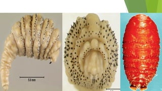
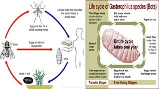
Botfly: Oestridae
 They are commonly known as bot flies, warble flies, heel flies, gadflies.
 The word "bot" means a maggot.
 A warble is a skin lump or callus which is caused by the presence of warble
fly maggot inside the skin.
 The infestation of any fly larvae inside the body is known as myiasis.
 The human bot fly, Dermatobia hominis is responsible for myiasis in human.
 The human bot fly is native to Central and South America.
 The fly do not transmit pathogens, but the larvae of D. hominis infest the skin
of mammals and live out the larval stage in the subcutaneous layer, causing
painful pustules (bumps) that secrete fluids.
 The larvae of some species grow in the flesh of their hosts, while others grow
within the hosts' alimentary tracts.
BODY CHARACTERISTICS AND LIFE CYCLE
Adults:
Hairy, 12-18 mm long in length with a wide array of colors. The face is yellow
with a metallic blue abdomen and orange legs. Adult have extremely sensitive
antennae which help male and female to find each other quickly. During the adult
stage, D. hominis does not feed.
Hosts: Hosts are warm-blooded animals including buffalo, cattle, cats, dogs,
humans, monkeys, pigs, rabbits, sheep, cattle and dogs.
Egg:
The egg of the bot fly is creamy colored and oval in shape.
Egg is attached to different species of blood-feeding insects (mosquito or a tick)
captured by the female bot fly. This behavior is known as phoresy.
The eggs, usually attached to the ventral side of the body, hatch when the insect
carrying the eggs begins to blood feed on a warm-blooded host.
Larva:
The larva feed on tissue exudates. It burrows into the skin through the bite
wound or hair follicles. It breath through 2 posterior spiracles.
A white maggot undergoes through three instars once in the mammalian host.
Each instar possesses backward projecting spines that encircle the thorax.
The first instar is worm-like with a bulbous end. The second instar larva has a
bottle-neck shape. The third instar is cylinder shaped.
After 7th
day of infestation, the larvae molt in to 2nd
instars, and then to 3rd
instar
after 18 days.
After 30 day interval, 3rd
instar larvae crawl out from the host body and pupate in
the soil. Generally, the life of the larvae inside the host takes 5-12 weeks.
Pupa: It have prominent anterior spiracles. Pupation takes place in the ground
and the pupae do not feed. Adults will emerge after two to three weeks.
SYMPTOMS
 Dermatobia hominis larvae cause a raised lesion in the skin that becomes hard 
and sometimes painful.
   In  some  cases  the  patients  can  feel  the  larvae  moving  when  they  shower  or 
cover the wound. 
 The  host  reacts  with  elevated  white  cell  counts  and  a  high  amount  of 
macrophages can be found around the wound. For this reason, the lesion often 
secretes pus.
TREATMENT
 Conventional Methods
 The  most  conventional  way  of  removing  the  larvae  is  with  a  simple  surgical 
procedure that includes local anesthesia. Using a scalpel to cut a slit to enlarge 
the wound, the larvae can be taken out.
 Dermatobia hominis survives in its host by breathing through spiracles that are 
flush  with  the  skin.  In  order  to  coax  the  larva  out,  the  spiracles  need  to  be 
covered. They can be covered with bacon, petroleum jelly, beeswax, or any other 
thick substance that prevents the larvae from breathing. The larvae will come up 
out of the lesion to breath. It can be removed with forceps.
 Modern Technique for treatment
 Larva  may  be  popped  out  by  applying  pressure  around  the  wound.  Use  two 
wooden spatulas to apply pressure to pop the larva out.
 Lidocaine injections can be used underneath the cyst. This creates pressure that 
pushes the larva out.
 After any of these 2 procedures, antibiotics are given to prevent further infection. 
The wound should heal in 1-2 weeks with little or no scarring.
 Management
 Travelers to botfly infestation regions and people residing in those areas need to 
take  preventive  measures,  including  applying  insect  repellent  and  wearing 
protective clothing
SCREW-WORM FLY
 Cochliomyia hominivorax, screw-worm fly,  or screw-worm  is 
parasitic fly whose larvae  (maggots) eat  the  living  tissue  of warm-
blooded animals.
 A female can lay up to 3,000 eggs and fly up to 200 km (120 mi) during her life.
 There are 5 species of Cochliomyia
  Infestation of a live vertebrate animal by a maggot is technically called myiasis.
 It  is  called  screw-worm  because  larvae  burrow  or  "screw" deeper  into  victim 
flesh.
 Screw-worm maggots only attack healthy tissue. It do not attack dead tissue in 
any case.
LIFE CYCLE
 Eggs
 Females  lay  250-500 eggs/batch in  exposed  flesh  of  warm-blooded  animals, 
including humans, such as in wounds and the navels of newly-born animals. 
 Larvae
 The larvae hatch and burrow into the surrounding tissue as they feed. 
 The maggots  are capable of causing severe tissue  damage or even death to the 
host. After 3-7 to seven days of hatching, the larvae fall to the ground to pupate.
 Pupa
 The pupae reach the adult stage about seven days later.
 Adult
 Female mate within 4-5 days after hatching. The entire life cycle takes 20 days. 
MANAGEMENT
 During the cold months when screwworm fly activity is negligiable. Breeding of
livestock must be timed in such away that newborn animals were born during the
cold months of the year.
 Human-induced wounds from branding, dehorning, castrating, earmarking and
shearing should be done only in cold months.
 Animals should be inspected routinely for the presence of wounds and
screwworm infestations. Wounds should be prophylactically treated.
 Use benzol and pine tar oil to treat screwworms infested wounds.
MYIASIS
 Frederick William Hope coined the term myiasis in 1840
 Myiasis is defined as the parasitic infestation of organs and tissues of humans or
animals by dipterous larvae for a period of time. The host my be living, necrotic
or dead.
 Some flies are attracted to open wounds and urine or feces soaked fur of victim.
 Some flies species (botfly, blowfly and screwfly) can infest unbroken skin and
have even been known to use moist soil.
 Myiasis is classified in various types. Such variations depend largely on the fly
species and where larvae are located. Some flies lay eggs in open wounds, other
larvae may invade unbroken skin or enter the body through the nose or ears, and
still others may be swallowed if the eggs are deposited on victim lips or food.
LIFE CYCLE
 The female flies lay their eggs on the sheep in damp, protected areas of the body
that are soaked with urine and feces, mainly the sheep's breech (buttocks).
 It takes 8 hrs 1 day for the eggs to hatch, depending on environmental conditions.
 Once hatched, the larvae then lacerate the skin with their mouthparts, causing
open sores.
 Once the skin has been breached, the larvae then tunnel through the sores into the
host's subcutaneous tissue, causing deep and irritating lesions highly subject to
infection.
 Bacterial infection starts after 2nd
day. If it is left untreated, toxemia (presence of
blood toxins) or septicemia  (infection in lungs, abdomen, and urinary tract)
results in victim body. This leads to anorexia (loss of appetite) and weakness and
is generally fatal if untreated.
CLASSIFICATION OF MYIASIS
 Classification according to the part of host that is infected
1. CUTANEOUS
Creeping myiasis: where larvae burrow through or under the skin. It
involves the migration of fly larvae underneath the skin.
Furuncular, where a larva remains in one spot, causing a boil-like lesion.
the “tumbu fly” Cordylobia anthropophaga penetrate healthy skin and
produce itchy sores that develop into painful boil-like lesions or furuncles,
hence the term furuncular myiasis.
1. DERMAL
2. SUB-DERMAL
 NASOPHARYNGEAL/ NASAL MYIASIS
 It is nasal cavity infestation by S. haemorrhoidalis which oviposit either directly
within the nasal cavity or in the vicinity while the patient is sleeping.
 The endoscopic use of forceps remove the maggots manually.
 Turpentine solution is also used to kill the maggots.
 AURICULAR/ AURAL MYIASIS, OR OTOMYIASIS.
 It involves infestation of external ear and/or middle ear. The eggs or larvae are
oviposited around the aural cavity. Deafness may occur and penetration within
the central nervous system results meningitis and eventually death.
 Aural myiasis must be treated by the manual extraction of the larvae irrigation of
the ear with saline, 70% ethanol, 10% chloroform, normal saline, oil drops, urea,
dextrose, creatine, topical ivermectin, or iodine saline has been used to help
remove the maggots.

 OPHTHALMIC MYIASIS/ OPHTHALMOMYIASIS
 It is infestation of anterior or posterior segment of the eyeball. The larva may be
seen in the anterior segment and the vitreous and subretinal space.
 The larva is destroyed by laser photocoagulation.
 GASTRIC/ RECTAL/ OR INTESTINAL/ Gastrointestinal MYIASIS
 It occurs by ingesting contaminated food and infestations may be persistant if left
untreated. 20 species of Sarcophaga cause gastrointestinal myiasis. Presence of
larvae in fresh stool and rectal area can diagnose it.
 UROGENITAL MYIASIS
 It is the infestation of genitourinary tract of males and females by dipteran larvae.
 Endoscopic imaging should be performed to diagnose and treatment. Broad
antibiotic treatment is recommended to prevent secondary infections.
 Classification on the basis of relationship between the host and the parasite
 Obligatory myiasis: where the parasite need a living animal/human host to
complete certain part of its life cycle e.g. screw worm. Obligatory myiasis may
be Specific, Semispecific, or Opportunistic.
 Facultative myiasis: Incidental, or accidental, a living host is not essential for
parasite to complete its life cycle. Normally a free-living parasitic larva
accidentally gain chance to enter the living host and results myiasis.
 Accidental myiasis/ Enteric myiasis/ pseudomyiasis: The invasion of body
orifices especially when the larvae cannot complete their development.
Accidental myiasis commonly is enteric, resulting from swallowing eggs or
larvae with one's food.
 One cause of pseudomyiasis is ingestion of cheese fly maggots. Pseudomyiasis
leeds to significant medical symptoms.
PREVENTION
 Poor sanitation is the most important risk factor for myiasis. Lack of basic
sanitation and inadequate garbage disposal, leaving organic material exposed,
attract insects and small animals. Adequate sanitation can be adapted by
combined effort of government, population, and education programs.
 Emptying and steam cleaning of dumpsters on regular basis must be adapted.
Washing food before consumption, storing food in adequate receptacles, wounds
must be cleaned and dressed regularly.
 In regions of endemicity, sleeping nude, outdoors, and on the floor should be
avoided. Use of screens and mosquito nets is essential to prevent flies.
 Application of insect repellents containing diethyltoluamide (DEET).
 Drying clothes in bright sunlight and ironing them are effective methods of
destroying occult eggs laid in clothing by C. anthropophaga.
 Sterile insect technique by ionizing radiation has been highly successful.

Botfly

  • 1.
    Botfly: Oestridae  Theyare commonly known as bot flies, warble flies, heel flies, gadflies.  The word "bot" means a maggot.  A warble is a skin lump or callus which is caused by the presence of warble fly maggot inside the skin.  The infestation of any fly larvae inside the body is known as myiasis.  The human bot fly, Dermatobia hominis is responsible for myiasis in human.  The human bot fly is native to Central and South America.  The fly do not transmit pathogens, but the larvae of D. hominis infest the skin of mammals and live out the larval stage in the subcutaneous layer, causing painful pustules (bumps) that secrete fluids.  The larvae of some species grow in the flesh of their hosts, while others grow within the hosts' alimentary tracts.
  • 2.
    BODY CHARACTERISTICS ANDLIFE CYCLE Adults: Hairy, 12-18 mm long in length with a wide array of colors. The face is yellow with a metallic blue abdomen and orange legs. Adult have extremely sensitive antennae which help male and female to find each other quickly. During the adult stage, D. hominis does not feed. Hosts: Hosts are warm-blooded animals including buffalo, cattle, cats, dogs, humans, monkeys, pigs, rabbits, sheep, cattle and dogs. Egg: The egg of the bot fly is creamy colored and oval in shape. Egg is attached to different species of blood-feeding insects (mosquito or a tick) captured by the female bot fly. This behavior is known as phoresy. The eggs, usually attached to the ventral side of the body, hatch when the insect carrying the eggs begins to blood feed on a warm-blooded host.
  • 3.
    Larva: The larva feedon tissue exudates. It burrows into the skin through the bite wound or hair follicles. It breath through 2 posterior spiracles. A white maggot undergoes through three instars once in the mammalian host. Each instar possesses backward projecting spines that encircle the thorax. The first instar is worm-like with a bulbous end. The second instar larva has a bottle-neck shape. The third instar is cylinder shaped. After 7th day of infestation, the larvae molt in to 2nd instars, and then to 3rd instar after 18 days. After 30 day interval, 3rd instar larvae crawl out from the host body and pupate in the soil. Generally, the life of the larvae inside the host takes 5-12 weeks. Pupa: It have prominent anterior spiracles. Pupation takes place in the ground and the pupae do not feed. Adults will emerge after two to three weeks.
  • 6.
    SYMPTOMS  Dermatobia hominis larvae cause a raised lesion in the skin that becomes hard  and sometimes painful.   In  some  cases  the  patients  can  feel  the  larvae  moving  when  they  shower  or  cover the wound.   The  host  reacts  with  elevated  white  cell  counts  and  a  high  amount  of  macrophages can be found around the wound. For this reason, the lesion often  secretes pus.
  • 7.
    TREATMENT  Conventional Methods The  most  conventional  way  of  removing  the  larvae  is  with  a  simple  surgical  procedure that includes local anesthesia. Using a scalpel to cut a slit to enlarge  the wound, the larvae can be taken out.  Dermatobia hominis survives in its host by breathing through spiracles that are  flush  with  the  skin.  In  order  to  coax  the  larva  out,  the  spiracles  need  to  be  covered. They can be covered with bacon, petroleum jelly, beeswax, or any other  thick substance that prevents the larvae from breathing. The larvae will come up  out of the lesion to breath. It can be removed with forceps.
  • 8.
     Modern Techniquefor treatment  Larva  may  be  popped  out  by  applying  pressure  around  the  wound.  Use  two  wooden spatulas to apply pressure to pop the larva out.  Lidocaine injections can be used underneath the cyst. This creates pressure that  pushes the larva out.  After any of these 2 procedures, antibiotics are given to prevent further infection.  The wound should heal in 1-2 weeks with little or no scarring.  Management  Travelers to botfly infestation regions and people residing in those areas need to  take  preventive  measures,  including  applying  insect  repellent  and  wearing  protective clothing
  • 9.
    SCREW-WORM FLY  Cochliomyiahominivorax, screw-worm fly,  or screw-worm  is  parasitic fly whose larvae  (maggots) eat  the  living  tissue  of warm- blooded animals.  A female can lay up to 3,000 eggs and fly up to 200 km (120 mi) during her life.  There are 5 species of Cochliomyia   Infestation of a live vertebrate animal by a maggot is technically called myiasis.  It  is  called  screw-worm  because  larvae  burrow  or  "screw" deeper  into  victim  flesh.  Screw-worm maggots only attack healthy tissue. It do not attack dead tissue in  any case.
  • 10.
    LIFE CYCLE  Eggs Females  lay  250-500 eggs/batch in  exposed  flesh  of  warm-blooded  animals,  including humans, such as in wounds and the navels of newly-born animals.   Larvae  The larvae hatch and burrow into the surrounding tissue as they feed.   The maggots  are capable of causing severe tissue  damage or even death to the  host. After 3-7 to seven days of hatching, the larvae fall to the ground to pupate.  Pupa  The pupae reach the adult stage about seven days later.  Adult  Female mate within 4-5 days after hatching. The entire life cycle takes 20 days. 
  • 12.
    MANAGEMENT  During thecold months when screwworm fly activity is negligiable. Breeding of livestock must be timed in such away that newborn animals were born during the cold months of the year.  Human-induced wounds from branding, dehorning, castrating, earmarking and shearing should be done only in cold months.  Animals should be inspected routinely for the presence of wounds and screwworm infestations. Wounds should be prophylactically treated.  Use benzol and pine tar oil to treat screwworms infested wounds.
  • 13.
    MYIASIS  Frederick WilliamHope coined the term myiasis in 1840  Myiasis is defined as the parasitic infestation of organs and tissues of humans or animals by dipterous larvae for a period of time. The host my be living, necrotic or dead.  Some flies are attracted to open wounds and urine or feces soaked fur of victim.  Some flies species (botfly, blowfly and screwfly) can infest unbroken skin and have even been known to use moist soil.  Myiasis is classified in various types. Such variations depend largely on the fly species and where larvae are located. Some flies lay eggs in open wounds, other larvae may invade unbroken skin or enter the body through the nose or ears, and still others may be swallowed if the eggs are deposited on victim lips or food.
  • 14.
    LIFE CYCLE  Thefemale flies lay their eggs on the sheep in damp, protected areas of the body that are soaked with urine and feces, mainly the sheep's breech (buttocks).  It takes 8 hrs 1 day for the eggs to hatch, depending on environmental conditions.  Once hatched, the larvae then lacerate the skin with their mouthparts, causing open sores.  Once the skin has been breached, the larvae then tunnel through the sores into the host's subcutaneous tissue, causing deep and irritating lesions highly subject to infection.  Bacterial infection starts after 2nd day. If it is left untreated, toxemia (presence of blood toxins) or septicemia  (infection in lungs, abdomen, and urinary tract) results in victim body. This leads to anorexia (loss of appetite) and weakness and is generally fatal if untreated.
  • 15.
    CLASSIFICATION OF MYIASIS Classification according to the part of host that is infected 1. CUTANEOUS Creeping myiasis: where larvae burrow through or under the skin. It involves the migration of fly larvae underneath the skin. Furuncular, where a larva remains in one spot, causing a boil-like lesion. the “tumbu fly” Cordylobia anthropophaga penetrate healthy skin and produce itchy sores that develop into painful boil-like lesions or furuncles, hence the term furuncular myiasis. 1. DERMAL 2. SUB-DERMAL
  • 16.
     NASOPHARYNGEAL/ NASALMYIASIS  It is nasal cavity infestation by S. haemorrhoidalis which oviposit either directly within the nasal cavity or in the vicinity while the patient is sleeping.  The endoscopic use of forceps remove the maggots manually.  Turpentine solution is also used to kill the maggots.  AURICULAR/ AURAL MYIASIS, OR OTOMYIASIS.  It involves infestation of external ear and/or middle ear. The eggs or larvae are oviposited around the aural cavity. Deafness may occur and penetration within the central nervous system results meningitis and eventually death.  Aural myiasis must be treated by the manual extraction of the larvae irrigation of the ear with saline, 70% ethanol, 10% chloroform, normal saline, oil drops, urea, dextrose, creatine, topical ivermectin, or iodine saline has been used to help remove the maggots. 
  • 17.
     OPHTHALMIC MYIASIS/OPHTHALMOMYIASIS  It is infestation of anterior or posterior segment of the eyeball. The larva may be seen in the anterior segment and the vitreous and subretinal space.  The larva is destroyed by laser photocoagulation.  GASTRIC/ RECTAL/ OR INTESTINAL/ Gastrointestinal MYIASIS  It occurs by ingesting contaminated food and infestations may be persistant if left untreated. 20 species of Sarcophaga cause gastrointestinal myiasis. Presence of larvae in fresh stool and rectal area can diagnose it.  UROGENITAL MYIASIS  It is the infestation of genitourinary tract of males and females by dipteran larvae.  Endoscopic imaging should be performed to diagnose and treatment. Broad antibiotic treatment is recommended to prevent secondary infections.
  • 18.
     Classification onthe basis of relationship between the host and the parasite  Obligatory myiasis: where the parasite need a living animal/human host to complete certain part of its life cycle e.g. screw worm. Obligatory myiasis may be Specific, Semispecific, or Opportunistic.  Facultative myiasis: Incidental, or accidental, a living host is not essential for parasite to complete its life cycle. Normally a free-living parasitic larva accidentally gain chance to enter the living host and results myiasis.  Accidental myiasis/ Enteric myiasis/ pseudomyiasis: The invasion of body orifices especially when the larvae cannot complete their development. Accidental myiasis commonly is enteric, resulting from swallowing eggs or larvae with one's food.  One cause of pseudomyiasis is ingestion of cheese fly maggots. Pseudomyiasis leeds to significant medical symptoms.
  • 19.
    PREVENTION  Poor sanitationis the most important risk factor for myiasis. Lack of basic sanitation and inadequate garbage disposal, leaving organic material exposed, attract insects and small animals. Adequate sanitation can be adapted by combined effort of government, population, and education programs.  Emptying and steam cleaning of dumpsters on regular basis must be adapted. Washing food before consumption, storing food in adequate receptacles, wounds must be cleaned and dressed regularly.  In regions of endemicity, sleeping nude, outdoors, and on the floor should be avoided. Use of screens and mosquito nets is essential to prevent flies.  Application of insect repellents containing diethyltoluamide (DEET).  Drying clothes in bright sunlight and ironing them are effective methods of destroying occult eggs laid in clothing by C. anthropophaga.  Sterile insect technique by ionizing radiation has been highly successful.