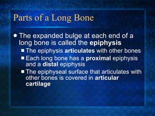
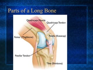
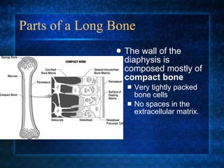
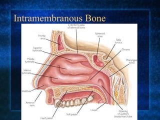
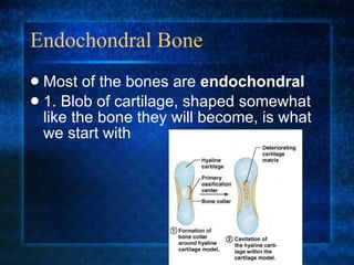
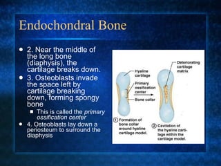
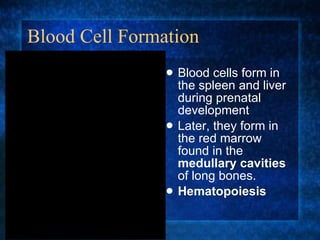
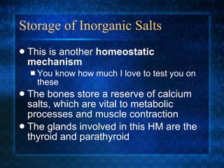
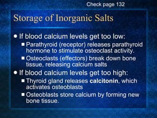
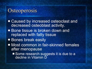
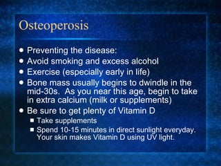
Bone Function and Formation Chapter 7 (Part 1)
Parts of a Long Bone Long bones are defined as any bone that is longer than it is wide.
Parts of a Long Bone The expanded bulge at each end of a long bone is called the  epiphysis The epiphysis  articulates  with other bones Each long bone has a  proximal  epiphysis and a  distal  epiphysis The epiphyseal surface that articulates with other bones is covered in  articular cartilage
Parts of a Long Bone
Parts of a Long Bone The long shaft of the bone is called the  diaphysis
Parts of a Long Bone Bones are covered in a thin, tough layer called the  periosteum . The periosteum covers the entire bone,  except  where there is  articular cartilage It is firmly attached to the bone and is continuous with ligaments and tendons connecting to the bone.
Parts of a Long Bone Periostium
Parts of a Long Bone
Parts of a Long Bone The wall of the diaphysis is composed mostly of  compact bone Very tightly packed bone cells No spaces in the extracellular matrix.
Parts of a Long Bone In the center of the diaphysis, is a tube called the  medullary cavity . Special layer of cells called the  endosteum  produce marrow inside the cavity.
Marrow Bone marrow comes in two different types Red marrow produces  red blood cells, white blood cells, and platelets Yellow marrow stores fat inside the bone As we age, red marrow is replaced with yellow marrow The two can be converted from one to the other, as needed.
Parts of a Long Bone The epiphysis is composed mostly of  spongy bone . Lots of space between bone cells Only a very thin layer of compact bone covers the epiphysis Helps to reduce the bone’s weight
Microscopic Anatomy Bone is composed of cells called  osteocytes   (os- =  bone ) Found in small chambers called  lacunae Surround osteocytes Made of hard extracellular material (bone matrix) Calcium carbonate Calcium phosphate
Microscopic Anatomy Bone matrix is deposited in concentric circles called  lamellae  (singular: lamella)
Microscopic Anatomy The lamellae surround a central canal Every central canal contains a blood vessel that supplies nutrients to the osteocytes in the surrounding lamellae
Microscopic Anatomy Osteocytes have  cytoplasmic extensions  called  canaliculi Used to help osteocytes obtain nutrients & communicate with surrounding osteocytes
Bone Formation During development, tissue that will become bone starts off as  cartilage There are two basic ways bone can form: Intramembranous Bone Endochondral Bone
Intramembranous Bone These bones develop into thin, sheet-like bones like those found in the cranium Originates between two sheet-like layers of connective tissue Bone forming cells called  osteoblasts  lay down spongy bone near the center between the membranes Cells of the membrane form the  periosteum Cells of the periosteum produce  osteoblasts  that form  compact   bone  to surround the spongy bone.
Intramembranous Bone
Endochondral Bone Most of the bones are  endochondral 1. Blob of cartilage, shaped somewhat like the bone they will become, is what we start with
Endochondral Bone 2. Near the middle of the long bone (diaphysis), the cartilage breaks down. 3. Osteoblasts invade the space left by cartilage breaking down, forming spongy bone This is called the  primary ossification center 4. Osteoblasts lay down a periosteum to surround the diaphysis
Endochondral Bone 5. After a time of growth and  ossification  in the diaphysis, the cartilage in the epiphyses begins to break down 6. Osteoblasts invade the areas in the epiphyses Create a  secondary ossificaiton center
Endochondral Bone
Endochondral Bone As the primary and secondary ossification centers grow toward each other, they form an  epiphyseal plate AKA:  Growth Plate
Endochondral Bone As toddlers and teenagers grow, bones lengthen by laying down new  bone matrix  from the  epiphyseal plate Damage = bad!
Homeostasis of Bones Throughout life, old bone tissue is broken down and “digested” by  osteoclasts New bone tissue is formed by  osteoblasts .
Bone Function Support the body Protect internal organs Allow for attachment of muscles that help up move Produce blood cells Store inorganic salts
Protection The skull protects the brain, eyes, nasal & oral passages The ribs protect the heart, lungs, windpipe, and esophagus Pelvis (hips) protect internal reproductive organs (female) and the urinary bladder.
Movement Bones (and their associated muscles) act as levers in the body Levers have three parts: A long, rigid rod or bar (the bone) A pivot point (a joint) Something that supplies a force to move the rod (muscle)
Blood Cell Formation Blood cells form in the spleen and liver during prenatal development Later, they form in the red marrow found in the  medullary cavities  of long bones. Hematopoiesis
Storage of Inorganic Salts This is another  homeostatic mechanism You know how much I love to test you on these The bones store a reserve of calcium salts, which are vital to metabolic processes and muscle contraction The glands involved in this HM are the thyroid and parathyroid
Storage of Inorganic Salts If blood calcium levels get too low: Parathyroid (receptor) releases parathyroid hormone to stimulate osteoclast activity. Osteoclasts (effectors) break down bone tissue, releasing calcium salts If blood calcium levels get too high: Thyroid gland releases  calcitonin , which activates osteoblasts Osteoblasts store calcium by forming new bone tissue. Check page 132
Storage of Inorganic Salts
Osteoperosis Caused by increased osteoclast and decreased osteoblast activity. Bone tissue is broken down and replaced with fatty tissue Bones break easily Most common in fair-skinned females after menopause Some research suggests it is due to a decline in Vitamin D
Osteoperosis
Osteoperosis
Osteoperosis Preventing the disease: Avoid smoking and excess alcohol Exercise (especially early in life) Bone mass usually begins to dwindle in the mid-30s.  As you near this age, begin to take in extra calcium (milk or supplements) Be sure to get plenty of Vitamin D Take supplements Spend 10-15 minutes in direct sunlight everyday.  Your skin makes Vitamin D using UV light.

Bone function and_formation_2009

  • 1.
    Bone Function andFormation Chapter 7 (Part 1)
  • 2.
    Parts of aLong Bone Long bones are defined as any bone that is longer than it is wide.
  • 3.
    Parts of aLong Bone The expanded bulge at each end of a long bone is called the epiphysis The epiphysis articulates with other bones Each long bone has a proximal epiphysis and a distal epiphysis The epiphyseal surface that articulates with other bones is covered in articular cartilage
  • 4.
    Parts of aLong Bone
  • 5.
    Parts of aLong Bone The long shaft of the bone is called the diaphysis
  • 6.
    Parts of aLong Bone Bones are covered in a thin, tough layer called the periosteum . The periosteum covers the entire bone, except where there is articular cartilage It is firmly attached to the bone and is continuous with ligaments and tendons connecting to the bone.
  • 7.
    Parts of aLong Bone Periostium
  • 8.
    Parts of aLong Bone
  • 9.
    Parts of aLong Bone The wall of the diaphysis is composed mostly of compact bone Very tightly packed bone cells No spaces in the extracellular matrix.
  • 10.
    Parts of aLong Bone The wall of the diaphysis is composed mostly of compact bone Very tightly packed bone cells No spaces in the extracellular matrix.
  • 11.
    Parts of aLong Bone In the center of the diaphysis, is a tube called the medullary cavity . Special layer of cells called the endosteum produce marrow inside the cavity.
  • 12.
    Marrow Bone marrowcomes in two different types Red marrow produces red blood cells, white blood cells, and platelets Yellow marrow stores fat inside the bone As we age, red marrow is replaced with yellow marrow The two can be converted from one to the other, as needed.
  • 13.
    Parts of aLong Bone The epiphysis is composed mostly of spongy bone . Lots of space between bone cells Only a very thin layer of compact bone covers the epiphysis Helps to reduce the bone’s weight
  • 14.
    Microscopic Anatomy Boneis composed of cells called osteocytes (os- = bone ) Found in small chambers called lacunae Surround osteocytes Made of hard extracellular material (bone matrix) Calcium carbonate Calcium phosphate
  • 15.
    Microscopic Anatomy Bonematrix is deposited in concentric circles called lamellae (singular: lamella)
  • 16.
    Microscopic Anatomy Thelamellae surround a central canal Every central canal contains a blood vessel that supplies nutrients to the osteocytes in the surrounding lamellae
  • 17.
    Microscopic Anatomy Osteocyteshave cytoplasmic extensions called canaliculi Used to help osteocytes obtain nutrients & communicate with surrounding osteocytes
  • 18.
    Bone Formation Duringdevelopment, tissue that will become bone starts off as cartilage There are two basic ways bone can form: Intramembranous Bone Endochondral Bone
  • 19.
    Intramembranous Bone Thesebones develop into thin, sheet-like bones like those found in the cranium Originates between two sheet-like layers of connective tissue Bone forming cells called osteoblasts lay down spongy bone near the center between the membranes Cells of the membrane form the periosteum Cells of the periosteum produce osteoblasts that form compact bone to surround the spongy bone.
  • 20.
  • 21.
  • 22.
    Endochondral Bone Mostof the bones are endochondral 1. Blob of cartilage, shaped somewhat like the bone they will become, is what we start with
  • 23.
    Endochondral Bone 2.Near the middle of the long bone (diaphysis), the cartilage breaks down. 3. Osteoblasts invade the space left by cartilage breaking down, forming spongy bone This is called the primary ossification center 4. Osteoblasts lay down a periosteum to surround the diaphysis
  • 24.
    Endochondral Bone 5.After a time of growth and ossification in the diaphysis, the cartilage in the epiphyses begins to break down 6. Osteoblasts invade the areas in the epiphyses Create a secondary ossificaiton center
  • 25.
  • 26.
    Endochondral Bone Asthe primary and secondary ossification centers grow toward each other, they form an epiphyseal plate AKA: Growth Plate
  • 27.
    Endochondral Bone Astoddlers and teenagers grow, bones lengthen by laying down new bone matrix from the epiphyseal plate Damage = bad!
  • 28.
    Homeostasis of BonesThroughout life, old bone tissue is broken down and “digested” by osteoclasts New bone tissue is formed by osteoblasts .
  • 29.
    Bone Function Supportthe body Protect internal organs Allow for attachment of muscles that help up move Produce blood cells Store inorganic salts
  • 30.
    Protection The skullprotects the brain, eyes, nasal & oral passages The ribs protect the heart, lungs, windpipe, and esophagus Pelvis (hips) protect internal reproductive organs (female) and the urinary bladder.
  • 31.
    Movement Bones (andtheir associated muscles) act as levers in the body Levers have three parts: A long, rigid rod or bar (the bone) A pivot point (a joint) Something that supplies a force to move the rod (muscle)
  • 32.
    Blood Cell FormationBlood cells form in the spleen and liver during prenatal development Later, they form in the red marrow found in the medullary cavities of long bones. Hematopoiesis
  • 33.
    Storage of InorganicSalts This is another homeostatic mechanism You know how much I love to test you on these The bones store a reserve of calcium salts, which are vital to metabolic processes and muscle contraction The glands involved in this HM are the thyroid and parathyroid
  • 34.
    Storage of InorganicSalts If blood calcium levels get too low: Parathyroid (receptor) releases parathyroid hormone to stimulate osteoclast activity. Osteoclasts (effectors) break down bone tissue, releasing calcium salts If blood calcium levels get too high: Thyroid gland releases calcitonin , which activates osteoblasts Osteoblasts store calcium by forming new bone tissue. Check page 132
  • 35.
  • 36.
    Osteoperosis Caused byincreased osteoclast and decreased osteoblast activity. Bone tissue is broken down and replaced with fatty tissue Bones break easily Most common in fair-skinned females after menopause Some research suggests it is due to a decline in Vitamin D
  • 37.
  • 38.
  • 39.
    Osteoperosis Preventing thedisease: Avoid smoking and excess alcohol Exercise (especially early in life) Bone mass usually begins to dwindle in the mid-30s. As you near this age, begin to take in extra calcium (milk or supplements) Be sure to get plenty of Vitamin D Take supplements Spend 10-15 minutes in direct sunlight everyday. Your skin makes Vitamin D using UV light.