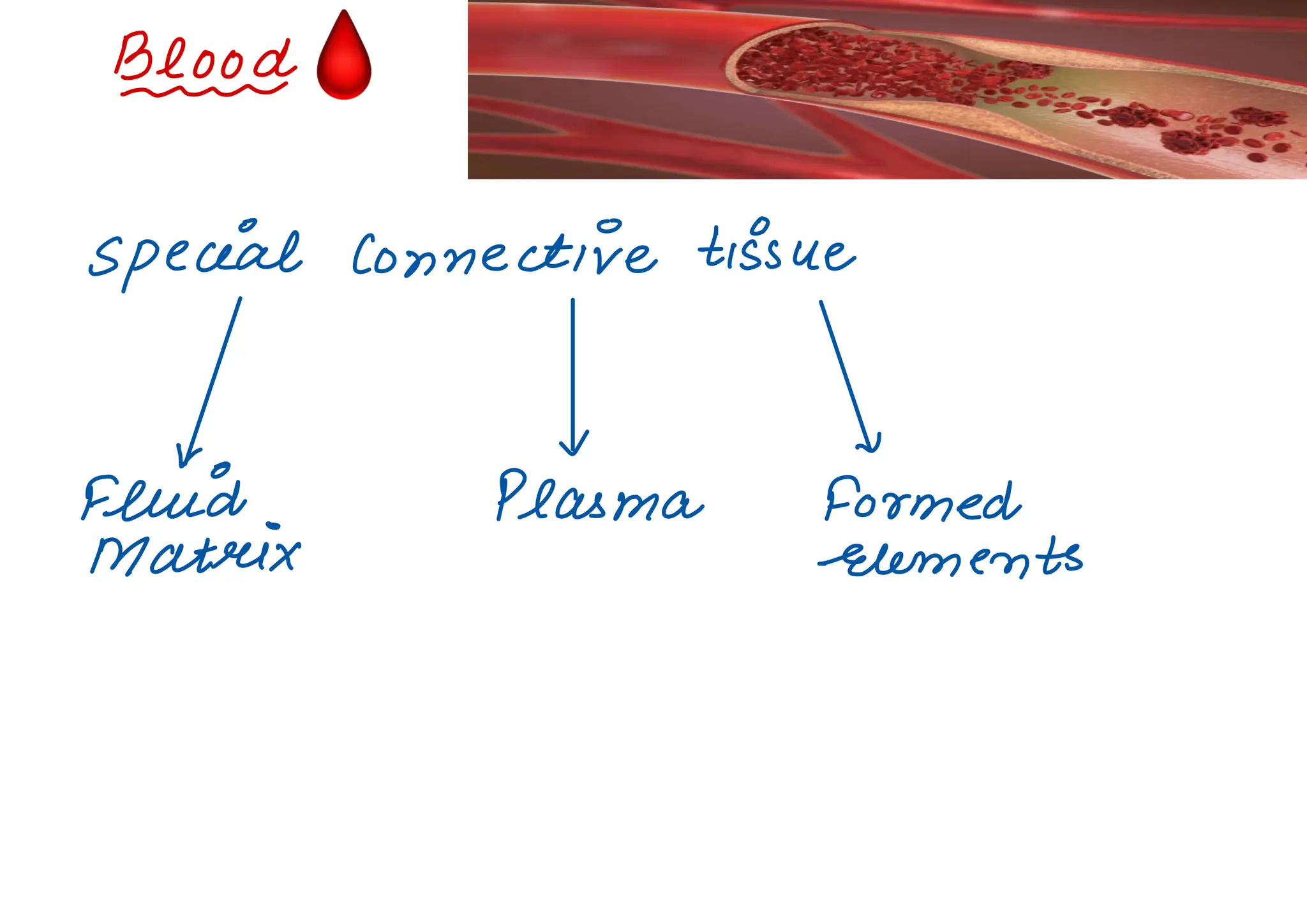
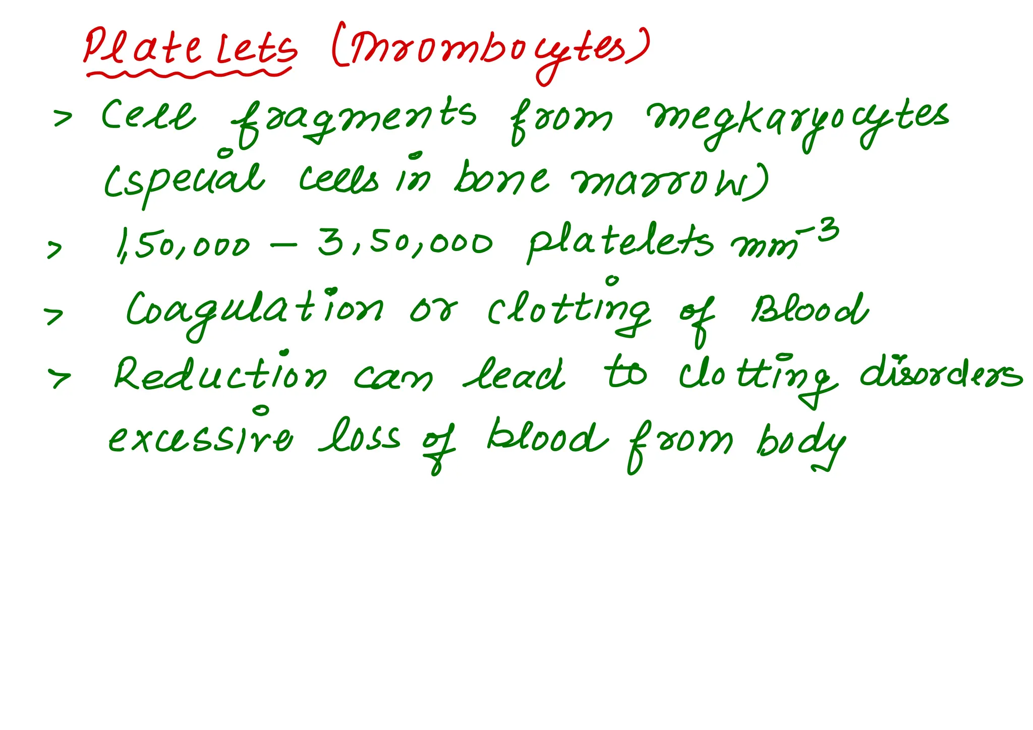
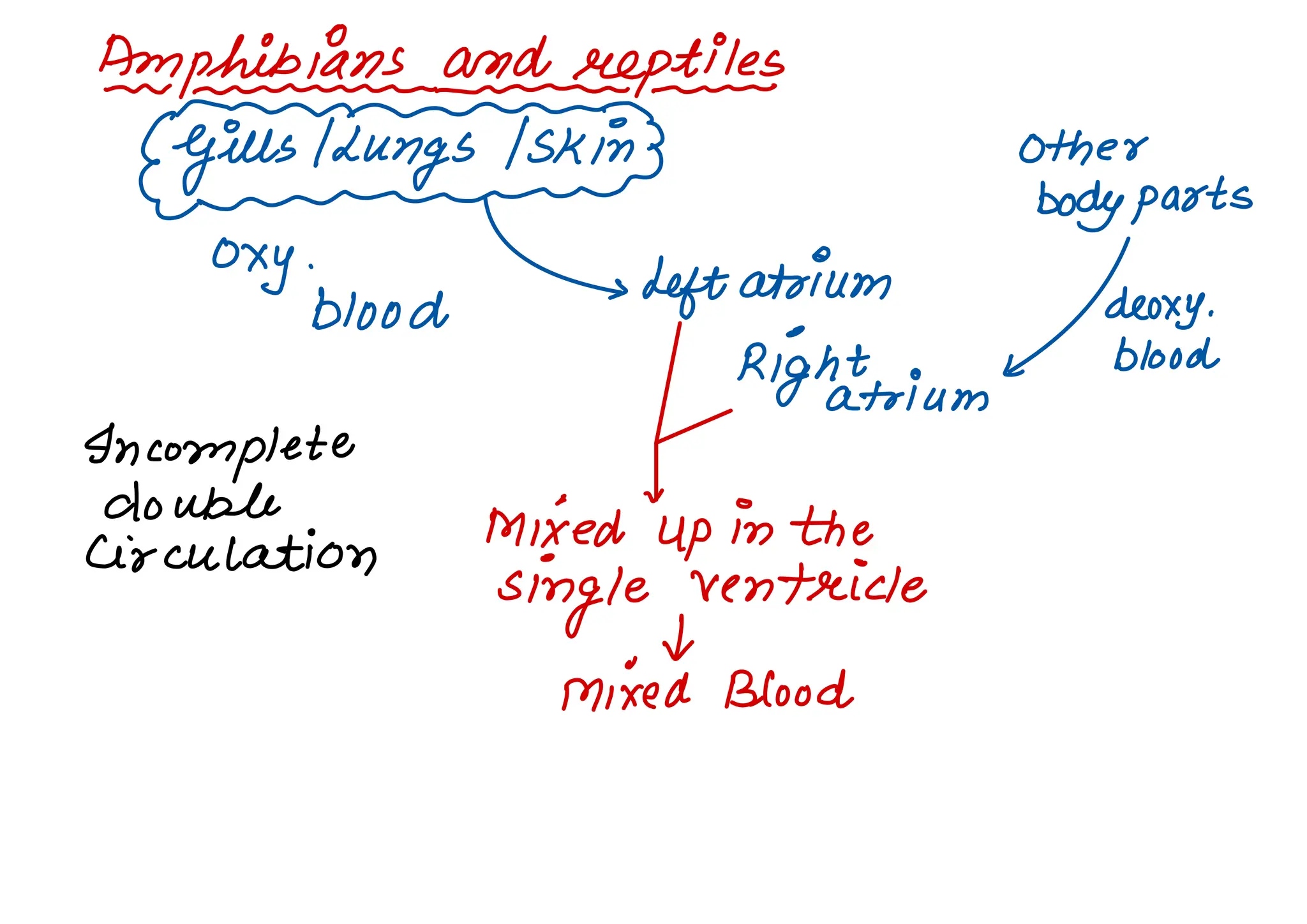
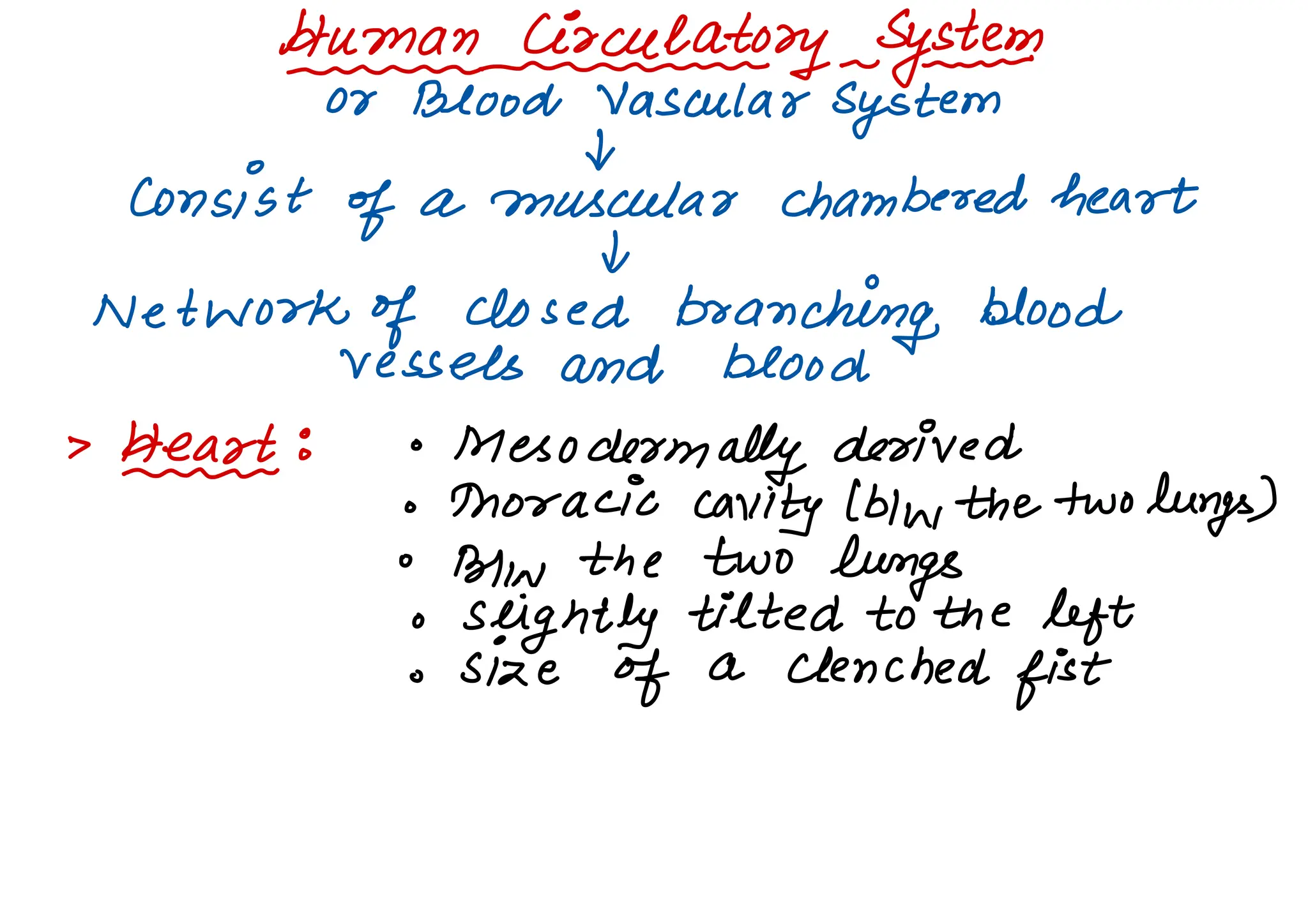
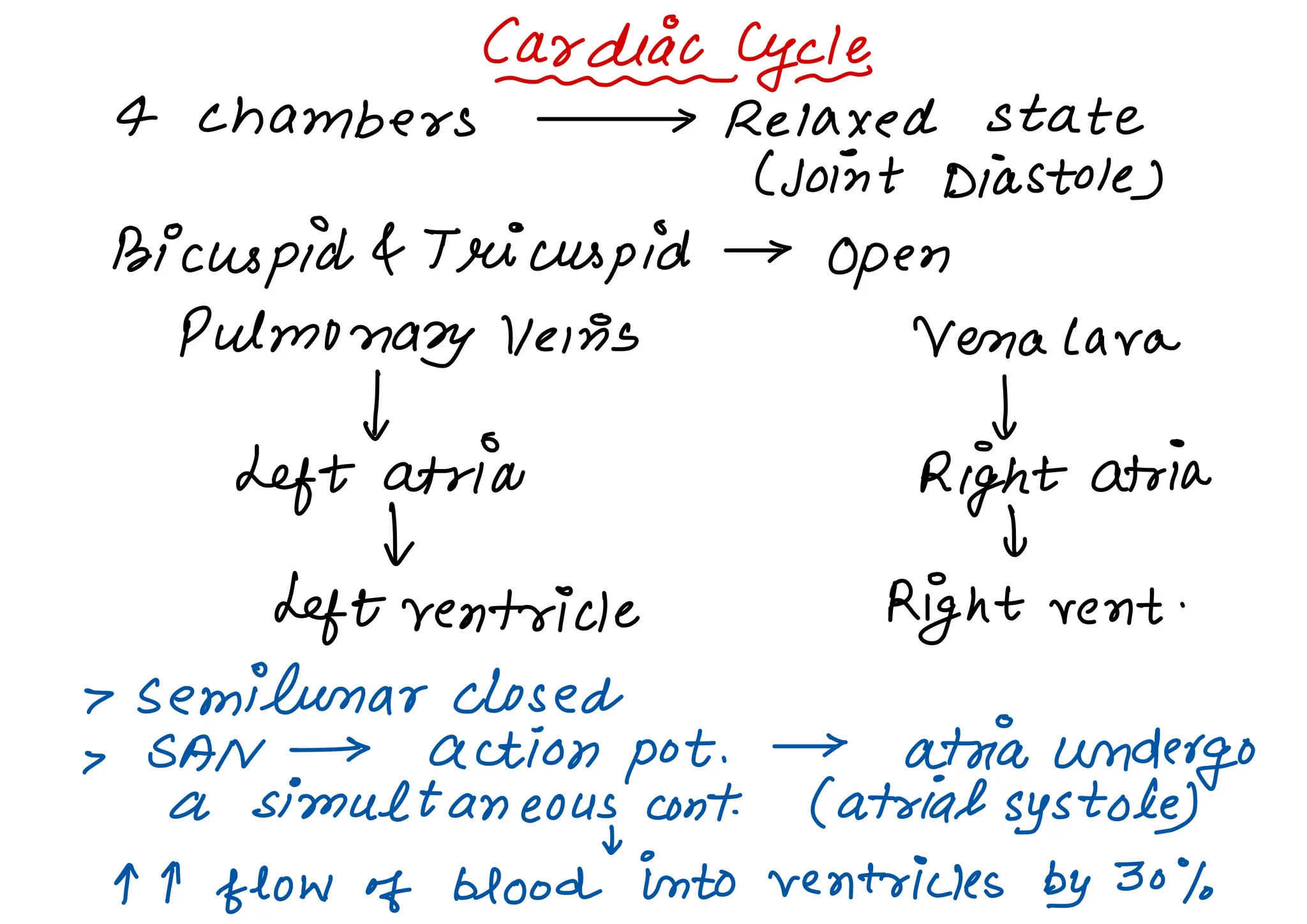
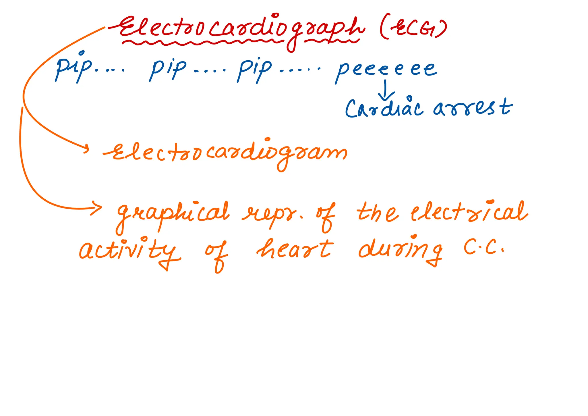
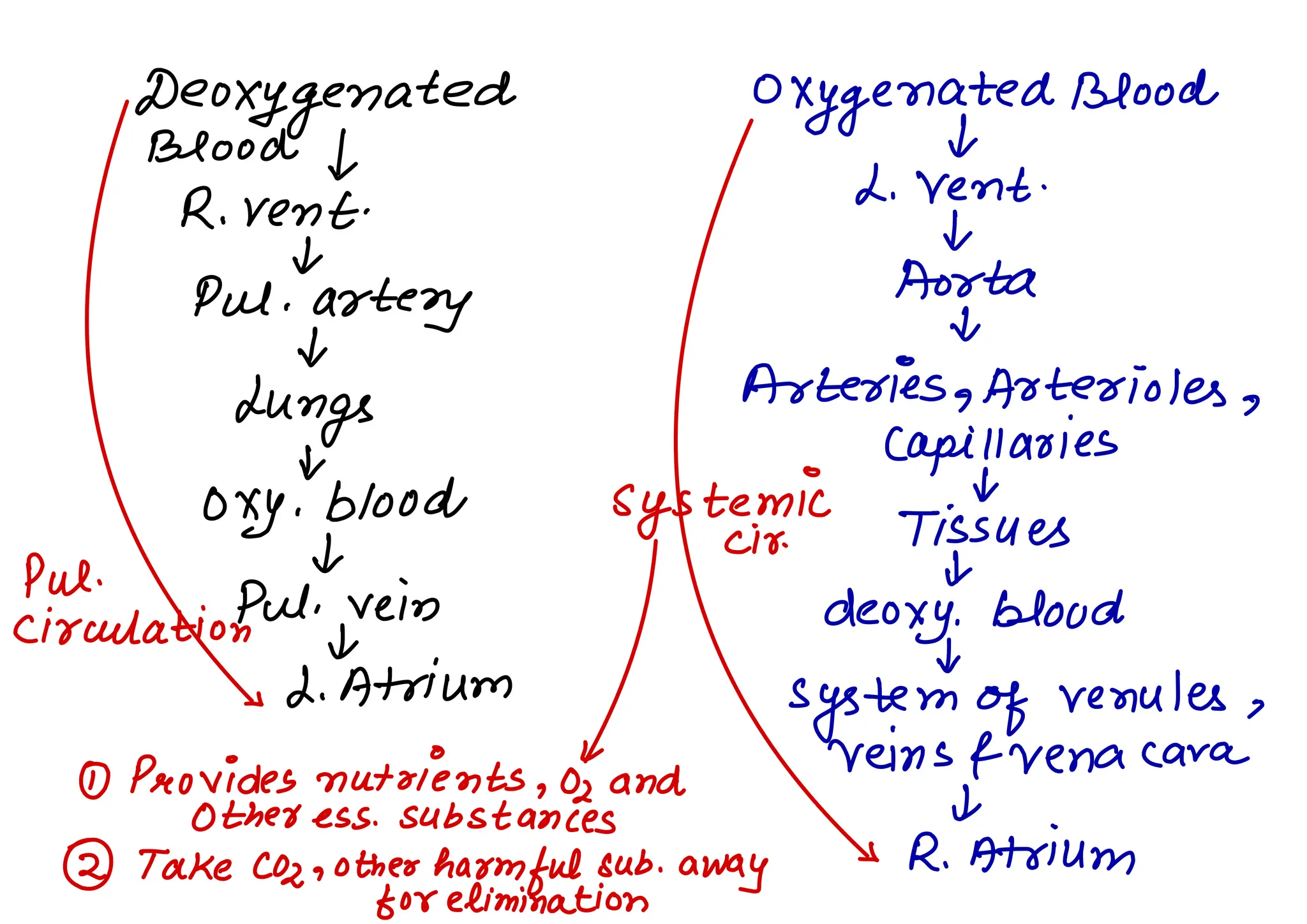
Body Fluids &
Circulation
Nutrients
Waste
Oz and other
>
ÑÑn& >
harmful
08
essential cells
substances
substances
> We need efficient mechanisms
1.
sponges , coelenterates
> circulate water
through their body
cavities .
2.
High organisms ,
Humans
> Blood , Lymph (tissue fluid)
🩸
Blotchy
special connective tissue
v
v v
Fluid Plasma Formed
Matrix elements
1. Plasmin s straw coloured
> viscous fluid
> 55010
of
blood
plasma > Plasma without
L ✓
,
clotting factors
Small
go -9201.
G- go/.
> Serum
Amt .
of lnlater Proteins > Albumin's
minerals to
Nat,
Cat,
osmotic
balance
Mg2+aHco5,
fibrinogen Vgeobulins
6-
<
&
Blood clotting
Glucose >
(
coagulation
defence
amino acids ,
presenting
mech
lipids .
inactive
form
T-02medelement.sn
,
erythrocytes
'
Leeecocytes
>
Platelets
> 45% of blood
4)
Eoyth=EyEEEdBloodCelb_
> Most abundant
> 5 to 505 millions
of RBC mm
-3
>
formed in seed bone marrow ( adults
> devoid
of nucleus ( most
of mammals
)
> biconcave in shape .
> red coloured ,
Fe containing Hb .
>
Healthy individual > 12-16
gm
Hb in
every 100Mt of blood
>
transport of respiratory gases
>
avg.
life span → 120
days
>
destroyed in
spleen (graveyard of RBCs
)
(2) LIEytesBs
> colourless
> lack
of Hb
> Nucleated
> 6000 -
Sooo mm
-3
> short lived
KLEES
Aigoanulocytes
y±azEytes - - -
Neutrophils
'
Eosinophils Basophils Lymphocytes Monocytes
> 60-654.
>
2-30/0 > Least 720-2501.
> 6- 8%
of total resist 10.5-1%1 L ✓
1^1134 infections> secretes
B T >
Phago
(Most
abundant
)
> also histamine > immune
>
Phagocytic
associated serotonin
response
-
attic
with heparin of
allergic to
body
reactions involved
in
inflamm
-
atony
rxn
Platelets (thrombocytes)
> Cele
fragments from megkaryocytes
(special cells in bone marrow
)
> 1,501000 -
3. 50,000 platelets mm
-3
>
Coagulation or clotting of Blood
> Reduction can lead to
clotting disorders
excessive loss
of blood
from body
> ABO
Bloodykeeps > an
•
Eberling :
> based on presence or absence of two
surfaces antigens.
375
> Plasma contains two natural antibodies
(proteins in response to
antigens)
> As Ba AB ,
O
> Blood
transfusion (safely)
• Rh
grouping :
rn rn -
> Present in 8001.
of human (Rmt)
> Rh
-
>
antigen absent
> Rht > Rh
-
> IN ill
form specific
antibodies
> Rh -
ve Rht
mother
Fqetus
First Pregnancy
d) Two bloods are kiell
separated by
placenta .
(2) During delivery
exp .
of maternal blood to small
amount of Rh
-1
blood from foetus .
> Mother > antibodies
against Rh
antigen
>
Subsequent pregnancies
antibodies > can destroy foetal RBCs ifrht
fatal sevreoe v
anaemia
Jaundice
erythroblastosis foetalis
> Avoid by administering anti -
Rh
anti-
bodies to mother
after delivery
of first child .
Eeggalantine ofBlood
Injury oo Trauma
Iv
coagulation Mechanism
dark reddish
"
brown scum
a
clot or
coagulation
(Network
of threads 4 a
fibrin s)
( dead &
damaged formed elements )
are
trapped
Inactive thrombin
>
Fibeeins
>
Fibrm0genp
thrombokinase
Prothrombin
> Cla cascade process
involving no .
of factors present in the
plasma in an inactive state .
Injury 00 Trauma
&
Platelets
&
Mech .
of coagulation
> certain
factors present at the site of
injury also imitate
coagulation.
Lymph ( Tissue
fluid)
~ ~ ur mum -
Blood >
IN ater + small water Sol. sub .
&
out into the spaces btw cells
of tissues
leaving larger proteins & most
of the
formed elements in blood vessels
&
Fluid is Cla Tissue or Interstitial Fluid
EEkkateyi.at#aysQhenn cloyed
Arthropods , Molluscs Annelids > Chordates
Heart to
I
closed network
Large vessels
of blood vessels
I t
open spaces or more
advantageous
body cavities
( sinuses)
Vertebrates
muscular chambered heart
C) Fishes (2 Chambered )
Atrium srentuicle
(2) Amphibians And reptiles (except coocodi
-
less
3 chambered heart
two atria ,
single ventricle
(3) Crocodiles ,
Birds , Mammals
4 chambered heart
Two atria ,
two ventricles
Fisher
Heart >
deoxygenated blood
I
oxygenated by gills
t single
Body parts CIRCU
(deoxygenated) -
LAMON
t
Heart
ttn-phinbhazsnaadnke.pt/9es-
{gÑÑungTskm3 other
new body parts
OXY -
wood
>
Left atrium
deoxy.
Right L blood
atrium
Incomplete
double
mixed
"
upinthe
circulation
single ventricle
I
mixed Blood
Bineknanmdmnammalsm
Oxy . blood deoxy.
blood
Left atria Right atria
v v
Left vent .
Right vent.
No Mixing Occurs
Double Circulation
Eumenides-
systems
or Blood Vascular
System
t
consist of a muscular chambered heart
a
Network of closed
branching blood
vessels and blood
>
Hearty : °
Mesoderm ally derived
☐ thoracic cavity (b)in,
the two
lungs)
°
Blin,
the two
lungs
o
slightly tilted to the left
o size of a clenched
fist
Pericardium
Protected by double walled membranous
bag .
>
enclosing the pericardial fluid
chambering
Two small uppers Two laager lower
chambers called chamber called
atria ventricles .
snter-atyfg.tn?enfwYm=auaoiaialh
separates night and left atria
Interventricular septum
u
m
m
m
m me
separates left and
eight ventricles .
Atrioventricular septum
nm--_
_rm
thick fibrous tissue
separates atrium and ventricle .
T
ricuspid Bicuspid or
Freyre
( three muscular
Cups) btw btw, left atrium
right atrium & and left ventricle
right ventricle
Semilunasrllalresnn
opening of right and left ventricles into
pulmonary artery and aorta .
> valves prevent any backflow
Heart > made
up of
cardiac muscles
walls
of ventricles are much thicker than
that
of atria .
Nodal tissue (special caodiac musculature
distributed in the heart
)
upper night corner lower left corner
of right atrium of right atrium close
Cla Sino -
atrial to atrioventricular
Node septum
Cla Atrio -
ventricular Node
> Bundles of Nodal fibres
D-trio -
Ventricular Bundle (AV
Passes through Bundles
AV v
Septa emerge on the top of Inter -
Ventricular Sep.
Left
"
Right
bundle bundle
> these branches gives minute
fibres
throughout the ventricular musculature
of respective sides & are ya Purkinje
Fibres
> Autotabe I
generate action pot .
without external
stimuli ]
No .
of Action pot .
> SAN > 70-75 beats 1min (Max.
)
leeesp.
for initiating & maintaining
rhythmic contractile activity of
heart
)
called as PACEMAKER .
> Heart > 70-75 b) min
avg .
72101min
cazcyc.is
4 chambers > Relaxed state
(joint Diastole)
Bicuspid & Tricuspid >
open
pulmonary veins Verna lava
I t
Left atria Right atria
to I
Left ventricle Right vent .
> semilunar closed
> SAN → action pot.
> atria undergo
a simultaneous cont
.
(atrial systole
I
1^9 flow of blood into ventricles by 30%
AYN and AY bundle
t,
Action
Bundle of His Pot .
u
to
to entire ventricular musculature
to
Ventricular muscles contract
( ventricular
systole)
jÑ& Atria >
relax (diastole)
ventricular systole
to
closure
of tricuspid and bicuspid
I
ventricular press.
TT
t
semilunar valves open
&
Yentaides relax ( diastole)
to
ventricular press . titi
&
Closure
of semilunar valves
to
ventricular press titi so
, tricuspid and
bis cuspid valves are
opened by
increasing atrial pressure
I
Blood moves
freely into ventricles
t
ventricles and atria again relaxed
(diastole)
clinically repeated → Cla cardiac
Both
-
systole <
cycle
atrial Diastole -
ventricles
> Cardiac
cycle > 72 times / min
t
Duration of one > 0.8sec .
Stroke Vol.
Blood pumped out
by each
ventricle during cardiac
cycle.
approx. 70mL
cardiac output →
stroke Vol ✗ heart rate
5000mL or 5L
Body can alter stroke vol.
,
heart rate
Hence , cardiac output
two prominent sounds
1 I
dub DUB
> Closure
of Tricuspid > Closure of
& Bicuspid semilunar
CLUB) Valves (DUB
)
>
Stethoscope
> Clinical
diagnostic significance
teeteringoast ceca>
Pip. . . .
pip . . . .
pip . . . . .
peeeeee
Caotdiac arrest
'
electrocardiogram
>
graphical reps .
of the electrical
activity of heart during c- c.
3 electrical leads
( one to each wrist & left ankle )
> For detailed evaluation →
Multiply
leads to the chest region
p-kiaveelecteei.cat excitation ( or depotaoisation)
of
atria
contraction
of both the atria
QRSComptexdepolaeeu.at
ion
of the ventricles initiates
ventricular contraction .
> Contraction starts after On and beginning
of systole .
> TINaxe
>
Return
of ventricles
excited to normal state
(Repolarisation)
> end
of T
ulare → End
of systole
> No .
of QRS complexes → Heart beat rate
of an individual
>
Any deviation in shape →
Abnormality
or disease
> Great clinical significance .
Double
circulation
Blood → Blood vessels
(arteries , reins
)
3
layers
(1) Tunica Intima → Inner lining of squamous
epithelium
(2) Tunica Media → middle
layer of smooth
comparatively muscle and elastic
fibres
thin in the
veins
(3) Tunica externa
→
text .
layer of fibrous
connective tissue .
Deoxygenated Oxygenated Blood
Blood
f I
d. Vent .
R . vent .
I
+
Put .
artery
Aorta
&
&
Lungs
Arteries , Arterioles ,
tr capillaries
&
oxy . blood systemic Tissues
Pul .
I cir.
µ
•
Put. vein
circulation y
deoxy.
blood
&
> 2. Atrium
system of venules ,
u
veinsfvenacara
① Provides nutrients , 02 and
otherness.
substances I
② T
ake cozaotheo harmful sub .
away
S R. Atrium
for elimination
Hepatic Portal
System
ummmm
- me
unique vascular conn . btw digestive tract
and liver .
>
Hepatic portal vein
t
carries blood from intestine to hier
t
then
systemic circulation
> A special coronary Sys .
of blood
vessels present in our body exclusively
for circulation of blood to & from
cardiac musculature .
-8=88
-0
A
Disorderly

Body_Fluids_And_Circulation class 11th biology

  • 1.
  • 2.
    Nutrients Waste Oz and other > ÑÑn&> harmful 08 essential cells substances substances > We need efficient mechanisms 1. sponges , coelenterates > circulate water through their body cavities . 2. High organisms , Humans > Blood , Lymph (tissue fluid)
  • 3.
    🩸 Blotchy special connective tissue v vv Fluid Plasma Formed Matrix elements
  • 4.
    1. Plasmin sstraw coloured > viscous fluid > 55010 of blood plasma > Plasma without L ✓ , clotting factors Small go -9201. G- go/. > Serum Amt . of lnlater Proteins > Albumin's minerals to Nat, Cat, osmotic balance Mg2+aHco5, fibrinogen Vgeobulins 6- < & Blood clotting Glucose > ( coagulation defence amino acids , presenting mech lipids . inactive form
  • 5.
    T-02medelement.sn , erythrocytes ' Leeecocytes > Platelets > 45% ofblood 4) Eoyth=EyEEEdBloodCelb_ > Most abundant > 5 to 505 millions of RBC mm -3 > formed in seed bone marrow ( adults > devoid of nucleus ( most of mammals ) > biconcave in shape . > red coloured , Fe containing Hb . > Healthy individual > 12-16 gm Hb in every 100Mt of blood > transport of respiratory gases
  • 6.
    > avg. life span →120 days > destroyed in spleen (graveyard of RBCs ) (2) LIEytesBs > colourless > lack of Hb > Nucleated > 6000 - Sooo mm -3 > short lived
  • 7.
    KLEES Aigoanulocytes y±azEytes - -- Neutrophils ' Eosinophils Basophils Lymphocytes Monocytes > 60-654. > 2-30/0 > Least 720-2501. > 6- 8% of total resist 10.5-1%1 L ✓ 1^1134 infections> secretes B T > Phago (Most abundant ) > also histamine > immune > Phagocytic associated serotonin response - attic with heparin of allergic to body reactions involved in inflamm - atony rxn
  • 8.
    Platelets (thrombocytes) > Cele fragmentsfrom megkaryocytes (special cells in bone marrow ) > 1,501000 - 3. 50,000 platelets mm -3 > Coagulation or clotting of Blood > Reduction can lead to clotting disorders excessive loss of blood from body
  • 9.
    > ABO Bloodykeeps >an • Eberling : > based on presence or absence of two surfaces antigens. 375 > Plasma contains two natural antibodies (proteins in response to antigens) > As Ba AB , O > Blood transfusion (safely)
  • 11.
    • Rh grouping : rnrn - > Present in 8001. of human (Rmt) > Rh - > antigen absent > Rht > Rh - > IN ill form specific antibodies > Rh - ve Rht mother Fqetus First Pregnancy d) Two bloods are kiell separated by placenta . (2) During delivery exp . of maternal blood to small amount of Rh -1 blood from foetus .
  • 12.
    > Mother >antibodies against Rh antigen > Subsequent pregnancies antibodies > can destroy foetal RBCs ifrht fatal sevreoe v anaemia Jaundice erythroblastosis foetalis > Avoid by administering anti - Rh anti- bodies to mother after delivery of first child .
  • 13.
    Eeggalantine ofBlood Injury ooTrauma Iv coagulation Mechanism dark reddish " brown scum a clot or coagulation (Network of threads 4 a fibrin s) ( dead & damaged formed elements ) are trapped
  • 14.
    Inactive thrombin > Fibeeins > Fibrm0genp thrombokinase Prothrombin > Clacascade process involving no . of factors present in the plasma in an inactive state .
  • 15.
    Injury 00 Trauma & Platelets & Mech. of coagulation > certain factors present at the site of injury also imitate coagulation. Lymph ( Tissue fluid) ~ ~ ur mum - Blood > IN ater + small water Sol. sub . & out into the spaces btw cells of tissues leaving larger proteins & most of the formed elements in blood vessels & Fluid is Cla Tissue or Interstitial Fluid
  • 16.
    EEkkateyi.at#aysQhenn cloyed Arthropods ,Molluscs Annelids > Chordates Heart to I closed network Large vessels of blood vessels I t open spaces or more advantageous body cavities ( sinuses)
  • 17.
    Vertebrates muscular chambered heart C)Fishes (2 Chambered ) Atrium srentuicle (2) Amphibians And reptiles (except coocodi - less 3 chambered heart two atria , single ventricle (3) Crocodiles , Birds , Mammals 4 chambered heart Two atria , two ventricles
  • 18.
    Fisher Heart > deoxygenated blood I oxygenatedby gills t single Body parts CIRCU (deoxygenated) - LAMON t Heart
  • 19.
    ttn-phinbhazsnaadnke.pt/9es- {gÑÑungTskm3 other new bodyparts OXY - wood > Left atrium deoxy. Right L blood atrium Incomplete double mixed " upinthe circulation single ventricle I mixed Blood
  • 20.
    Bineknanmdmnammalsm Oxy . blooddeoxy. blood Left atria Right atria v v Left vent . Right vent. No Mixing Occurs Double Circulation
  • 21.
    Eumenides- systems or Blood Vascular System t consistof a muscular chambered heart a Network of closed branching blood vessels and blood > Hearty : ° Mesoderm ally derived ☐ thoracic cavity (b)in, the two lungs) ° Blin, the two lungs o slightly tilted to the left o size of a clenched fist
  • 23.
    Pericardium Protected by doublewalled membranous bag . > enclosing the pericardial fluid chambering Two small uppers Two laager lower chambers called chamber called atria ventricles . snter-atyfg.tn?enfwYm=auaoiaialh separates night and left atria
  • 24.
    Interventricular septum u m m m m me separatesleft and eight ventricles . Atrioventricular septum nm--_ _rm thick fibrous tissue separates atrium and ventricle . T ricuspid Bicuspid or Freyre ( three muscular Cups) btw btw, left atrium right atrium & and left ventricle right ventricle
  • 25.
    Semilunasrllalresnn opening of rightand left ventricles into pulmonary artery and aorta . > valves prevent any backflow Heart > made up of cardiac muscles walls of ventricles are much thicker than that of atria . Nodal tissue (special caodiac musculature distributed in the heart ) upper night corner lower left corner of right atrium of right atrium close Cla Sino - atrial to atrioventricular Node septum Cla Atrio - ventricular Node
  • 26.
    > Bundles ofNodal fibres D-trio - Ventricular Bundle (AV Passes through Bundles AV v Septa emerge on the top of Inter - Ventricular Sep. Left " Right bundle bundle > these branches gives minute fibres throughout the ventricular musculature of respective sides & are ya Purkinje Fibres
  • 27.
    > Autotabe I generateaction pot . without external stimuli ] No . of Action pot . > SAN > 70-75 beats 1min (Max. ) leeesp. for initiating & maintaining rhythmic contractile activity of heart ) called as PACEMAKER . > Heart > 70-75 b) min avg . 72101min
  • 28.
    cazcyc.is 4 chambers >Relaxed state (joint Diastole) Bicuspid & Tricuspid > open pulmonary veins Verna lava I t Left atria Right atria to I Left ventricle Right vent . > semilunar closed > SAN → action pot. > atria undergo a simultaneous cont . (atrial systole I 1^9 flow of blood into ventricles by 30%
  • 29.
    AYN and AYbundle t, Action Bundle of His Pot . u to to entire ventricular musculature to Ventricular muscles contract ( ventricular systole) jÑ& Atria > relax (diastole)
  • 30.
    ventricular systole to closure of tricuspidand bicuspid I ventricular press. TT t semilunar valves open & Yentaides relax ( diastole) to ventricular press . titi & Closure of semilunar valves to ventricular press titi so , tricuspid and bis cuspid valves are opened by increasing atrial pressure
  • 31.
    I Blood moves freely intoventricles t ventricles and atria again relaxed (diastole) clinically repeated → Cla cardiac Both - systole < cycle atrial Diastole - ventricles
  • 32.
    > Cardiac cycle >72 times / min t Duration of one > 0.8sec . Stroke Vol. Blood pumped out by each ventricle during cardiac cycle. approx. 70mL cardiac output → stroke Vol ✗ heart rate 5000mL or 5L Body can alter stroke vol. , heart rate Hence , cardiac output two prominent sounds 1 I dub DUB
  • 33.
    > Closure of Tricuspid> Closure of & Bicuspid semilunar CLUB) Valves (DUB ) > Stethoscope > Clinical diagnostic significance
  • 34.
    teeteringoast ceca> Pip. .. . pip . . . . pip . . . . . peeeeee Caotdiac arrest ' electrocardiogram > graphical reps . of the electrical activity of heart during c- c.
  • 35.
    3 electrical leads (one to each wrist & left ankle ) > For detailed evaluation → Multiply leads to the chest region p-kiaveelecteei.cat excitation ( or depotaoisation) of atria contraction of both the atria
  • 36.
    QRSComptexdepolaeeu.at ion of the ventriclesinitiates ventricular contraction . > Contraction starts after On and beginning of systole . > TINaxe > Return of ventricles excited to normal state (Repolarisation) > end of T ulare → End of systole > No . of QRS complexes → Heart beat rate of an individual > Any deviation in shape → Abnormality or disease > Great clinical significance .
  • 38.
    Double circulation Blood → Bloodvessels (arteries , reins ) 3 layers (1) Tunica Intima → Inner lining of squamous epithelium (2) Tunica Media → middle layer of smooth comparatively muscle and elastic fibres thin in the veins (3) Tunica externa → text . layer of fibrous connective tissue .
  • 39.
    Deoxygenated Oxygenated Blood Blood fI d. Vent . R . vent . I + Put . artery Aorta & & Lungs Arteries , Arterioles , tr capillaries & oxy . blood systemic Tissues Pul . I cir. µ • Put. vein circulation y deoxy. blood & > 2. Atrium system of venules , u veinsfvenacara ① Provides nutrients , 02 and otherness. substances I ② T ake cozaotheo harmful sub . away S R. Atrium for elimination
  • 40.
    Hepatic Portal System ummmm - me uniquevascular conn . btw digestive tract and liver . > Hepatic portal vein t carries blood from intestine to hier t then systemic circulation > A special coronary Sys . of blood vessels present in our body exclusively for circulation of blood to & from cardiac musculature .
  • 42.
  • 43.