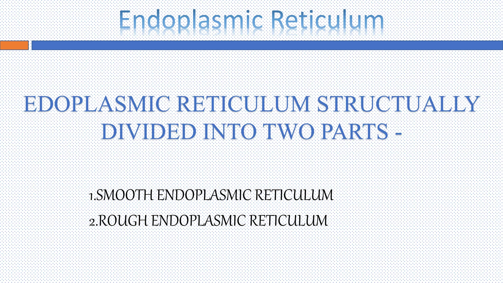
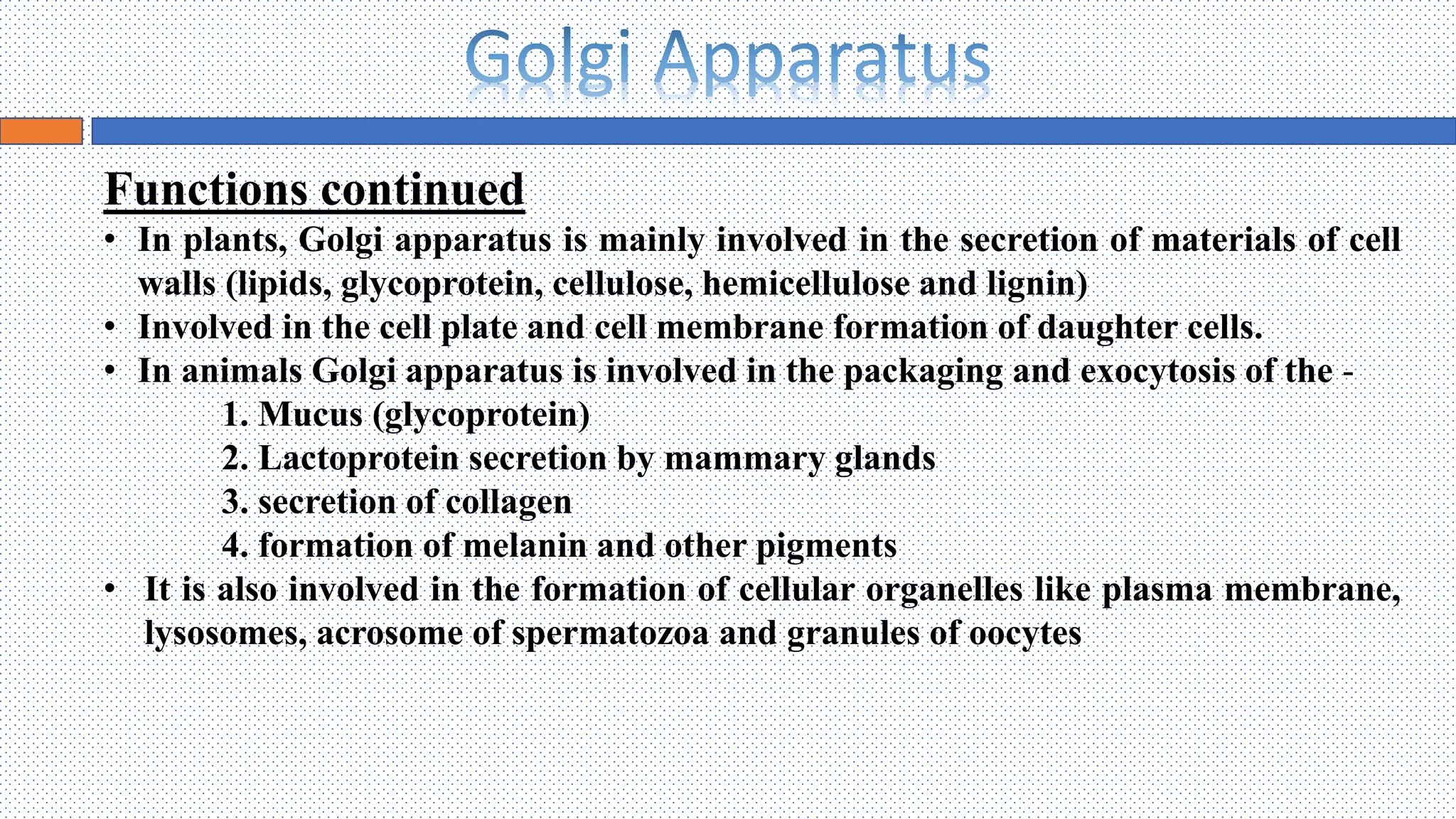
Faculty of Biosciences,
Institute of Biosciences and Technology,
Shri Ramswaroop Memorial University
INTRODUCTION
• The Endoplasmic Reticulum (ER) is arguably the most dynamic and
morphologically variable of all membranous organelles. The ER
utilizes a cytoskeleton scaffold, associated motor proteins, and less well
characterized mechanisms to undergo constant rearrangement while
maintaining the characteristic forms of a continuous network of
interconnected tubules, cisternae, and highly organized lamellar sheets.
• In the year 1945 - The lace like membranes of the endoplasmic
reticulum were first seen in the cytoplasm of chick embryo cells.
• These are membrane bound channels, seen in the form of a network of
delicate strands and vesicles in the cytoplasm.
• These are single membrane cell organelles, and exclusively present in
Eukaryotic cells.
• These form an interconnected network of tubules, vesicles and
cisternae with in cells.
DISCOVERY
• Endoplasmic Reticulum was discovered by Emilio Veratti in the year of 1902 as Sarcoplasmic Reticulum in
muscle fibers which is similar to Endoplasmic Reticulum in other cells.
• Fifty years later in 1953, this new organelle was first visualized through electron microscopy by Keith
Porter and termed it as “Endoplasmic Reticulum”
BIOGENESIS
• It is important to distinguish the concepts of de novo creation of the ER from ER biogenesis. In
the strictest sense, de novo biogenesis of the ER cannot occur. Cells do not lose the ER at some
stage and then reform it. There are examples of reversible fragmentation of the ER, but no
recorded instance of a cell that lacks an ER or outer Nuclear Envelope spontaneously generating
a complete ER.
• The ER’s role in protein translocation accounts for why the ER can only form from pre-existing
ER. As we know, protein insertion in the ER occurs co- and post-translationally. Most protein
insertion into the ER membrane and lumen occurs co-translationally via the translocon. Many
essential translocon components, such as Sec 61α and the signal recognition particle receptor α
subunit (SRP receptor), are co-translationally inserted into the ER membrane through pre-
existing translocons. The requirement of preexisting translocons to form translocons is the critical
factor that renders de novo ER biogenesis impossible. In practical terms, ER “Biogenesis” refers
to proliferation and differentiation of existing ER.
Diagram showing changes in Endoplasmic Reticulum during Cell
Proliferation
STRUCTURE
ENDOPLASMIC RETICULUM CONTAINS THREE DIFFERENT STRUCTURE –
1.CISTERNAE
2.TUBULES
3.VESICLES
 Cisternae - These are long, flat and unbranched plates
or lamellae arranged in parallel rows.
 Tubules - These are irregularly branched tube like
structures having a diameter of 50-100nm.These are
surrounded by this unit membrane of 50-60thickness
and their lumen is filled with the secretary products of
the cell.
 Vesicles - These are usually round or ovoid sacs. They
often occur isolated in the cytoplasm.
1.SMOOTH ENDOPLASMIC RETICULUM
2.ROUGH ENDOPLASMIC RETICULUM
EDOPLASMIC RETICULUM STRUCTUALLY
DIVIDED INTO TWO PARTS -
STRUCTURE OF SER
SMOOTH ENDOPLASMIC RETICULUM
• It is also known as Agranular Endoplasmic Reticulum
• Smooth ER found in smooth and straited muscle.
• The SER consist of tubules that are near the cell periphery.
• The smooth endoplasmic reticulum, does not have ribosomes .
• It participates in the production of phospholipids, the chief
lipids in cell membranes and are essential in the process of
metabolism.
STRUCTURE OF RER
ROUGH ENDOPLASMIC RETICULUM
• It is also known as Granular Endoplasmic Reticulum
• Rough ER is well developed in cells active in protein
synthesis.
• It has rough appearance due to presence of
ribosomes on its surface.
FUNCTIONS
• Transport of materials- The ER facilitates transport of materials from one part of the cell to
another, thus forming the cell’s circulatory system.
FUNCTIONS
Illustration of the plasmodesmata structure spanning the cell wall (CW).The desmotubule
(DM) is shown as a continuation of the endoplasmic reticulum (ER), with various membrane
proteins linking it to the plasma membrane (PM)
FUNCTIONS continued
• Support - the ER acts as an intracellular supporting framework, the cytoskeleton, that also
maintains the shape of the cell.
• ER act as passage for transportation of genetic information from nucleus to various cell
organelles to control the biosynthesis of protein, fats carbohydrates.
• Storage of materials - the ER provides space for temporary storage of synthetic products
such as glycogen.
• Photoreceptor - ER of pigmented cells of retina act as a photoreceptor.
• Helps information of primary lysosome with hydrolytic enzymes.
• Rough ER - surface for protein synthesis.
• Smooth ER – detoxification, convert harmful materials (drugs and poisons) into harmless
ones for excretion by the cell.
Diagram showing the functioning of Endoplasmic Reticulum
Introduction
• A Golgi apparatus, also known as a Golgi body or
complex, is a cell organelle that helps process and
package of proteins and lipid molecules, especially
proteins destined to be exported from the cell.
• Membrane bound organelles, which are sac-like.
• Found in cytoplasm of most eukaryotic cells and
absent in prokaryotes, Mammalian RBC’s and sperm
cells of bryophytes.
• Ranges from one to several within a cell.
• In plant cells there are several small golgi complex
known as Dictyosomes.
• The Golgi Apparatus is also know as the Post Office of
the cell due to its property to process , package and
delivery of molecules.
Discovery
• The Golgi Complex was discovered by an Italian physician and Nobel Laureate Camillo Golgi
in 1898 during an investigation of the nervous system.
• It’s electron microscopic structure was described by Dalton and Felix in 1954.
Biogenesis
• Method of origin of Golgi Apparatus is de novo formation i.e. new golgi
complexes arise from pre-existing Golgi bodies.
• Individual stacks of cisternae may arise from pre-existing stacks by division or
fragmentation.
• Membrane of Golgi apparatus originates from the membrane of smooth
endoplasmic reticulum.
• Cells of dormant seeds of higher plants generally lack Golgi Apparatuses.
Morphology of the Golgi during the cell cycle in mammalian cells
Schematic illustration of the mitotic division of the mammalian Golgi. During interphase, the Golgi stacks (green) are interconnected
into a ribbon-like structure near the centrosomes (red) and next to the nucleus (blue). In late G2, before the cells enter mitosis, the
connections between the stacks are severed. In early mitosis, the Golgi cisternae unstack and vesiculate, leading to the disassembly of
the Golgi. By metaphase, mitotic Golgi membranes cluster at the spindle poles, associate with astral microtubules, and are dispersed
throughout the cytoplasm. The membranes are then partitioned as the spindle elongates and segregates the chromosomes during
anaphase. During telophase and cytokinesis, the Golgi membranes reassemble in each daughter cell into a larger ribbon next to the
centrosome and a smaller ribbon adjacent to the cleavage furrow. In the final stage, the smaller ribbon moves to the opposite side of
the nucleus to merge with the larger ribbon
Structure
The Golgi is made of 5-8 folds called cisternae. The cisternae
contain specific enzymes creating five functional regions which
modify proteins passing through them in a stereotypical way, as
follows:
• Cis-Golgi network: faces the nucleus, forms a connection
with the endoplasmic reticulum and is the entry point into
the Golgi apparatus.
• Cis-Golgi: major processing area allowing biochemical
modifications.
• Medial-Golgi: major processing area allowing
biochemical modifications.
Structure continued
• Trans-Golgi: major processing area allowing
biochemical modifications.
• Trans-Golgi network: exit point for vesicles
budding off the Golgi surface, packages and
sorts biochemicals into the vesicles according
to their destination.
Functions
• Golgi vesicles are often , referred to as the "traffic police" of the cell. They play a key role in
sorting many of the cells proteins and membrane constituents and in directing them to their
proper destinations
• Golgi enzymes adds signal or tag such as carbohydrates or phosphate residues to certain
proteins to direct them to their proper destination.
Functions continued
• In plants, Golgi apparatus is mainly involved in the secretion of materials of cell
walls (lipids, glycoprotein, cellulose, hemicellulose and lignin)
• Involved in the cell plate and cell membrane formation of daughter cells.
• In animals Golgi apparatus is involved in the packaging and exocytosis of the -
1. Mucus (glycoprotein)
2. Lactoprotein secretion by mammary glands
3. secretion of collagen
4. formation of melanin and other pigments
• It is also involved in the formation of cellular organelles like plasma membrane,
lysosomes, acrosome of spermatozoa and granules of oocytes
Diagram showing functioning of Golgi Apparatus
• Endoplasmic Reticulum Biogenesis March 2007 DOI:10.1007/0-387-26867-7_4 In book: The
Biogenesis of Cellular Organelles (pp.63-95)
• Knox, K.; Wang, P.; Kriechbaumer, V.; Tilsner, J.; Frigerio, L.; Sparkes, I.; Hawes, C.; Oparka, K.
(2015). "Putting the Squeeze on Plasmodesmata: A Role for Reticulons in Primary Plasmodesmata
Formation". Plant Physiology. 168 (4): 1563 -
1572. Doi:10.1104/pp.15.00668. PMC 4528765. PMID 26084919
• The structure and function of the Golgi apparatus: A hundred years of questions September 1998
Journal of Experimental Botany 49(325):1281-1291 DOI:10.1093/jxb/49.325.1281
• Golgi biogenesis by Yanzhuang Wang 1, Joachim Seemann
PMID: 21690214 PMCID: PMC3179335 DOI: 10.1101/cshperspect.a005330
Endoplasmic Reticulum and Golgi Apparatus.pptx

Endoplasmic Reticulum and Golgi Apparatus.pptx

  • 1.
    Faculty of Biosciences, Instituteof Biosciences and Technology, Shri Ramswaroop Memorial University
  • 3.
    INTRODUCTION • The EndoplasmicReticulum (ER) is arguably the most dynamic and morphologically variable of all membranous organelles. The ER utilizes a cytoskeleton scaffold, associated motor proteins, and less well characterized mechanisms to undergo constant rearrangement while maintaining the characteristic forms of a continuous network of interconnected tubules, cisternae, and highly organized lamellar sheets. • In the year 1945 - The lace like membranes of the endoplasmic reticulum were first seen in the cytoplasm of chick embryo cells. • These are membrane bound channels, seen in the form of a network of delicate strands and vesicles in the cytoplasm. • These are single membrane cell organelles, and exclusively present in Eukaryotic cells. • These form an interconnected network of tubules, vesicles and cisternae with in cells.
  • 4.
    DISCOVERY • Endoplasmic Reticulumwas discovered by Emilio Veratti in the year of 1902 as Sarcoplasmic Reticulum in muscle fibers which is similar to Endoplasmic Reticulum in other cells. • Fifty years later in 1953, this new organelle was first visualized through electron microscopy by Keith Porter and termed it as “Endoplasmic Reticulum”
  • 5.
    BIOGENESIS • It isimportant to distinguish the concepts of de novo creation of the ER from ER biogenesis. In the strictest sense, de novo biogenesis of the ER cannot occur. Cells do not lose the ER at some stage and then reform it. There are examples of reversible fragmentation of the ER, but no recorded instance of a cell that lacks an ER or outer Nuclear Envelope spontaneously generating a complete ER. • The ER’s role in protein translocation accounts for why the ER can only form from pre-existing ER. As we know, protein insertion in the ER occurs co- and post-translationally. Most protein insertion into the ER membrane and lumen occurs co-translationally via the translocon. Many essential translocon components, such as Sec 61α and the signal recognition particle receptor α subunit (SRP receptor), are co-translationally inserted into the ER membrane through pre- existing translocons. The requirement of preexisting translocons to form translocons is the critical factor that renders de novo ER biogenesis impossible. In practical terms, ER “Biogenesis” refers to proliferation and differentiation of existing ER.
  • 6.
    Diagram showing changesin Endoplasmic Reticulum during Cell Proliferation
  • 7.
    STRUCTURE ENDOPLASMIC RETICULUM CONTAINSTHREE DIFFERENT STRUCTURE – 1.CISTERNAE 2.TUBULES 3.VESICLES  Cisternae - These are long, flat and unbranched plates or lamellae arranged in parallel rows.  Tubules - These are irregularly branched tube like structures having a diameter of 50-100nm.These are surrounded by this unit membrane of 50-60thickness and their lumen is filled with the secretary products of the cell.  Vesicles - These are usually round or ovoid sacs. They often occur isolated in the cytoplasm.
  • 8.
    1.SMOOTH ENDOPLASMIC RETICULUM 2.ROUGHENDOPLASMIC RETICULUM EDOPLASMIC RETICULUM STRUCTUALLY DIVIDED INTO TWO PARTS -
  • 9.
  • 10.
    SMOOTH ENDOPLASMIC RETICULUM •It is also known as Agranular Endoplasmic Reticulum • Smooth ER found in smooth and straited muscle. • The SER consist of tubules that are near the cell periphery. • The smooth endoplasmic reticulum, does not have ribosomes . • It participates in the production of phospholipids, the chief lipids in cell membranes and are essential in the process of metabolism.
  • 11.
  • 12.
    ROUGH ENDOPLASMIC RETICULUM •It is also known as Granular Endoplasmic Reticulum • Rough ER is well developed in cells active in protein synthesis. • It has rough appearance due to presence of ribosomes on its surface.
  • 13.
    FUNCTIONS • Transport ofmaterials- The ER facilitates transport of materials from one part of the cell to another, thus forming the cell’s circulatory system.
  • 14.
    FUNCTIONS Illustration of theplasmodesmata structure spanning the cell wall (CW).The desmotubule (DM) is shown as a continuation of the endoplasmic reticulum (ER), with various membrane proteins linking it to the plasma membrane (PM)
  • 15.
    FUNCTIONS continued • Support- the ER acts as an intracellular supporting framework, the cytoskeleton, that also maintains the shape of the cell. • ER act as passage for transportation of genetic information from nucleus to various cell organelles to control the biosynthesis of protein, fats carbohydrates. • Storage of materials - the ER provides space for temporary storage of synthetic products such as glycogen. • Photoreceptor - ER of pigmented cells of retina act as a photoreceptor. • Helps information of primary lysosome with hydrolytic enzymes. • Rough ER - surface for protein synthesis. • Smooth ER – detoxification, convert harmful materials (drugs and poisons) into harmless ones for excretion by the cell.
  • 16.
    Diagram showing thefunctioning of Endoplasmic Reticulum
  • 18.
    Introduction • A Golgiapparatus, also known as a Golgi body or complex, is a cell organelle that helps process and package of proteins and lipid molecules, especially proteins destined to be exported from the cell. • Membrane bound organelles, which are sac-like. • Found in cytoplasm of most eukaryotic cells and absent in prokaryotes, Mammalian RBC’s and sperm cells of bryophytes. • Ranges from one to several within a cell. • In plant cells there are several small golgi complex known as Dictyosomes. • The Golgi Apparatus is also know as the Post Office of the cell due to its property to process , package and delivery of molecules.
  • 19.
    Discovery • The GolgiComplex was discovered by an Italian physician and Nobel Laureate Camillo Golgi in 1898 during an investigation of the nervous system. • It’s electron microscopic structure was described by Dalton and Felix in 1954.
  • 20.
    Biogenesis • Method oforigin of Golgi Apparatus is de novo formation i.e. new golgi complexes arise from pre-existing Golgi bodies. • Individual stacks of cisternae may arise from pre-existing stacks by division or fragmentation. • Membrane of Golgi apparatus originates from the membrane of smooth endoplasmic reticulum. • Cells of dormant seeds of higher plants generally lack Golgi Apparatuses. Morphology of the Golgi during the cell cycle in mammalian cells
  • 21.
    Schematic illustration ofthe mitotic division of the mammalian Golgi. During interphase, the Golgi stacks (green) are interconnected into a ribbon-like structure near the centrosomes (red) and next to the nucleus (blue). In late G2, before the cells enter mitosis, the connections between the stacks are severed. In early mitosis, the Golgi cisternae unstack and vesiculate, leading to the disassembly of the Golgi. By metaphase, mitotic Golgi membranes cluster at the spindle poles, associate with astral microtubules, and are dispersed throughout the cytoplasm. The membranes are then partitioned as the spindle elongates and segregates the chromosomes during anaphase. During telophase and cytokinesis, the Golgi membranes reassemble in each daughter cell into a larger ribbon next to the centrosome and a smaller ribbon adjacent to the cleavage furrow. In the final stage, the smaller ribbon moves to the opposite side of the nucleus to merge with the larger ribbon
  • 22.
    Structure The Golgi ismade of 5-8 folds called cisternae. The cisternae contain specific enzymes creating five functional regions which modify proteins passing through them in a stereotypical way, as follows: • Cis-Golgi network: faces the nucleus, forms a connection with the endoplasmic reticulum and is the entry point into the Golgi apparatus. • Cis-Golgi: major processing area allowing biochemical modifications. • Medial-Golgi: major processing area allowing biochemical modifications.
  • 23.
    Structure continued • Trans-Golgi:major processing area allowing biochemical modifications. • Trans-Golgi network: exit point for vesicles budding off the Golgi surface, packages and sorts biochemicals into the vesicles according to their destination.
  • 24.
    Functions • Golgi vesiclesare often , referred to as the "traffic police" of the cell. They play a key role in sorting many of the cells proteins and membrane constituents and in directing them to their proper destinations • Golgi enzymes adds signal or tag such as carbohydrates or phosphate residues to certain proteins to direct them to their proper destination.
  • 25.
    Functions continued • Inplants, Golgi apparatus is mainly involved in the secretion of materials of cell walls (lipids, glycoprotein, cellulose, hemicellulose and lignin) • Involved in the cell plate and cell membrane formation of daughter cells. • In animals Golgi apparatus is involved in the packaging and exocytosis of the - 1. Mucus (glycoprotein) 2. Lactoprotein secretion by mammary glands 3. secretion of collagen 4. formation of melanin and other pigments • It is also involved in the formation of cellular organelles like plasma membrane, lysosomes, acrosome of spermatozoa and granules of oocytes
  • 26.
    Diagram showing functioningof Golgi Apparatus
  • 27.
    • Endoplasmic ReticulumBiogenesis March 2007 DOI:10.1007/0-387-26867-7_4 In book: The Biogenesis of Cellular Organelles (pp.63-95) • Knox, K.; Wang, P.; Kriechbaumer, V.; Tilsner, J.; Frigerio, L.; Sparkes, I.; Hawes, C.; Oparka, K. (2015). "Putting the Squeeze on Plasmodesmata: A Role for Reticulons in Primary Plasmodesmata Formation". Plant Physiology. 168 (4): 1563 - 1572. Doi:10.1104/pp.15.00668. PMC 4528765. PMID 26084919 • The structure and function of the Golgi apparatus: A hundred years of questions September 1998 Journal of Experimental Botany 49(325):1281-1291 DOI:10.1093/jxb/49.325.1281 • Golgi biogenesis by Yanzhuang Wang 1, Joachim Seemann PMID: 21690214 PMCID: PMC3179335 DOI: 10.1101/cshperspect.a005330