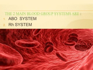
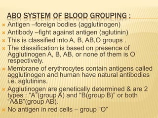
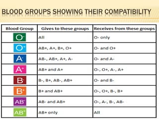
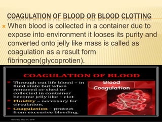
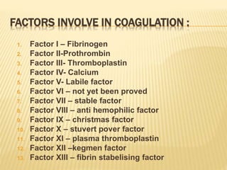
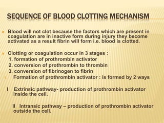
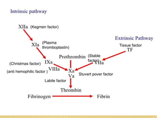
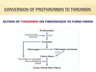
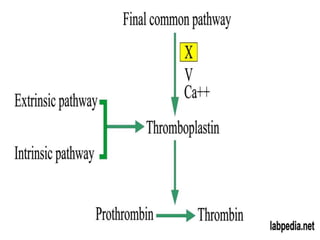
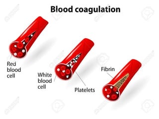
Blood is a fluid tissue with a complex structure.
Blood consists of plasma (55%) and blood cells (45%).
Blood cells are of three types. i.e.
RBC, WBC & Platelets.
WBC: WBC also known as Leucocytes.
Do not possess any pigment, they appear colorless.
Provide immunity to the body, is the main function of WBC.
For More Medicine Free PPT - http://playnever.blogspot.com/
For Health benefits and medicine videos Subscribe youtube channel - https://www.youtube.com/playlist?list=PLKg-H-sMh9G01zEg4YpndngXODW2bq92w
Blood is a body fluid in humans and other animals that delivers necessary substances such as nutrients and oxygen to the cells and transports metabolic waste products away from those same cells. In vertebrates, it is composed of blood cells suspended in blood plasma.
BLOOD composition&functions of blood,plasma proteins.(The Guyton and Hall...Maryam Fida
BLOOD composition&functions of blood,plasma proteins
Blood is a connective tissue in fluid form.
It is considered as the ‘fluid of life’ because it carries oxygen from lungs to all parts of the body and carbon dioxide from all parts of the body to the lungs.
It is known as ‘fluid of growth’ because it carries nutritive substances from the digestive system and hormones from endocrine gland to all the tissues.
The blood is also called the ‘fluid of health’ because it protects the body against the diseases and gets rid of the waste products and unwanted substances by transporting them to the excretory organs like kidneys
function
1. Nutrient Function:
Supplies nutrients like glucose, amino acids, lipids.
2. Respiratory:
carries oxygen to the tissues and transport CO² to lungs for expiration.
3. Excretory:
removes waste products from tissues and carried to the excretory organs like kidneys, skin, liver etc.
4. Regulation of body temperature
5. Defensive function:
WBCs are responsible for this function (Neutophils and monocytes) engulf the bacteria by phagocytosis.
Lymphocytes are involved in immunity.
Eosinophils are responsible for detoxification, disintegration( to break or separate into constituent elements or parts) and removal of foreign proteins.
6. Storage function
Blood cells:
1) Red blood cells or Erythroctes.
2) White blood cells or leukocytes.
3) Platelets or Thrombocytes.
Plasma is the intercellular matrix of the blood
Composition of plasma
Plasma Proteins 7-9%
Albumin
Globulin
Fibrinogen
Dissolved solutes 3%
Water 90%
albumin
Smallest in molecular weight
Synthesized by liver
Provide osmotic pressure. This is needed to maintain volume and pressure of blood
GLOBULIN
Transport and storage proteins
TYPES :
Alpha & beta : transport lipids and fat soluble vitamins (liver)
Gamma globulins : antibodies produced by lymphocytes
(IgD) belong to the group of gamma globulins and serve as defense proteins (antibodies).
IgG is the most abundant immunoglobulin, can cross the placental barrier (maternofetal transmission).
fibrinogen
Imp. clotting factor
Largest molecular weight
Helps in coagulation of blood
All the plasma proteins are synthesized in liver except ?? Gamma globulins (which are derived from B cells)
What is difference between plasma and serum ??
Plasma – fibrinogen = serum
Platelets also called thrombocytes are tiny blood cells that help your body form clots to stop bleeding. If one of your blood vessels gets damaged, it sends out signals to the platelets. The platelets then rush to the site of damage. they form a plug (clot) to fix the damage.
Normal Blood count: 1.5‐4lakh/ μL of blood
What is blood..
Its composition??
Plasma
Blood Cells (RBC, WBC, Platelets)
Its types
Its functions
FACTS AND FIGURES
Disorders of blood
Sequential Arrangement
Function of blood
Disorders of blood??
Blood is essential to life. Blood circulates through our body and delivers essential substances like oxygen and nutrients to the body’s cells. It also transports metabolic waste products away from those same cells. There is no substitute for blood. It cannot be made or manufactured. Generous blood donors are the only source of blood for patients in need of a blood transfusion.
(May 29th, 2024) Advancements in Intravital Microscopy- Insights for Preclini...Scintica Instrumentation
Intravital microscopy (IVM) is a powerful tool utilized to study cellular behavior over time and space in vivo. Much of our understanding of cell biology has been accomplished using various in vitro and ex vivo methods; however, these studies do not necessarily reflect the natural dynamics of biological processes. Unlike traditional cell culture or fixed tissue imaging, IVM allows for the ultra-fast high-resolution imaging of cellular processes over time and space and were studied in its natural environment. Real-time visualization of biological processes in the context of an intact organism helps maintain physiological relevance and provide insights into the progression of disease, response to treatments or developmental processes.
In this webinar we give an overview of advanced applications of the IVM system in preclinical research. IVIM technology is a provider of all-in-one intravital microscopy systems and solutions optimized for in vivo imaging of live animal models at sub-micron resolution. The system’s unique features and user-friendly software enables researchers to probe fast dynamic biological processes such as immune cell tracking, cell-cell interaction as well as vascularization and tumor metastasis with exceptional detail. This webinar will also give an overview of IVM being utilized in drug development, offering a view into the intricate interaction between drugs/nanoparticles and tissues in vivo and allows for the evaluation of therapeutic intervention in a variety of tissues and organs. This interdisciplinary collaboration continues to drive the advancements of novel therapeutic strategies.
Multi-source connectivity as the driver of solar wind variability in the heli...Sérgio Sacani
The ambient solar wind that flls the heliosphere originates from multiple
sources in the solar corona and is highly structured. It is often described
as high-speed, relatively homogeneous, plasma streams from coronal
holes and slow-speed, highly variable, streams whose source regions are
under debate. A key goal of ESA/NASA’s Solar Orbiter mission is to identify
solar wind sources and understand what drives the complexity seen in the
heliosphere. By combining magnetic feld modelling and spectroscopic
techniques with high-resolution observations and measurements, we show
that the solar wind variability detected in situ by Solar Orbiter in March
2022 is driven by spatio-temporal changes in the magnetic connectivity to
multiple sources in the solar atmosphere. The magnetic feld footpoints
connected to the spacecraft moved from the boundaries of a coronal hole
to one active region (12961) and then across to another region (12957). This
is refected in the in situ measurements, which show the transition from fast
to highly Alfvénic then to slow solar wind that is disrupted by the arrival of
a coronal mass ejection. Our results describe solar wind variability at 0.5 au
but are applicable to near-Earth observatories.
Observation of Io’s Resurfacing via Plume Deposition Using Ground-based Adapt...Sérgio Sacani
Since volcanic activity was first discovered on Io from Voyager images in 1979, changes
on Io’s surface have been monitored from both spacecraft and ground-based telescopes.
Here, we present the highest spatial resolution images of Io ever obtained from a groundbased telescope. These images, acquired by the SHARK-VIS instrument on the Large
Binocular Telescope, show evidence of a major resurfacing event on Io’s trailing hemisphere. When compared to the most recent spacecraft images, the SHARK-VIS images
show that a plume deposit from a powerful eruption at Pillan Patera has covered part
of the long-lived Pele plume deposit. Although this type of resurfacing event may be common on Io, few have been detected due to the rarity of spacecraft visits and the previously low spatial resolution available from Earth-based telescopes. The SHARK-VIS instrument ushers in a new era of high resolution imaging of Io’s surface using adaptive
optics at visible wavelengths.
Slide 1: Title Slide
Extrachromosomal Inheritance
Slide 2: Introduction to Extrachromosomal Inheritance
Definition: Extrachromosomal inheritance refers to the transmission of genetic material that is not found within the nucleus.
Key Components: Involves genes located in mitochondria, chloroplasts, and plasmids.
Slide 3: Mitochondrial Inheritance
Mitochondria: Organelles responsible for energy production.
Mitochondrial DNA (mtDNA): Circular DNA molecule found in mitochondria.
Inheritance Pattern: Maternally inherited, meaning it is passed from mothers to all their offspring.
Diseases: Examples include Leber’s hereditary optic neuropathy (LHON) and mitochondrial myopathy.
Slide 4: Chloroplast Inheritance
Chloroplasts: Organelles responsible for photosynthesis in plants.
Chloroplast DNA (cpDNA): Circular DNA molecule found in chloroplasts.
Inheritance Pattern: Often maternally inherited in most plants, but can vary in some species.
Examples: Variegation in plants, where leaf color patterns are determined by chloroplast DNA.
Slide 5: Plasmid Inheritance
Plasmids: Small, circular DNA molecules found in bacteria and some eukaryotes.
Features: Can carry antibiotic resistance genes and can be transferred between cells through processes like conjugation.
Significance: Important in biotechnology for gene cloning and genetic engineering.
Slide 6: Mechanisms of Extrachromosomal Inheritance
Non-Mendelian Patterns: Do not follow Mendel’s laws of inheritance.
Cytoplasmic Segregation: During cell division, organelles like mitochondria and chloroplasts are randomly distributed to daughter cells.
Heteroplasmy: Presence of more than one type of organellar genome within a cell, leading to variation in expression.
Slide 7: Examples of Extrachromosomal Inheritance
Four O’clock Plant (Mirabilis jalapa): Shows variegated leaves due to different cpDNA in leaf cells.
Petite Mutants in Yeast: Result from mutations in mitochondrial DNA affecting respiration.
Slide 8: Importance of Extrachromosomal Inheritance
Evolution: Provides insight into the evolution of eukaryotic cells.
Medicine: Understanding mitochondrial inheritance helps in diagnosing and treating mitochondrial diseases.
Agriculture: Chloroplast inheritance can be used in plant breeding and genetic modification.
Slide 9: Recent Research and Advances
Gene Editing: Techniques like CRISPR-Cas9 are being used to edit mitochondrial and chloroplast DNA.
Therapies: Development of mitochondrial replacement therapy (MRT) for preventing mitochondrial diseases.
Slide 10: Conclusion
Summary: Extrachromosomal inheritance involves the transmission of genetic material outside the nucleus and plays a crucial role in genetics, medicine, and biotechnology.
Future Directions: Continued research and technological advancements hold promise for new treatments and applications.
Slide 11: Questions and Discussion
Invite Audience: Open the floor for any questions or further discussion on the topic.
3. THE 2 MAIN BLOOD GROUP SYSTEMS ARE :
1. ABO SYSTEM
2. Rh SYSTEM
4. ABO SYSTEM OF BLOOD GROUPING :
Antigen –foreign bodies (agglutinogen)
Antibody –fight against antigen (aglutinin)
This is classified into A, B, AB,O groups .
The classification is based on presence of
Agglutinogen A, B, AB, or none of them is O
respectively.
Membrane of erythrocytes contain antigens called
agglutinogen and human have natural antibodies
i.e. aglutinins.
Agglutinogen are genetically determined & are 2
types : “A”(group A) and “B(group B)” or both
“A&B”(group AB).
No antigen in red cells – group “O”
7. RH SYSTEM OF BLOOD GROUP
There is another antigen called Rh antigen or Rh
factor found in the RBC’s .
As A,B,&O these Rh factor also inherited from one’s
parents.
Rh factor is a blood protien that plays a critical role in
some pregnancies.
People without Rh factor are known as Rh negative,
while people with Rh factor are known as positive.
If Rh factor present in blood is +ve .
If Rh factor absent in blood is –ve.
Rh = Rhesus factor
8. Rh factor is discovered in 1937 by Karl Landsteiner and
Alexander wiener .
Incompatibilities of Rh factor :
When a woman and her unborn baby carry different Rhesus
(Rh) protien factors, their condition is called as Rh-
incompatibility.
If a women who is Rh negative is pregnant with a fetus who is
Rh positive , her body will make antibodeis against the fetus
‘s blood .
This can cause Rh disease also known as hemolytic disease of
new born baby & in severe cases Rh desease leads to brain
damage and even death.
Since 1968,a vaccine has existed to prevent the mother’s body
from making anti-bodies against the fetus blood .
AB is universal accepter.
O is universal doner.
9.
10.
11. COAGULATION OF BLOOD OR BLOOD CLOTTING
When blood is collected in a container due to
expose into environment it looses its purity and
converted onto jelly like mass is called as
coagulation as a result form
fibrinogen(glycoprotien).
12. FACTORS INVOLVE IN COAGULATION :
1. Factor I – Fibrinogen
2. Factor II-Prothrombin
3. Factor III- Thromboplastin
4. Factor IV- Calcium
5. Factor V- Labile factor
6. Factor VI – not yet been proved
7. Factor VII – stable factor
8. Factor VIII – anti hemophilic factor
9. Factor IX – christmas factor
10. Factor X – stuvert pover factor
11. Factor XI – plasma thromboplastin
12. Factor XII –kegmen factor
13. Factor XIII – fibrin stabelising factor
13. SEQUENCE OF BLOOD CLOTTING MECHANISM
Blood will not clot because the factors which are present in
coagulation are in inactive form during injury they become
activated as a result fibrin will form i.e. blood is clotted.
Clotting or coagulation occur in 3 stages :
1. formation of prothrombin activator
2. conversion of prothrombin to thrombin
3. conversion of fibrinogen to fibrin
1. Formation of prothrombin activator : is formed by 2 ways
I Extrinsic pathway- production of prothrombin activator
inside the cell.
II Intransic pathway – production of prothrombin activator
outside the cell.
19. ANTICOAGULANTS
Anticoagulants are commonly known as
blood thinners, are chemical substances that
prevent or reduce coagulation or clotting of
blood , prolonging the clotting time .
Ex : heparin (inactivate IX,X,XI,XII)
coumarin derivative (inactivete factors
II,VII,IX,X & inhibit vit.K)
EDTA(remove calcium from blood )
20. MAJOR DISORDERS OF BLOOD
I )Anemia : it is the condition in which the oxygen
carrying capacity of blood is reduced or is less than
the normal quantity of heamoglobin in the blood .
Symptoms of Anemia :
• Weakness
• Tiredness
• Pale skin
• Fast heartbeat
• Shortness of breath
• Fatigue
• Chest pain
23. Causes of Anaemia :
Anemia can be caused by blood loss, decreased red blood cell
production, and increased red blood cell breakdown.
Causes of blood loss include trauma and gastrointestinal bleeding.
Causes of decreased production include iron deficiency, vitamin B12
deficiency, thalassemia, and a number of neoplasms of the bone
marrow.
Causes of increased breakdown include genetic conditions such
as sickle cell anemia, infections such as malaria, and certain
autoimmune diseases.
Anemia can also be classified based on the size of the red blood
cells and amount of hemoglobin in each cell.
i. If the cells are small, it is called microcytic anemia
ii. if they are large, it is called macrocytic anemia and
iii. if they are normal sized, it is called normocytic anemia.
The diagnosis of anemia
in men is based on a hemoglobin of less than 130 to 140 g/L (13
to 14 g/dL);
24. TYPES OF ANEMIA
Impaired RBCs production could be impaired
due to the following reasons :
a) Iron deficiency anaemia
b) Megaloblastic anaemia
c) Pernicious anaemia
d) Hypoplastic anaemia
Increased RBCs loss :
a) heamolytic anaemia
b) Haemorrhagic anaemia
25. a) Iron deficiency Anaemia :
• It is a common type of anaemia results due to inadequate absorption
of iron, excessive loss of iron increased iron requirement or
insufficient intake of iron.
• Women are at greater risk for anaemia due to menstrual blood loss
and increased iron demands of the growing foetus during pregnancy.
• Causes if body does not make enough RBC. Bleeding causes
loss of RBC more quickly. Common cause of this is infestation
due to parasitic worms, round worms etc. Malaria and
vit A deficiency can also cause IDA.
b) Megaloblastic Anaemia :
It is also called as folic acid anaemia or vitamin B12 deficiency anaemia.
It is a condition in which the bone marrow produces unusually large,
structurally abnormal, immature RBC or megaloblasts.
Deficiency of vitamin B12(cobalamine) and vitamin B9(folate ) are 2
most common causes for megaloblastic anaemia.
These 2 are serve as building blocks and essential for production of
healthy cells.
26. SYMPTOMS :
Fatigue
Paleness of skin
Shortness of breath
Dizziness
Irregular heartbeat
Muscle weakness
Treatment
• Deficiency can be treated with apprpriate changes to the diet
and the administration of suppliments.
C) Pernicious Anaemia :
It is the condition in which decrease in RBC that occures
when the intestine cannot properly absorb vitamin B12.
D) Aplastic Anaemia :
• It is the condition that occures when yoour body stops
producing enough new blood cells.
27. HEAMOLYTIC ANAEMIA :
It is a disorder in which RBCs are destroyed
faster than they can be made.
Heamorrhagic Anaemia :
excessive loss of RBCs resulting from large
wound, stomach ulcer, and heavy
menstruation leads to heamorrhagic
anaemia.
28. SICKLE CELL ANAEMIA :
It is a serious disorder in which body makes sickle
shaped(crescent shaped) RBCs.
Sickle cells contain abnormal heamoglobin called as sickled
heamoglobin or heamoglobin S.
This heamoglobin causes the cells to develop sickle or crescent
shape.
Sickle cells are stiff and sticky .
They tend to block blood flow in the bood vessels of the limbs
and organ, it is due to genetic mutation that causes substitution of
wrong amino acid in particular portion of globin.
At low oxygen concentration in many cappillaries, heamoglobin
molecules combine with each other to formfibre like structures to
form sickle shape.
Sickle cell Anaemia can leads to stroke, organ damage,
blindness, leg ulcer, etc.
29.
30.
31. TREATMENT
Rehydration with intravenous fluid helps RBC
to return its normal shape .
Blood transfusion can improve transport of
oxygen and nutrients as needed.
Supplemental oxygen is given through the
mask .