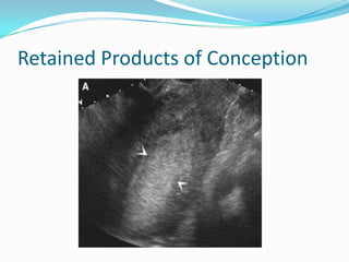
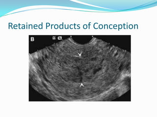
DR NURULHUDA
O&G DEPARTMENT
           SGH
 MISCARRIAGE
    THREATENED
    SILENT
    INEVITABLE
    INCOMPLETE
 TROPHOBLASTIC DISEASES
 ECTOPIC PREGNANCY
 LOCAL CAUSE
Clinical presentation
Clinical Presentation
  Amount of bleeding


  Pain


  Passage of products
MISCARRIAGE
 SPORADIC MISCARRIAGE

Definition

In UK : loss of an intrauterine pregnancy before 24
  completed weeks of gestation.

   WHO : expulsion of fetus / embryo wt < 500 g & gestation limit of <
    22 completed wks of pregnancy.
MISCARRIAGE
 SPORADIC MISCARRIAGE

 A distressing complication of pregnancy

   Occur ~ 20% of pregnancies

   Majority of sporadic miscarriage occur in 1st trimester = early
    pregnancy loses

       Only 2-5% of pregnancy miscarry after FH activity has been
        detected by USG
Spontaneous Miscarriage
Incidence
 10-15% in clinically evident pregnancy
 30% in chemically evident pregnancy


 80% of spontaneous miscarriage occurred in
 gestational age less than 14 weeks.
Miscarriage
 Threatened - 25% of all pregnancies
   ~ uterine bleeding prior to 24 w of pregnancy

 Inevitable - complete or incomplete depending on
 whether all or not POC expelled from the uterus.

 Early fetal/embryonic demise
 (missed/anembryonic/blighted ovum)
   ~ failure of pregnancy is identified before the
     expulsion of fetal and placental tissue.
Miscarriage
   Septic
   Recurrent miscarriage
     ~ primary - no previous live birth
     ~ secondary - at least one previous successful
        pregnancy.
AETIOLOGY
   No demonstrable cause – commonest

   MORPHOLOGIC AND GENETIC ABNORMALITY

   Abnormal karyotype~

     50% in first trimester
     20 - 30% in 2nd trimester
     5- 10% in 3rd trimester
AETIOLOGY
   50% of spontaneous miscarriage is due to aneuploidy

   50% of aneuploidy is autosomal trisomies eg: trisomy 16

    during first trimester- commonly trisomy 16 or
     monosomy X (45XO Turner Syndrome - 20%)

    polyploidy (triploidy) 20%,

        producing blighted ovum or partial mole
AETIOLOGY
MATERNAL FACTORS
 underlying systemic diseases:
     ~ endocrine
     ~ CVS - HPT
     ~ renal disease
     ~ CTD - SLE
   maternal infection
   uterine defects
   malnutrition
   emotional disturbance - no valid evidence
Infective Causes of Miscarriage
 Mechanisms – unclear but postulated due to maternal
 pyrexia/ bacteraemia

 Recent prospective study found :
   Women experience 1st trimester miscarriage – no more
    likely to have a clinical infection than those having a
    successful pregnancy

 In recurrent miscarriage :
   Infective causes (> genital tract infection) is unclear
Infective Causes of Miscarriage
 HIV
   Remains unknown

 Syphillis & Parvovirus B19

   Commonly cause late 2nd trimester miscarriage &
    stillbirth

 Group B Streptococcus (GBS)

   In late miscarriages & preterm labour
Structural causes of Miscarriage
 Mullerian duct defects - Anatomical uterine abn
   Prevalence : 3% (in lap sterilisation)
   Prevalence in recurrent miscarriers: 3 – 27%
   Most sensitive method of Ix :
     Hysteroscopy > HSG > ? USG


   In early preg loss,
     embryo must be presumed to implant in an avascular area of
      endometrial cavity – lead to arrested dev & early preg failure.
     This process is unlikely in recurrent preg loss
Structural causes of Miscarriage
   Open surgical / abd resection :
     Risk of pelvic & intrauterine adhesion formation(Asherman’s
      Syndrome)
     myomectomy
   Hysteroscopic resection of intrauterine septa
     More promising but lack of randomised studies


 Cervical incompetence
   Def :
     Painless dilatation of the cx lead to miscarriage in the absence
      of uterine contractions / haemorrhage
   Aetiology of 2nd trimester loss
Other causes of miscarriage
 Fetal sex
    More in males, but sex ratio remain unknown
 Multiple pregnancy
    Assoc with an increased risk of fetal loss (either by resorption, post-
     implantation loss / 2nd trimester miscarriage)
    Risk of miscarriage 2x singleton preg
    Monochorionic twin preg – 12% risk
 Parity
    Rises risk with parity
    Results of reproductive compensation & related with maternal age
     (assoc with trisomic pregnancies)
Maternal health in miscarriage
 Cigarete smoking
      Correlated with miscarriage
      Nicotine has adverse effect on trophoblastic invasion
   Cocaine – increased risk of miscarriage
   Alcohol - higher in women ended up with miscarriage
   Caffeine – high level ass. with miscarriage
   Chemicals : lead,ethylene oxide, solvents, pestisides, vinyl chloride &
    anaesthetic gases – ass. with fetal loss
 Radiotherapy & Chemotherapy
      May cause miscarriage & fetal abnormality in a dose of > 25 rads –
       0.1% risk
CLINICAL FINDINGS
   Threatened
    Minimal vaginal bleeding + pain (usually painless)

   Closed cervical os,

   Without expulsion of POC

   Viable pregnancy by USG

     soft, non-tender abdomen, uterus=POA
     Continued vomiting ass. with increased chance of life birth
CLINICAL FINDINGS
    Inevitable
      fresh vaginal bleeding with abdominal/back pain with cx
       dilatation and effacement

      50% will miscarry

      Diagnosis determined by confirmation of cervical dilatation
       at VE

      Occasionally severe shock – may be due to massive
       haemorrhage / vasovagal reaction – cervical shock syndrome
       due to distension of the cervix by POC (Treatment is by
       quick removal of the POC from os by bedside)
CLINICAL and USG FINDINGS
 Incomplete
   heavy bleeding & abd cramps with open cx os
   POC partially expulsed & USG showed hyperechoic material in the
    uterine cavity
 Complete
   A hx of pain + bleeding + POCs seen
   bleeding and pain cease afterwards as POCs completely expelled.
    USG ~ empty uterus
   Recent studies of women showed around 20-30% of all miscarriage
    is complete(Chung et al 1994; Mansur 1992)
   Expectant mx of early fetal demise has demonstrated that, with
    time, up to 25% women go on to have complete miscarriage
    (Jurkovic et al 1998)
CLINICAL FINDINGS
    Early Embryonic / Fetal Demise* (Missed /
    Silent)

      Failure of preg is identified before any
       expulsion of POC occur
      Disappearance of sn/sx of pregnancy with ut <
       gest age & closed cervical os
      An USG dx of non-viable preg in the absence of
       PV bleeding / pain(accidental finding)
      A fetal pole > 5 mm w/out FH activity or when
       USG I / 2 wks apart have shown no growth / no
       FH activity: Missed / silent miscarriage
CLINICAL FINDINGS w/out a fetal pole :
      A sac > 20 mm in diameter
        a blighted ovum*

        Goldstein - most of anembryonic pregnancy
        are not truly without embryos. They lose
        viability before our ability to image them.

        Many initially had early embryonic dev. with
        subsequent loss of viability, followed by
        embryonic resorption and thus ,appearance of
        empty sac.

         * An embryonic pregnancy / blighted ovum has been
           replaced with early embryo/ fetal demise in UK
CLINICAL FINDINGS
  Septic
    A complication of incomplete miscarriage where the
       remaining POCs become infected by ascending
       organisms + instrumentation of the uterus
      Presented with suprapubic pain, malaise, fever + PV
       bleeding
      O/E : Fever, suprapubic pain/abd rigidity, uterine &
       adnexal tenderness and closed cervix.
      Around 3-6% following termination of pregnancy
      Common organisms : E.Coli,Bacteroides, streptococci
       Clostridium welchii
      Complications : Septicaemia → bactaremic shock →
       maternal death
INVESTIGATIONS
    USG
      ~ is essential in the diagnosis - usually TVS.
      ~ will determine on-going pregnancy/failing
     pregnancy/rule out ectopic and trophoblastic
     disease.
    Pregnancy test - by urinary or serum hCG to
     distinguish an early complete miscarriage or on-
     going ectopic pregnancy.
    Blood grouping and Rh typing - Rh neg. should
     receive anti-D Ig regardless of gestational age.
Management of miscarriage
 Tx aim : to reduce the potential complication of
  miscarriage (i.e prolonged pain & bleeding, sepsis & rh-
  iso-sensitisation)

 Conservative
    Expectant Mx ~ avoids surgical procedure &
    anaesthetic.
        Appropriate for pts with an incomplete miscarriage with
         POC < 50mm in diameter on TVS
            In cases without haemodynamic compromise /
             maternal anaemia, spontaneous resolution
             occurred within 3 days in up to 80% of cases with
             minimal retained POC
        No evidence of impairment of future fetility
Management of miscarriage
     Conservative Management
     less useful for blighted ovum

          because bleeding appears to be heavier & more
           prolonged than surgical management

          60% which treatment conservatively require
           ERPOC at some stage
Management of miscarriage
 Surgical
    Evacuation of retained POC (ERPOC) – most common form of tx
     for miscarriage
    Cx is dilated & retained POC removed by S+C
    Cpx :
      Perforation of the uterus (by dilator / currete), infection
        (commonest) & incomplete emptying of the cavity ~ in up to 6%
        of cases, tearing of cervix
          Incidence of serious morbidity : 2.1% (RCOG 1985)

      Potential anaesthetic cpx
      Asherman syndrome (intrauterine adhesions)
      Incidence of mortality : 0.5 / 100 000 (Lawson et al 1994)
Threatened Miscarriage Mx
 97-98% chance of a live birth
    Women > 40s, miscarriage rate : 15-30% - even after FH present by
     USG
 Tx : reassurance & continued medical & emotional support
 Advise bedrest & avoiding SI
    Bedrest ↓pressure & improve outcome – but no clinical evidence
 Progesterone supplementation
    However, several meta-analysis trial unable to demonstrate
     beneficial effect of progesterone tx (Goldstein et al 1989)
Incomplete Miscarriage Mx
 Tx : Surgical evacuation of POC
 Without tx : maternal mortality of 1.6%
    Due to haemodynamic compromise of cervical shock.
 Suction evacuation >safer technique than sharp curettage
    Lower rate of perforation, blood loss & subsequent intrauterine
     adhesion formation (Edmonds 1992, Verkuyl & Crowther 1993)
 Routine use of syntocinon / ergometrine – no benefits in
  ↓blood loss during surgical tx of 1st trimester miscarriage (Beeby et al
  1984)
 Screening for chlamydia infection is recommended
Early Fetal Demise Mx
 Same for incomplete miscarriage
 Tx :
    Surgical tx after cervical ripening (with mifepristone/
      prostaglandin)
       To ↓ risk of cx trauma / ut perforation assoc with forced cx
        dilatation
 Expectant mx :
    < effective than cases of incomplete miscarriage
       With only 25% proceeding to complete miscarriage
 Efficacy of medical tx also < inc miscarriage
    Complete miscarriage rate can be up to 90% with higher dose of
      mifepristone & misoprostol
Coronal TVUS of the uterus shows a gestational sac with
hyperechoic margins (arrow) and endometrial cavity (curved
arrow).
Double Decidual Sac Sign. Coronal TVUS of the uterus reveals an
intrauterine gestational sac (straight arrow), decidua capsularis
(curved arrow), decidua parietalis (arrowhead), and effaced
endometrial cavity (asterisks)
Yolk sac (thin arrow) outside the amniotic membrane
(arrowhead), which has not yet fused with the chorion (curved
arrow). Embryo (thick arrow) is seen within the amniotic sac
Anembryonic Pregnancy
Abnormal Shape of the GS
Abortion in Progress
Missed Abortion
Subchorionic Haemorrhage
Retained Products of Conception
Introduction

 Leading cause of death of pregnancy related deaths during
  first trimester
 13 maternal deaths resulting from ectopic pregnancy in the
  UK in 1997–99.
 Incidence of ectopic pregnancy has remained static in
  recent years (11.1/1000 pregnancies)
 32000 ectopic pregnancies are diagnosed in the UK within
  a three year period.
Clinical presentation

 Clinical suspicion – positive pregnancy test, pain, bleeding
  and adnexal mass
 Clinical triad – pain, bleeding, adnexal mass
  ~ 45% of patients         Pain


                       Clinical Triad


            Bleeding                    Adnexal mass
Clinical presentation

 Location of pain
      lower abdominal ~ 74%
      generalised abdominal
      ipsilateral lower quadrant
      contralateral lower abdomen
      shoulder tip
      back pain
      vaginal
Risk Factors for Ectopic pregnancy
 High Risk
 Tubal surgery
 Sterilization
 Previous ectopic pregnancy
 Use of IUCD
 Documented tubal disease
 In utero exposure to diethylstilbesterol
Risk Factors for Ectopic pregnancy
 High Risk

 Moderate Risk
  Infertility
  Multiple sexual partners
  Previous genital infections


 Slight Risk
  Cigarrette smoking
  Previous pelvic / abdominal
  surgery
Pregnancy testing


 Urine pregnancy test
 - Sensitive radioimmunoassays are widely available
 - +ve at approximately 23 menstrual days (9 days
 postconception)
Pregnancy testing
 Beta hCG quantitation (serum B hCG)
 Normal intrauterine pregnancy
     - hCG doubling time of 2 days (66%)
     - Intrauterine GS (US Scan) with hCG 1000 – 2000
     mIU/ml
Pregnancy testing


 Beta hCG quantitation (serum B hCG)
     Ectopic pregnancy
     - hCG doubling time is different
     - hCG doubling time increased
     - disproportionately high level of hCG in correlation
     ultrasound findings
Diagnosis of Ectopic Pregnancy
 Clinical
 Biochemical
 Ultrasound
 Diagnostic laparoscopy ~ GOLD
                       STANDARD
SONOGRAPHY
                   Early intrauterine pregnancy
 Earliest sign ~ small fluid collection in the
 endometrium
SONOGRAPHY
 Gestational sac
                  Early intrauterine pregnancy
 intradecidual sign with echogenic ring formed by chorionic villi
 double decidua sac sign (DSS)
 eccentric to the endometrial cavity


                    Uterus
 Bladder
SONOGRAPHY
 Gestational sac
                  Early intrauterine pregnancy
     presence of yolk sac & the embryo


               Yolk Sac
No identifiable intrauterine GS
 One of three possibilities
  - Very early intrauterine pregnancy
  - recent spontaneous miscarriage
  - Ectopic pregnancy
                                        Bladder

                         Cervix
                                            Uterus
Sites of Ectopic Pregnancy
SONOGRAPHY
                                Ectopic Pregnancy

            Empty uterus

                                   Normal ovary


**Normal pelvic sonogram does
not exclude Ectopic pregnancy
~ 26%
SONOGRAPHY
                           Ectopic Pregnancy
Pseudogestational sac
20% of ectopic pregnancy
SONOGRAPHY
                  Ectopic Pregnancy

 Uterus


                              Uterus


Adnexal ring
“doughnut sign”
SONOGRAPHY
               Ectopic Pregnancy


                    Uterus
Adnexal mass


                 Mass
SONOGRAPHY
                     Ectopic Pregnancy

                 LIVER


LIVER
                                   Kidney

        Kidney


                 Free fluid at Morrison’s
                 Pouch
  Normal
MANAGEMENT OF ECTOPIC PREGNANCY
 Surgical option
 - Laparotomy VS Laparoscopy
 - Salpingotomy VS salpingectomy
• Nonsurgical treatment
 - Expectant management
 - Metrotrexate
• Combined medical-surgical treatment
MANAGEMENT OF TUBAL ECTOPIC PREGNANCY
          Laparoscopy VS Laparotomy
   A laparoscopic should be the surgical management of
   tubal pregnancy, in the haemodynamically stable
   patient.
MANAGEMENT OF TUBAL ECTOPIC PREGNANCY
                Laparoscopy VS Laparotomy


shorter operation times,
less intraoperative blood loss,
shorter hospital stay
lower analgesic requirements
MANAGEMENT OF TUBAL ECTOPIC PREGNANCY
       Laparoscopy VS Laparotomy
   no difference in overall tubal patency rates
   Similar subsequent intrauterine pregnancy rates
   Lower repeat ectopic pregnancy rates in laparoscopic
    approach
   laparoscopic salpingotomy was less successful than an
    open approach in elimination of the tubal pregnancy
    (higher rates of persistent trophoblast)
MANAGEMENT OF TUBAL ECTOPIC PREGNANCY
In haemodynamically unstable, management should be
by the most expedient method.
In most cases, this will be laparotomy.
MANAGEMENT OF TUBAL ECTOPIC
          Medical Management – Methotrexate

             Patient selection :
       Compliant
       Adnexal mass < 3.5 cm
       Beta hCG < 3000 mIU/ml
       Absent fetal heart activity
       Minimal symptom

  15% of women will require more than one dose of
   methotrexate
  7% will experience tubal rupture during follow up.
  75% will experience abdominal pain following treatment
MANAGEMENT OF TUBAL ECTOPIC
            Medical Management – Methotrexate

 im methotrexate as a single dose
  calculated from patient body surface area (50 mg/m2)
   - 75 mg and 90 mg.
 Serum hCG levels checked on days four and seven
 a further dose is given if hCG levels have failed to fall > than
  15% between day four and day seven.
 < 10% of women treated with this regimen will require
  surgical intervention
MANAGEMENT OF TUBAL ECTOPIC
           Medical Management – Methotrexate

 2 X weekly hCG measurements
  (ideally < than 50% of its initial level within seven days)
 weekly transvaginal US examinations (reduction in the size
  of adnexal mass by seven days)
 Thereafter, weekly hCG and transvaginal US examinations
  until serum hCG levels are < than 20 mIU/ml
Pregnancy of unknown location
                 Conservative Management

  clinically stable women with minimal symptoms
  Beta hCG level < 1500 to 2000 mIU/ml
  44–69% of pregnancies of unknown location resolve
   spontaneously with expectant management
        - small ectopic pregnancies which were :
        spontaneously absorbed or resolved by tubal
        abortion.
        - early intrauterine pregnancies that miscarried.
  14-28% lead to ectopic pregnancy
Remember !!!
Rhesus Negative women
  Nonsensitised women who are rhesus negative with a
   confirmed or suspected ectopic pregnancy should receive
   anti-D immunoglobulin.
WORK-UP FOR ECTOPIC PREGNANCY
                                  Qualitative Beta HCG (UPT)
             negative                           positive

                                                   TVS
 Pregnancy excluded


Intrauterine pregnancy
(normal / abnormal)       No IUP & Tubal mass


                                                      No IUP &
                 stable      unstable
                                                 No adnexal mass /
                                                     Free Fluid
         Laparoscopy /        Laparotomy
           Medical
                                                           Follow up
WORK-UP FOR ECTOPIC PREGNANCY
                           Empty uterus & no
                        adnexal mass / Free fluid


                        Serum Beta HCG


                                           Serum Beta HCG < 1500
Serum Beta HCG > 1500

                                               Repeat 48 hours later
Laparoscopy

                                                 66% or more rise
                     Rise by < 66% or
                    condition worsen

                                         Repeat scan in 1 week unless
                                              condition worsen
Bleeding in Early Pregnancy

Bleeding in Early Pregnancy

  • 1.
  • 2.
     MISCARRIAGE  THREATENED  SILENT  INEVITABLE  INCOMPLETE  TROPHOBLASTIC DISEASES  ECTOPIC PREGNANCY  LOCAL CAUSE
  • 3.
  • 4.
    Clinical Presentation Amount of bleeding  Pain  Passage of products
  • 5.
    MISCARRIAGE  SPORADIC MISCARRIAGE Definition InUK : loss of an intrauterine pregnancy before 24 completed weeks of gestation.  WHO : expulsion of fetus / embryo wt < 500 g & gestation limit of < 22 completed wks of pregnancy.
  • 6.
    MISCARRIAGE  SPORADIC MISCARRIAGE A distressing complication of pregnancy  Occur ~ 20% of pregnancies  Majority of sporadic miscarriage occur in 1st trimester = early pregnancy loses  Only 2-5% of pregnancy miscarry after FH activity has been detected by USG
  • 7.
    Spontaneous Miscarriage Incidence  10-15%in clinically evident pregnancy  30% in chemically evident pregnancy  80% of spontaneous miscarriage occurred in gestational age less than 14 weeks.
  • 8.
    Miscarriage  Threatened -25% of all pregnancies ~ uterine bleeding prior to 24 w of pregnancy  Inevitable - complete or incomplete depending on whether all or not POC expelled from the uterus.  Early fetal/embryonic demise (missed/anembryonic/blighted ovum) ~ failure of pregnancy is identified before the expulsion of fetal and placental tissue.
  • 9.
    Miscarriage Septic  Recurrent miscarriage ~ primary - no previous live birth ~ secondary - at least one previous successful pregnancy.
  • 10.
    AETIOLOGY No demonstrable cause – commonest MORPHOLOGIC AND GENETIC ABNORMALITY  Abnormal karyotype~  50% in first trimester  20 - 30% in 2nd trimester  5- 10% in 3rd trimester
  • 11.
    AETIOLOGY 50% of spontaneous miscarriage is due to aneuploidy  50% of aneuploidy is autosomal trisomies eg: trisomy 16 during first trimester- commonly trisomy 16 or monosomy X (45XO Turner Syndrome - 20%) polyploidy (triploidy) 20%, producing blighted ovum or partial mole
  • 12.
    AETIOLOGY MATERNAL FACTORS  underlyingsystemic diseases: ~ endocrine ~ CVS - HPT ~ renal disease ~ CTD - SLE  maternal infection  uterine defects  malnutrition  emotional disturbance - no valid evidence
  • 13.
    Infective Causes ofMiscarriage  Mechanisms – unclear but postulated due to maternal pyrexia/ bacteraemia  Recent prospective study found :  Women experience 1st trimester miscarriage – no more likely to have a clinical infection than those having a successful pregnancy  In recurrent miscarriage :  Infective causes (> genital tract infection) is unclear
  • 14.
    Infective Causes ofMiscarriage  HIV  Remains unknown  Syphillis & Parvovirus B19  Commonly cause late 2nd trimester miscarriage & stillbirth  Group B Streptococcus (GBS)  In late miscarriages & preterm labour
  • 15.
    Structural causes ofMiscarriage  Mullerian duct defects - Anatomical uterine abn  Prevalence : 3% (in lap sterilisation)  Prevalence in recurrent miscarriers: 3 – 27%  Most sensitive method of Ix :  Hysteroscopy > HSG > ? USG  In early preg loss,  embryo must be presumed to implant in an avascular area of endometrial cavity – lead to arrested dev & early preg failure.  This process is unlikely in recurrent preg loss
  • 16.
    Structural causes ofMiscarriage  Open surgical / abd resection :  Risk of pelvic & intrauterine adhesion formation(Asherman’s Syndrome)  myomectomy  Hysteroscopic resection of intrauterine septa  More promising but lack of randomised studies  Cervical incompetence  Def :  Painless dilatation of the cx lead to miscarriage in the absence of uterine contractions / haemorrhage  Aetiology of 2nd trimester loss
  • 17.
    Other causes ofmiscarriage  Fetal sex  More in males, but sex ratio remain unknown  Multiple pregnancy  Assoc with an increased risk of fetal loss (either by resorption, post- implantation loss / 2nd trimester miscarriage)  Risk of miscarriage 2x singleton preg  Monochorionic twin preg – 12% risk  Parity  Rises risk with parity  Results of reproductive compensation & related with maternal age (assoc with trisomic pregnancies)
  • 18.
    Maternal health inmiscarriage  Cigarete smoking  Correlated with miscarriage  Nicotine has adverse effect on trophoblastic invasion  Cocaine – increased risk of miscarriage  Alcohol - higher in women ended up with miscarriage  Caffeine – high level ass. with miscarriage  Chemicals : lead,ethylene oxide, solvents, pestisides, vinyl chloride & anaesthetic gases – ass. with fetal loss  Radiotherapy & Chemotherapy  May cause miscarriage & fetal abnormality in a dose of > 25 rads – 0.1% risk
  • 19.
    CLINICAL FINDINGS  Threatened Minimal vaginal bleeding + pain (usually painless) Closed cervical os, Without expulsion of POC Viable pregnancy by USG  soft, non-tender abdomen, uterus=POA  Continued vomiting ass. with increased chance of life birth
  • 20.
    CLINICAL FINDINGS  Inevitable  fresh vaginal bleeding with abdominal/back pain with cx dilatation and effacement  50% will miscarry  Diagnosis determined by confirmation of cervical dilatation at VE  Occasionally severe shock – may be due to massive haemorrhage / vasovagal reaction – cervical shock syndrome due to distension of the cervix by POC (Treatment is by quick removal of the POC from os by bedside)
  • 21.
    CLINICAL and USGFINDINGS  Incomplete heavy bleeding & abd cramps with open cx os  POC partially expulsed & USG showed hyperechoic material in the uterine cavity  Complete  A hx of pain + bleeding + POCs seen  bleeding and pain cease afterwards as POCs completely expelled. USG ~ empty uterus  Recent studies of women showed around 20-30% of all miscarriage is complete(Chung et al 1994; Mansur 1992)  Expectant mx of early fetal demise has demonstrated that, with time, up to 25% women go on to have complete miscarriage (Jurkovic et al 1998)
  • 22.
    CLINICAL FINDINGS  Early Embryonic / Fetal Demise* (Missed / Silent)  Failure of preg is identified before any expulsion of POC occur  Disappearance of sn/sx of pregnancy with ut < gest age & closed cervical os  An USG dx of non-viable preg in the absence of PV bleeding / pain(accidental finding)  A fetal pole > 5 mm w/out FH activity or when USG I / 2 wks apart have shown no growth / no FH activity: Missed / silent miscarriage
  • 23.
    CLINICAL FINDINGS w/outa fetal pole :  A sac > 20 mm in diameter a blighted ovum*  Goldstein - most of anembryonic pregnancy are not truly without embryos. They lose viability before our ability to image them.  Many initially had early embryonic dev. with subsequent loss of viability, followed by embryonic resorption and thus ,appearance of empty sac. * An embryonic pregnancy / blighted ovum has been replaced with early embryo/ fetal demise in UK
  • 24.
    CLINICAL FINDINGS Septic  A complication of incomplete miscarriage where the remaining POCs become infected by ascending organisms + instrumentation of the uterus  Presented with suprapubic pain, malaise, fever + PV bleeding  O/E : Fever, suprapubic pain/abd rigidity, uterine & adnexal tenderness and closed cervix.  Around 3-6% following termination of pregnancy  Common organisms : E.Coli,Bacteroides, streptococci Clostridium welchii  Complications : Septicaemia → bactaremic shock → maternal death
  • 25.
    INVESTIGATIONS  USG ~ is essential in the diagnosis - usually TVS. ~ will determine on-going pregnancy/failing pregnancy/rule out ectopic and trophoblastic disease.  Pregnancy test - by urinary or serum hCG to distinguish an early complete miscarriage or on- going ectopic pregnancy.  Blood grouping and Rh typing - Rh neg. should receive anti-D Ig regardless of gestational age.
  • 26.
    Management of miscarriage Tx aim : to reduce the potential complication of miscarriage (i.e prolonged pain & bleeding, sepsis & rh- iso-sensitisation)  Conservative  Expectant Mx ~ avoids surgical procedure & anaesthetic.  Appropriate for pts with an incomplete miscarriage with POC < 50mm in diameter on TVS  In cases without haemodynamic compromise / maternal anaemia, spontaneous resolution occurred within 3 days in up to 80% of cases with minimal retained POC  No evidence of impairment of future fetility
  • 27.
    Management of miscarriage Conservative Management less useful for blighted ovum  because bleeding appears to be heavier & more prolonged than surgical management  60% which treatment conservatively require ERPOC at some stage
  • 28.
    Management of miscarriage Surgical  Evacuation of retained POC (ERPOC) – most common form of tx for miscarriage  Cx is dilated & retained POC removed by S+C  Cpx :  Perforation of the uterus (by dilator / currete), infection (commonest) & incomplete emptying of the cavity ~ in up to 6% of cases, tearing of cervix  Incidence of serious morbidity : 2.1% (RCOG 1985)  Potential anaesthetic cpx  Asherman syndrome (intrauterine adhesions)  Incidence of mortality : 0.5 / 100 000 (Lawson et al 1994)
  • 29.
    Threatened Miscarriage Mx 97-98% chance of a live birth  Women > 40s, miscarriage rate : 15-30% - even after FH present by USG  Tx : reassurance & continued medical & emotional support  Advise bedrest & avoiding SI  Bedrest ↓pressure & improve outcome – but no clinical evidence  Progesterone supplementation  However, several meta-analysis trial unable to demonstrate beneficial effect of progesterone tx (Goldstein et al 1989)
  • 30.
    Incomplete Miscarriage Mx Tx : Surgical evacuation of POC  Without tx : maternal mortality of 1.6%  Due to haemodynamic compromise of cervical shock.  Suction evacuation >safer technique than sharp curettage  Lower rate of perforation, blood loss & subsequent intrauterine adhesion formation (Edmonds 1992, Verkuyl & Crowther 1993)  Routine use of syntocinon / ergometrine – no benefits in ↓blood loss during surgical tx of 1st trimester miscarriage (Beeby et al 1984)  Screening for chlamydia infection is recommended
  • 31.
    Early Fetal DemiseMx  Same for incomplete miscarriage  Tx :  Surgical tx after cervical ripening (with mifepristone/ prostaglandin)  To ↓ risk of cx trauma / ut perforation assoc with forced cx dilatation  Expectant mx :  < effective than cases of incomplete miscarriage  With only 25% proceeding to complete miscarriage  Efficacy of medical tx also < inc miscarriage  Complete miscarriage rate can be up to 90% with higher dose of mifepristone & misoprostol
  • 32.
    Coronal TVUS ofthe uterus shows a gestational sac with hyperechoic margins (arrow) and endometrial cavity (curved arrow).
  • 33.
    Double Decidual SacSign. Coronal TVUS of the uterus reveals an intrauterine gestational sac (straight arrow), decidua capsularis (curved arrow), decidua parietalis (arrowhead), and effaced endometrial cavity (asterisks)
  • 34.
    Yolk sac (thinarrow) outside the amniotic membrane (arrowhead), which has not yet fused with the chorion (curved arrow). Embryo (thick arrow) is seen within the amniotic sac
  • 35.
  • 36.
  • 37.
  • 38.
  • 39.
  • 40.
  • 41.
  • 43.
    Introduction  Leading causeof death of pregnancy related deaths during first trimester  13 maternal deaths resulting from ectopic pregnancy in the UK in 1997–99.  Incidence of ectopic pregnancy has remained static in recent years (11.1/1000 pregnancies)  32000 ectopic pregnancies are diagnosed in the UK within a three year period.
  • 44.
    Clinical presentation  Clinicalsuspicion – positive pregnancy test, pain, bleeding and adnexal mass  Clinical triad – pain, bleeding, adnexal mass ~ 45% of patients Pain Clinical Triad Bleeding Adnexal mass
  • 45.
    Clinical presentation  Locationof pain lower abdominal ~ 74% generalised abdominal ipsilateral lower quadrant contralateral lower abdomen shoulder tip back pain vaginal
  • 46.
    Risk Factors forEctopic pregnancy  High Risk Tubal surgery Sterilization Previous ectopic pregnancy Use of IUCD Documented tubal disease In utero exposure to diethylstilbesterol
  • 47.
    Risk Factors forEctopic pregnancy  High Risk  Moderate Risk Infertility Multiple sexual partners Previous genital infections  Slight Risk Cigarrette smoking Previous pelvic / abdominal surgery
  • 48.
    Pregnancy testing  Urinepregnancy test - Sensitive radioimmunoassays are widely available - +ve at approximately 23 menstrual days (9 days postconception)
  • 49.
    Pregnancy testing  BetahCG quantitation (serum B hCG) Normal intrauterine pregnancy - hCG doubling time of 2 days (66%) - Intrauterine GS (US Scan) with hCG 1000 – 2000 mIU/ml
  • 50.
    Pregnancy testing  BetahCG quantitation (serum B hCG) Ectopic pregnancy - hCG doubling time is different - hCG doubling time increased - disproportionately high level of hCG in correlation ultrasound findings
  • 51.
    Diagnosis of EctopicPregnancy  Clinical  Biochemical  Ultrasound  Diagnostic laparoscopy ~ GOLD STANDARD
  • 52.
    SONOGRAPHY Early intrauterine pregnancy  Earliest sign ~ small fluid collection in the endometrium
  • 53.
    SONOGRAPHY  Gestational sac Early intrauterine pregnancy intradecidual sign with echogenic ring formed by chorionic villi double decidua sac sign (DSS) eccentric to the endometrial cavity Uterus Bladder
  • 54.
    SONOGRAPHY  Gestational sac Early intrauterine pregnancy presence of yolk sac & the embryo Yolk Sac
  • 55.
    No identifiable intrauterineGS  One of three possibilities - Very early intrauterine pregnancy - recent spontaneous miscarriage - Ectopic pregnancy Bladder Cervix Uterus
  • 56.
  • 57.
  • 58.
    SONOGRAPHY Ectopic Pregnancy Empty uterus Normal ovary **Normal pelvic sonogram does not exclude Ectopic pregnancy ~ 26%
  • 59.
    SONOGRAPHY Ectopic Pregnancy Pseudogestational sac 20% of ectopic pregnancy
  • 60.
    SONOGRAPHY Ectopic Pregnancy Uterus Uterus Adnexal ring “doughnut sign”
  • 61.
    SONOGRAPHY Ectopic Pregnancy Uterus Adnexal mass Mass
  • 62.
    SONOGRAPHY Ectopic Pregnancy LIVER LIVER Kidney Kidney Free fluid at Morrison’s Pouch Normal
  • 63.
    MANAGEMENT OF ECTOPICPREGNANCY  Surgical option - Laparotomy VS Laparoscopy - Salpingotomy VS salpingectomy • Nonsurgical treatment - Expectant management - Metrotrexate • Combined medical-surgical treatment
  • 64.
    MANAGEMENT OF TUBALECTOPIC PREGNANCY Laparoscopy VS Laparotomy A laparoscopic should be the surgical management of tubal pregnancy, in the haemodynamically stable patient.
  • 65.
    MANAGEMENT OF TUBALECTOPIC PREGNANCY Laparoscopy VS Laparotomy shorter operation times, less intraoperative blood loss, shorter hospital stay lower analgesic requirements
  • 66.
    MANAGEMENT OF TUBALECTOPIC PREGNANCY Laparoscopy VS Laparotomy  no difference in overall tubal patency rates  Similar subsequent intrauterine pregnancy rates  Lower repeat ectopic pregnancy rates in laparoscopic approach  laparoscopic salpingotomy was less successful than an open approach in elimination of the tubal pregnancy (higher rates of persistent trophoblast)
  • 67.
    MANAGEMENT OF TUBALECTOPIC PREGNANCY In haemodynamically unstable, management should be by the most expedient method. In most cases, this will be laparotomy.
  • 68.
    MANAGEMENT OF TUBALECTOPIC Medical Management – Methotrexate Patient selection : Compliant Adnexal mass < 3.5 cm Beta hCG < 3000 mIU/ml Absent fetal heart activity Minimal symptom  15% of women will require more than one dose of methotrexate  7% will experience tubal rupture during follow up.  75% will experience abdominal pain following treatment
  • 69.
    MANAGEMENT OF TUBALECTOPIC Medical Management – Methotrexate  im methotrexate as a single dose calculated from patient body surface area (50 mg/m2) - 75 mg and 90 mg.  Serum hCG levels checked on days four and seven  a further dose is given if hCG levels have failed to fall > than 15% between day four and day seven.  < 10% of women treated with this regimen will require surgical intervention
  • 70.
    MANAGEMENT OF TUBALECTOPIC Medical Management – Methotrexate  2 X weekly hCG measurements (ideally < than 50% of its initial level within seven days)  weekly transvaginal US examinations (reduction in the size of adnexal mass by seven days)  Thereafter, weekly hCG and transvaginal US examinations until serum hCG levels are < than 20 mIU/ml
  • 71.
    Pregnancy of unknownlocation Conservative Management  clinically stable women with minimal symptoms  Beta hCG level < 1500 to 2000 mIU/ml  44–69% of pregnancies of unknown location resolve spontaneously with expectant management - small ectopic pregnancies which were : spontaneously absorbed or resolved by tubal abortion. - early intrauterine pregnancies that miscarried.  14-28% lead to ectopic pregnancy
  • 72.
    Remember !!! Rhesus Negativewomen  Nonsensitised women who are rhesus negative with a confirmed or suspected ectopic pregnancy should receive anti-D immunoglobulin.
  • 73.
    WORK-UP FOR ECTOPICPREGNANCY Qualitative Beta HCG (UPT) negative positive TVS Pregnancy excluded Intrauterine pregnancy (normal / abnormal) No IUP & Tubal mass No IUP & stable unstable No adnexal mass / Free Fluid Laparoscopy / Laparotomy Medical Follow up
  • 74.
    WORK-UP FOR ECTOPICPREGNANCY Empty uterus & no adnexal mass / Free fluid Serum Beta HCG Serum Beta HCG < 1500 Serum Beta HCG > 1500 Repeat 48 hours later Laparoscopy 66% or more rise Rise by < 66% or condition worsen Repeat scan in 1 week unless condition worsen