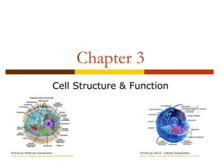
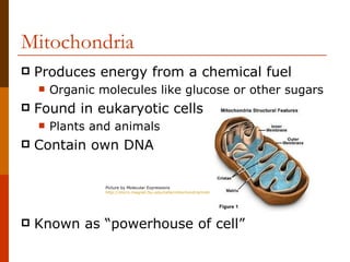
Chapter 3
                                Cell Structure & Function


Picture by Molecular Expressions                            Picture by Cell II: Cellular Organization
http://micro.magnet.fsu.edu/cells/animalcell.html           http://ridge.icu.ac.jp/biobk/BioBookCELL2.html
3.1: Microscopes & Cells
   Objectives

       1. Define the cell theory

       2. Identify the magnification powers of
        different types of microscopes

       3. Observe how the compound microscope
        changes an image
Early Microscopes
   Anton von Leeuwenhoek
       (1600s / Dutch)                                                   Picture by Answers.com
                                                                          http://www.answers.com/topic/anton-van-leeuwenhoek


       Used glass lenses to create instrument that
        magnified images of very small objects

            Light rays bend to make larger image

            Earliest light microscope


                        Picture by Mr. Cantor’s Biology Blog
                        http://cantorsbiologyblog.blogspot.com/2012/01/second-semester-begins-with-cells.html
Discovery of Cells
   Leeuwenhoek used microscope to look at
    drops of pond water
       Filled with tiny living things
            Called them “animalcules”


                                         Picture by Lens On Leeuwenhoek
                                         http://lensonleeuwenhoek.net/lenses.htm
Robert Hooke
   English physicist

   Used microscope to observe             Picture by History of the Microscope
                                           http://www.history-of-the-microscope.org/robert-hooke-m


       Flowers
       Insects
       Spider webs
       Slices of cork                                Picture by Dare To Unravel
                                                      http://daretounravel.blogspot.com/2011/01/mi


   Concluded
       Wood parts of plants had rectangular chambers
            Called them cells
Theodor Schwann
   1839, German Biologist
                                                                      Picture by Biographical Outlines
                                                                      http://www.merke.ch/biografien/biologen_en/schwann.php


   Found some animal tissues resemble
    cellular tissue of plants

   Eventually concluded
       Animals are made up of cells


                  Picture by Biographical Outlines
                  http://www.merke.ch/biografien/biologen_en/schwann.php
The Cell Theory
   Robert Brown (Scottish Biologist)
       Found object in center of cell (nucleus)
                                                                         Picture by Wikipedia
                                                                         http://en.wikipedia.org/wiki/Robert_Brown_(botanist)


   Matthias Schleiden (German Biologist)
       Suggested nucleus plays role in cell
        reproduction


              Picture by Biographical Outlines
              http://www.merke.ch/biografien/biologen_en/schleiden.php
The Cell Theory
   Rudolf Virchow
       1855 German Physician
                                                                       Picture by Wikipedia
                                                                       http://en.wikipedia.org/wiki/Rudolf_Virchow


   Proposed that animal and plant cells are
    produced only by the division of cells that
    already exist


         Picture by The Encyclopedia of Science
         http://www.daviddarling.info/encyclopedia/P/plant_cell.html
The Cell Theory
      1. All living things are composed of cells

      2. Cells are the smallest working units of
       living things

      3. All cells come from preexisting cells


Picture by Mrs. Olsen’s 5th Grade Class Page
http://www.ruediger.leon.k12.fl.us/olsonmd/Class%20Pictures/Forms/DispForm.aspx?ID=2&RootFolder=%2Folsonmd%2FClass%20Pictures%2Fclip%20art
Modern Microscopes
   1) Compound Light Microscope
       Contains more than 1 lens
       Magnifies up to 1000 times


   Parts of Microscope Handout


                                 Picture by Wikipedia
                                 http://wiki.district87.org/index.php/Compound_Light_Microscopes
Modern Microscopes
   2. Electron Microscope
           Uses magnets to focus beam of electrons to
            examine sample
           Magnifies 1000 times more than light
            microscope


                                             Picture by Ego TV
                                             http://egotvonline.com/2012/03/13/25-everyday-objects-under-an-electron-microscope/


Picture by UK Electron Microscope Facility
http://www.engr.uky.edu/~bjhinds/facil/emf
Types of Electron Microscopes
   Transmission Electron Microscope (TEM)
       Sends electrons through sample
       Puts image on fluorescent screen

   Scanning Electron Microscope (SEM)
       Electron scans sample’s surface
       Puts image on TV screen


                  Picture by Electron Microscope
                  http://egotvonline.com/2012/03/13/25-everyday-objects-under-an-electron-microscope/
Electron Microscope Limitations
       Sample must be in vacuum
               Be nonliving
   Samples for TEM need to be thin slices
   SEM doesn’t show internal structure


Picture by Electron Microscope
http://egotvonline.com/2012/03/13/25-everyday-objects-under-an-electron-microscope/
Scanning Probe Microscope
   Trace surface of sample with tiny tip
    (probe)
       Very tiny objects like atoms and molecules


           Picture by Principles of Scanning Probe Microscopy
           http://www.physics.leidenuniv.nl/sections/cm/ip/group/Principle_of_SPM.htm
3.2: Cell Boundaries
   Objectives

       1. Discuss the roles of the cell membrane and
        cell wall


       2. Describe passive transport and active
        transport
Cell Membrane
   Outer boundary that separates and
    protects cell from its surroundings
       Must allow certain substances to come in
       Must allow waste to exit


   Descried as selectively permeable
       “The bouncer” or “Main gate”


                     Picture by The Biology Corner
                     http://www.biologycorner.com/bio1/cell.html
Lipid Bilayer
   Cell membrane is made up of lipid molecules
       Known as phospholipids


   Polar end = head (attract water)
   Nonpolar end = tail (repel water)


                                 Picture by TutorVista.com
                                 http://www.tutorvista.com/biology/membrane-lipid-bilayer
Lipid Bilayer
   Line up in double-layer pattern
       Known as lipid bilayer


   Lipid Bilayer
       Provides cell membrane with tough flexible
        barrier
            Protects cells


                     Picture by TutorVista.com
                     http://www.tutorvista.com/biology/membrane-lipid-bilayer
Other Cell Membrane Components
   Most cell membranes have
    proteins in lipid bilayer
       Have carbohydrates
        attached

   Protein purpose
       Move material across cell
        membrane
                                    Picture by Cell Membrane Wiki
       Protect membrane            http://torresbioclan.pbworks.com/w/page/22377072/cell%20m


       Carbohydrates act as “ID”
        cards
Cell Wall
   Porous membrane located outside cell membrane
       Found in plant cells, algae, and bacteria
       NOT found in animal cells

   Supports and protects cells
       Allow substance to pass in and out

   Made of:
       Carbohydrates called cellulose
       Give plants their strength
       Proteins

                  Picture by Molecular Expressions
                  http://micro.magnet.fsu.edu/cells/plantcell.html
Passive Transport
   Process of moving substances in and out
    of a cell
       Does NOT use energy


           Picture by Cell Organization & Functions
           http://www.williamsclass.com/SeventhScienceWork/CellsOrganization.htm
Diffusion
   Process by which substances spread
    through a liquid or gas
       Move from region of high concentration to area
        of low concentration
       Example: food coloring in water

   In cells…
       Liquids and small lipids diffuse directly across
        cell membrane

           Picture by Biology Corner
           http://www.biologycorner.com/bio1/diffusion.html
Facilitated Diffusion
      Cell membrane also contains protein channels
              Allows larger substance to pass through


      Facilitated Diffusion
              Diffusion that occurs with the aided help of a protein
               channel
                     Each protein channel is specified for a certain substance


Picture by Our Virtual Classroom
http://ccaoscience.wordpress.com/notes/transport-protein/
Osmosis
   The diffusion of water through a
    selectively permeable membrane

   Water diffuses from areas of high
    concentration to areas of low
    concentration


        Picture by Free Drinking Water
        http://www.freedrinkingwater.com/resource-a-complete-resource-guide-to-osmosis.htm
Osmotic Pressure
   When water moves by osmosis, it
    produces pressure
       Enough to destroy cell


   Three ways to control:
       1) cell wall
       2) pump out water
       3) bathe cells in blood   Picture by Non Ideal Solutions
                                  http://www.chem.ufl.edu/~itl/4411/colligative/lec_i.html
Osmotic Pressure
   Cell Wall
       Strong tough wall prevents
        cell from expanding
            Counter acts osmotic pressure


   Pump Out
       Cells use contractile vacuole
        to pump out water


                                    Picture by Non Ideal Solutions
                                    http://www.chem.ufl.edu/~itl/4411/colligative/lec_i.html
Osmotic Pressure
 Bathe   Cells in Blood
   Mostly in large animals
   Blood cells have same concentration of
    dissolved substances
   Help absorb excess water


               Picture by A Sweet Life
               http://asweetlife.org/a-sweet-life-staff/articles/interview-dr-zachary-bloomgarden-on-the-hba1c-assay/8020/
Active Transport
   Movement of a substance against a
    concentration difference

   Requires A LOT of energy
       Transports:
            Large molecules
            Food
            Whole cells


                    Picture by G11-BioA -2011
                    http://g11-bioa-2011-12.wikispaces.com/(d)+Active+transport
3.3: Inside The Cell
   Objectives

       1. Describe the composition and function of
        the nucleus

       2. List and describe the organelles of the
        cytoplasm

       3. Interpret the changes observed when a
        paramecium takes in food
Nucleus
      Large, dense structure contained in cells
              Known as control center
              “Main office of factory”


Picture by Plant Cell
http://library.thinkquest.org/06aug/01942/plcells/nuclues.htm
Nucleus
   Organisms can be classified into 2 categories
        1. Eukaryotes
               Organisms cells HAVE nuclei
               Include unicellular and multicellular

        2. Prokaryotes
               Organisms that do NOT contain nuclei
               Small single celled organisms


    Picture by Bacterial/Prokaryotic Phylogeny
    http://www.bacterialphylogeny.info/eukaryotes.html
Role of Nucleus
   Contains nearly all of cell’s DNA
       Has instructions to make proteins / molecules
            Form material called chromatin
               Spreads through nucleus


   When cell divides
       Chromatin condense into chromosomes
            Acts as “blueprint of factory”


                       Picture by Animal Port
                       http://www.animalport.com/animal-cells.html
Structures In The Nucleus
   Nucleolus
       Small dense region inside nucleus
       Where ribosomes are made


                 Picture by Molecular Expressions
                 http://micro.magnet.fsu.edu/cells/nucleus/nucleolus.html


   Nuclear Envelope
       Membrane that surrounds the nucleus
       Has pores to transport materials in and out
Cytoplasm
   Fluid outside nucleus held in by cell
    membrane
       Holds all other cell organelles
            These are small structures that perform specialized
             functions in the cell
       Allows for movement


   Known as the “factory floor”


                       Picture by Daylilies
                       http://www.daylilies.org/ahs_dictionary/cytoplasm.html
Ribosomes
      Tiny particles made of RNA and protein
              Site for protein synthesis
              Gets instructions from nucleus

      Known as “the workers”


Picture by Cellupedia
http://library.thinkquest.org/C004535/eukaryotic_cells.html
Endoplasmic Reticulum (ER)
      Processing and transporting of proteins
       and other macromolecules
              Network of membranes
                     2 types
                           1. Smooth ER… NO ribosomes attached to surface
                           2. Rough ER… ribosomes attached to surface


              Act as the subway


Picture by Molecular Expressions
http://micro.magnet.fsu.edu/cells/endoplasmicreticulum/endoplasmicreticulum.html
Golgi Apparatus
      Act as packaging center
              Modify and add components to proteins
                      Attach carbohydrates or lipids


Picture by Molecular Expressions
http://micro.magnet.fsu.edu/cells/golgi/golgiapparatus.html
Lysosomes
      Sac-like membrane that gets rid of waste
              Filled with chemicals and enzymes
      Can also break down and gets rid of
       damaged organelles

      Known as the “garbage man”


Picture by Molecular Expressions
http://publications.nigms.nih.gov/insidethecell/images/ch1_lysosome.jpg
Cytoskeleton
   Act as a supporting framework for cell
       Found in eukaryotic cells


   Components
       Microtubules
       Mircofillaments
                                    Picture by Cellupedia
                                    http://library.thinkquest.org/C004535/cytoskeleton.html


       These are hollow tubes of protein that provide
        framework to support cell
Vacuoles
   Sac-like structure used for storage
       Animals
            Store proteins, fats, and carbohydrates

       Plants
            Store water and dissolved salts
            Provide support

       Known as “the warehouse”


                     Picture by Molecular Expressions
                     http://micro.magnet.fsu.edu/cells/plants/vacuole.html
Mitochondria
   Produces energy from a chemical fuel
       Organic molecules like glucose or other sugars
   Found in eukaryotic cells
       Plants and animals
   Contain own DNA

                 Picture by Molecular Expressions
                 http://micro.magnet.fsu.edu/cells/mitochondria/mitochondria.html


   Known as “powerhouse of cell”
Chloroplasts
   Organelle that produces energy from
    sunlight
       Found in plants ONLY and some algae
 Aids in the process of photosynthesis
 Green due to chlorophyll pigment
 Also contains own DNA


             Picture by eTeaching Program
             https://www.etap.org/demo/grade7_science/instruction2tutor.html
3.4: The Origin of the Eukaryotic Cell
   Objectives

       1. Define the endosymbiont hypothesis
Eukaryotic vs. Prokaryotic
      Eukaryotes                                                             Prokaryotes
             Nuclues                                                             Have NONE of these
             Mitochondria
             Chloroplasts
             Other organelles                                                    WHY???


Picture by BioCoach Activity
http://www.phschool.com/science/biology_place/biocoach/cells/common.html
The Work of Lynn Margulis
   Focused on mitochondria and chloroplasts
       Used on DNA to make compounds
       Both surrounded by two membranes
       Both reproduced separately from rest of cell
            Mitochondria from mitochondria
            Chloroplasts from chloroplasts


            Why did this happen???


                         Picture by The Alien Next Door Blog
                         http://sfgirl-thealiennextdoor.blogspot.com/2010/06/celebrating-womanhood-i-am-woman-i-am.html
The Endosymbiont Hypothesis
   Billions of years
    ago…
         Eukaryotic cells arose
          as a combination of
          different prokaryotic
          cells
                 Cells consumed other
                  cells but still each
                  survived


Picture by Molecular Expressions
http://www.tokresource.org/tok_classes/biobiobio/biomenu/options_folder/D1_life_origins/index.htm
Margulis’s Model
      Stated mitochondria and chloroplasts had
       ancestors that were free-living organisms
             Were consumed by larger cells
                    Became organelles in larger cells


Picture by The Endosymbiotic Hypothesis
http://endosymbiotichypothesis.wordpress.com/
Further Evidence
   Chloroplast DNA
       Similar to DNA in prokaryotic cells

   In Mitochondria & Chloroplasts
       Contain own ribosomes to make own proteins
              Ribosomes similar to prokaryotic ribosomes


        Picture by Molecular Expressions
        http://micro.magnet.fsu.edu/cells/mitochondria/mitochondria.html

Biology - Cell Organization & Function

  • 1.
    Chapter 3 Cell Structure & Function Picture by Molecular Expressions Picture by Cell II: Cellular Organization http://micro.magnet.fsu.edu/cells/animalcell.html http://ridge.icu.ac.jp/biobk/BioBookCELL2.html
  • 2.
    3.1: Microscopes &Cells  Objectives  1. Define the cell theory  2. Identify the magnification powers of different types of microscopes  3. Observe how the compound microscope changes an image
  • 3.
    Early Microscopes  Anton von Leeuwenhoek  (1600s / Dutch) Picture by Answers.com http://www.answers.com/topic/anton-van-leeuwenhoek  Used glass lenses to create instrument that magnified images of very small objects  Light rays bend to make larger image  Earliest light microscope Picture by Mr. Cantor’s Biology Blog http://cantorsbiologyblog.blogspot.com/2012/01/second-semester-begins-with-cells.html
  • 4.
    Discovery of Cells  Leeuwenhoek used microscope to look at drops of pond water  Filled with tiny living things  Called them “animalcules” Picture by Lens On Leeuwenhoek http://lensonleeuwenhoek.net/lenses.htm
  • 5.
    Robert Hooke  English physicist  Used microscope to observe Picture by History of the Microscope http://www.history-of-the-microscope.org/robert-hooke-m  Flowers  Insects  Spider webs  Slices of cork Picture by Dare To Unravel http://daretounravel.blogspot.com/2011/01/mi  Concluded  Wood parts of plants had rectangular chambers  Called them cells
  • 6.
    Theodor Schwann  1839, German Biologist Picture by Biographical Outlines http://www.merke.ch/biografien/biologen_en/schwann.php  Found some animal tissues resemble cellular tissue of plants  Eventually concluded  Animals are made up of cells Picture by Biographical Outlines http://www.merke.ch/biografien/biologen_en/schwann.php
  • 7.
    The Cell Theory  Robert Brown (Scottish Biologist)  Found object in center of cell (nucleus) Picture by Wikipedia http://en.wikipedia.org/wiki/Robert_Brown_(botanist)  Matthias Schleiden (German Biologist)  Suggested nucleus plays role in cell reproduction Picture by Biographical Outlines http://www.merke.ch/biografien/biologen_en/schleiden.php
  • 8.
    The Cell Theory  Rudolf Virchow  1855 German Physician Picture by Wikipedia http://en.wikipedia.org/wiki/Rudolf_Virchow  Proposed that animal and plant cells are produced only by the division of cells that already exist Picture by The Encyclopedia of Science http://www.daviddarling.info/encyclopedia/P/plant_cell.html
  • 9.
    The Cell Theory  1. All living things are composed of cells  2. Cells are the smallest working units of living things  3. All cells come from preexisting cells Picture by Mrs. Olsen’s 5th Grade Class Page http://www.ruediger.leon.k12.fl.us/olsonmd/Class%20Pictures/Forms/DispForm.aspx?ID=2&RootFolder=%2Folsonmd%2FClass%20Pictures%2Fclip%20art
  • 10.
    Modern Microscopes  1) Compound Light Microscope  Contains more than 1 lens  Magnifies up to 1000 times  Parts of Microscope Handout Picture by Wikipedia http://wiki.district87.org/index.php/Compound_Light_Microscopes
  • 11.
    Modern Microscopes  2. Electron Microscope  Uses magnets to focus beam of electrons to examine sample  Magnifies 1000 times more than light microscope Picture by Ego TV http://egotvonline.com/2012/03/13/25-everyday-objects-under-an-electron-microscope/ Picture by UK Electron Microscope Facility http://www.engr.uky.edu/~bjhinds/facil/emf
  • 12.
    Types of ElectronMicroscopes  Transmission Electron Microscope (TEM)  Sends electrons through sample  Puts image on fluorescent screen  Scanning Electron Microscope (SEM)  Electron scans sample’s surface  Puts image on TV screen Picture by Electron Microscope http://egotvonline.com/2012/03/13/25-everyday-objects-under-an-electron-microscope/
  • 13.
    Electron Microscope Limitations  Sample must be in vacuum  Be nonliving  Samples for TEM need to be thin slices  SEM doesn’t show internal structure Picture by Electron Microscope http://egotvonline.com/2012/03/13/25-everyday-objects-under-an-electron-microscope/
  • 14.
    Scanning Probe Microscope  Trace surface of sample with tiny tip (probe)  Very tiny objects like atoms and molecules Picture by Principles of Scanning Probe Microscopy http://www.physics.leidenuniv.nl/sections/cm/ip/group/Principle_of_SPM.htm
  • 15.
    3.2: Cell Boundaries  Objectives  1. Discuss the roles of the cell membrane and cell wall  2. Describe passive transport and active transport
  • 16.
    Cell Membrane  Outer boundary that separates and protects cell from its surroundings  Must allow certain substances to come in  Must allow waste to exit  Descried as selectively permeable  “The bouncer” or “Main gate” Picture by The Biology Corner http://www.biologycorner.com/bio1/cell.html
  • 17.
    Lipid Bilayer  Cell membrane is made up of lipid molecules  Known as phospholipids  Polar end = head (attract water)  Nonpolar end = tail (repel water) Picture by TutorVista.com http://www.tutorvista.com/biology/membrane-lipid-bilayer
  • 18.
    Lipid Bilayer  Line up in double-layer pattern  Known as lipid bilayer  Lipid Bilayer  Provides cell membrane with tough flexible barrier  Protects cells Picture by TutorVista.com http://www.tutorvista.com/biology/membrane-lipid-bilayer
  • 19.
    Other Cell MembraneComponents  Most cell membranes have proteins in lipid bilayer  Have carbohydrates attached  Protein purpose  Move material across cell membrane Picture by Cell Membrane Wiki  Protect membrane http://torresbioclan.pbworks.com/w/page/22377072/cell%20m  Carbohydrates act as “ID” cards
  • 20.
    Cell Wall  Porous membrane located outside cell membrane  Found in plant cells, algae, and bacteria  NOT found in animal cells  Supports and protects cells  Allow substance to pass in and out  Made of:  Carbohydrates called cellulose  Give plants their strength  Proteins Picture by Molecular Expressions http://micro.magnet.fsu.edu/cells/plantcell.html
  • 21.
    Passive Transport  Process of moving substances in and out of a cell  Does NOT use energy Picture by Cell Organization & Functions http://www.williamsclass.com/SeventhScienceWork/CellsOrganization.htm
  • 22.
    Diffusion  Process by which substances spread through a liquid or gas  Move from region of high concentration to area of low concentration  Example: food coloring in water  In cells…  Liquids and small lipids diffuse directly across cell membrane Picture by Biology Corner http://www.biologycorner.com/bio1/diffusion.html
  • 23.
    Facilitated Diffusion  Cell membrane also contains protein channels  Allows larger substance to pass through  Facilitated Diffusion  Diffusion that occurs with the aided help of a protein channel  Each protein channel is specified for a certain substance Picture by Our Virtual Classroom http://ccaoscience.wordpress.com/notes/transport-protein/
  • 24.
    Osmosis  The diffusion of water through a selectively permeable membrane  Water diffuses from areas of high concentration to areas of low concentration Picture by Free Drinking Water http://www.freedrinkingwater.com/resource-a-complete-resource-guide-to-osmosis.htm
  • 25.
    Osmotic Pressure  When water moves by osmosis, it produces pressure  Enough to destroy cell  Three ways to control:  1) cell wall  2) pump out water  3) bathe cells in blood Picture by Non Ideal Solutions http://www.chem.ufl.edu/~itl/4411/colligative/lec_i.html
  • 26.
    Osmotic Pressure  Cell Wall  Strong tough wall prevents cell from expanding  Counter acts osmotic pressure  Pump Out  Cells use contractile vacuole to pump out water Picture by Non Ideal Solutions http://www.chem.ufl.edu/~itl/4411/colligative/lec_i.html
  • 27.
    Osmotic Pressure  Bathe Cells in Blood  Mostly in large animals  Blood cells have same concentration of dissolved substances  Help absorb excess water Picture by A Sweet Life http://asweetlife.org/a-sweet-life-staff/articles/interview-dr-zachary-bloomgarden-on-the-hba1c-assay/8020/
  • 28.
    Active Transport  Movement of a substance against a concentration difference  Requires A LOT of energy  Transports:  Large molecules  Food  Whole cells Picture by G11-BioA -2011 http://g11-bioa-2011-12.wikispaces.com/(d)+Active+transport
  • 29.
    3.3: Inside TheCell  Objectives  1. Describe the composition and function of the nucleus  2. List and describe the organelles of the cytoplasm  3. Interpret the changes observed when a paramecium takes in food
  • 30.
    Nucleus  Large, dense structure contained in cells  Known as control center  “Main office of factory” Picture by Plant Cell http://library.thinkquest.org/06aug/01942/plcells/nuclues.htm
  • 31.
    Nucleus  Organisms can be classified into 2 categories  1. Eukaryotes  Organisms cells HAVE nuclei  Include unicellular and multicellular  2. Prokaryotes  Organisms that do NOT contain nuclei  Small single celled organisms Picture by Bacterial/Prokaryotic Phylogeny http://www.bacterialphylogeny.info/eukaryotes.html
  • 32.
    Role of Nucleus  Contains nearly all of cell’s DNA  Has instructions to make proteins / molecules  Form material called chromatin  Spreads through nucleus  When cell divides  Chromatin condense into chromosomes  Acts as “blueprint of factory” Picture by Animal Port http://www.animalport.com/animal-cells.html
  • 33.
    Structures In TheNucleus  Nucleolus  Small dense region inside nucleus  Where ribosomes are made Picture by Molecular Expressions http://micro.magnet.fsu.edu/cells/nucleus/nucleolus.html  Nuclear Envelope  Membrane that surrounds the nucleus  Has pores to transport materials in and out
  • 34.
    Cytoplasm  Fluid outside nucleus held in by cell membrane  Holds all other cell organelles  These are small structures that perform specialized functions in the cell  Allows for movement  Known as the “factory floor” Picture by Daylilies http://www.daylilies.org/ahs_dictionary/cytoplasm.html
  • 35.
    Ribosomes  Tiny particles made of RNA and protein  Site for protein synthesis  Gets instructions from nucleus  Known as “the workers” Picture by Cellupedia http://library.thinkquest.org/C004535/eukaryotic_cells.html
  • 36.
    Endoplasmic Reticulum (ER)  Processing and transporting of proteins and other macromolecules  Network of membranes  2 types  1. Smooth ER… NO ribosomes attached to surface  2. Rough ER… ribosomes attached to surface  Act as the subway Picture by Molecular Expressions http://micro.magnet.fsu.edu/cells/endoplasmicreticulum/endoplasmicreticulum.html
  • 37.
    Golgi Apparatus  Act as packaging center  Modify and add components to proteins  Attach carbohydrates or lipids Picture by Molecular Expressions http://micro.magnet.fsu.edu/cells/golgi/golgiapparatus.html
  • 38.
    Lysosomes  Sac-like membrane that gets rid of waste  Filled with chemicals and enzymes  Can also break down and gets rid of damaged organelles  Known as the “garbage man” Picture by Molecular Expressions http://publications.nigms.nih.gov/insidethecell/images/ch1_lysosome.jpg
  • 39.
    Cytoskeleton  Act as a supporting framework for cell  Found in eukaryotic cells  Components  Microtubules  Mircofillaments Picture by Cellupedia http://library.thinkquest.org/C004535/cytoskeleton.html  These are hollow tubes of protein that provide framework to support cell
  • 40.
    Vacuoles  Sac-like structure used for storage  Animals  Store proteins, fats, and carbohydrates  Plants  Store water and dissolved salts  Provide support  Known as “the warehouse” Picture by Molecular Expressions http://micro.magnet.fsu.edu/cells/plants/vacuole.html
  • 41.
    Mitochondria  Produces energy from a chemical fuel  Organic molecules like glucose or other sugars  Found in eukaryotic cells  Plants and animals  Contain own DNA Picture by Molecular Expressions http://micro.magnet.fsu.edu/cells/mitochondria/mitochondria.html  Known as “powerhouse of cell”
  • 42.
    Chloroplasts  Organelle that produces energy from sunlight  Found in plants ONLY and some algae  Aids in the process of photosynthesis  Green due to chlorophyll pigment  Also contains own DNA Picture by eTeaching Program https://www.etap.org/demo/grade7_science/instruction2tutor.html
  • 43.
    3.4: The Originof the Eukaryotic Cell  Objectives  1. Define the endosymbiont hypothesis
  • 44.
    Eukaryotic vs. Prokaryotic  Eukaryotes  Prokaryotes  Nuclues  Have NONE of these  Mitochondria  Chloroplasts  Other organelles  WHY??? Picture by BioCoach Activity http://www.phschool.com/science/biology_place/biocoach/cells/common.html
  • 45.
    The Work ofLynn Margulis  Focused on mitochondria and chloroplasts  Used on DNA to make compounds  Both surrounded by two membranes  Both reproduced separately from rest of cell  Mitochondria from mitochondria  Chloroplasts from chloroplasts  Why did this happen??? Picture by The Alien Next Door Blog http://sfgirl-thealiennextdoor.blogspot.com/2010/06/celebrating-womanhood-i-am-woman-i-am.html
  • 46.
    The Endosymbiont Hypothesis  Billions of years ago…  Eukaryotic cells arose as a combination of different prokaryotic cells  Cells consumed other cells but still each survived Picture by Molecular Expressions http://www.tokresource.org/tok_classes/biobiobio/biomenu/options_folder/D1_life_origins/index.htm
  • 47.
    Margulis’s Model  Stated mitochondria and chloroplasts had ancestors that were free-living organisms  Were consumed by larger cells  Became organelles in larger cells Picture by The Endosymbiotic Hypothesis http://endosymbiotichypothesis.wordpress.com/
  • 48.
    Further Evidence  Chloroplast DNA  Similar to DNA in prokaryotic cells  In Mitochondria & Chloroplasts  Contain own ribosomes to make own proteins  Ribosomes similar to prokaryotic ribosomes Picture by Molecular Expressions http://micro.magnet.fsu.edu/cells/mitochondria/mitochondria.html