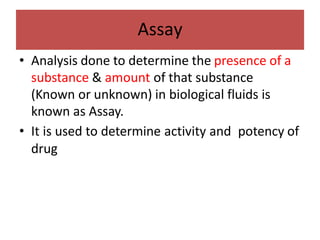
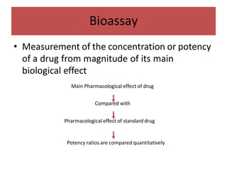
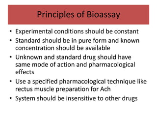
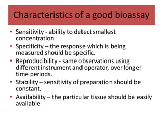
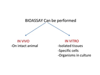
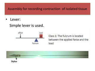
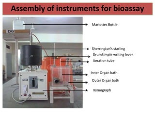
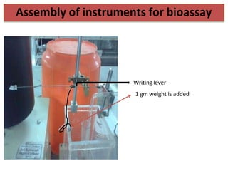
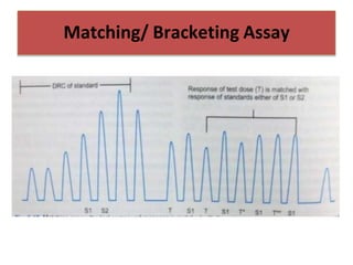
Dr Agboola, Samuel S.
BIOASSAY
• Analysis done to determine the presence of a
substance & amount of that substance
(Known or unknown) in biological fluids is
known as Assay.
• It is used to determine activity and potency of
drug
Assay
• Biological Method (Bioassay)
• Physico-chemical methods
– Chromatography, spectrophotometry
• Radioimmunoassay
• Microbiological methods
Types of Assays
• Measurement of the concentration or potency
of a drug from magnitude of its main
biological effect
Main Pharmacological effect of drug
Compared with
Pharmacological effect of standard drug
Potency ratios are compared quantitatively
Bioassay
• Measure pharmacological activity of new/
chemically undefined substance
• Measure concentration of known substance in
tissue extract or body fluid e.g Ach, histamine
• Measure ED50 or LD50 of a drug
• Biological standardization of natural drugs
which cannot be obtained in chemically pure
form e.g Vasopressin, oxytocin
Indications(Uses) of Bioassay
• Blood pressure (Dogs and cats):
Sympathomimetics
• Ach: contraction of rabbit duodenum or frog
rectus abdominis
• Skeletal muscle relaxants: rabbit head drop
method
• Histamine: Guinea pig ileum
• Serotonin on rat colon
Parameters recorded
• Experimental conditions should be constant
• Standard should be in pure form and known
concentration should be available
• Unknown and standard drug should have
same mode of action and pharmacological
effects
• Use a specified pharmacological technique like
rectus muscle preparation for Ach
• System should be insensitive to other drugs
Principles of Bioassay
• Sensitivity - ability to detect smallest
concentration
• Specificity – the response which is being
measured should be specific.
• Reproducibility - same observations using
different instrument and operator, over longer
time periods.
• Stability – sensitivity of preparation should be
constant.
• Availability – the particular tissue should be easily
available
Characteristics of a good bioassay
BIOASSAY Can be performed
IN VIVO
-On intact animal
IN VITRO
-Isolated tissues
-Specific cells
-Organisms in culture
Several preparations can be obtained from a
single animal.
Small amount of the test material is required.
Cheap, less time consuming.
Interference due to pharmacokinetic factors
and compensatory reflexes is avoided.
Advantages of isolated tissue experiments over
intact animal experiments
Organ Bath:
• Rudolph Magnus was first to design organ bath.
Outer organ bath: made up of perplex with electrical
thermostat and stirrer for uniform temperature.
Inner organ bath: made of glass, capacity of bath is
30ml.
Assembly for recording contraction of isolated
tissue
• Lever:
Simple lever is used.
Stylus
Assembly for recording contraction of isolated tissue
5-10
A
B
Magnification and Adjustment
A B
Magnification for Frog rectus is
Fast contracting- low
Slow contracting- high
1) Provides oxygen/ air to mounted tissue.
2)Allows uniform distribution of drug in organ bath
through bubbles.
3)Aeration tube helps in mounting tissue in organ
bath.
Aeration – 30-40 bubbles / min (air)
Role of Aeration / Aeration tube
Mariottes Bottle
Sherrington’s starling
DrumSimple writing lever
Aeration tube
Inner Organ bath
Outer Organ bath
Kymograph
Assembly of instruments for bioassay
Writing lever
1 gm weight is added
Assembly of instruments for bioassay
Procedure:
 A pithed frog is laid on its dorsal surface in a dissection tray.
 The skin of the abdomen cut to expose the whole abdomen.
 The rectus muscle is identified, and the border of the muscle
dissected out from adjacent abdominal wall.
 A thread is passed with the help of needle through the pubic
attachment of muscle and tied ,muscle is cut.
 The upper end of muscle i.e, xiphisternum part also tied and cut.
 The lower end of the muscle is tied to aeration tube at ‘s’ bent.
Dissection and mounting the tissue
PithingandD
issectionofFrog
 Before mounting the tissue make sure the blood vessels are
removed.
 Aeration tube with mounted tissue is placed in inner organ bath
filled with PSS.
 The upper end of muscle is tied to lever with thread.
 A load of 1gm is attached to the other side of lever at same
distance as that of tissue from the fulcrum. This helps the muscle
to relax in between contraction.
 Tissue is aerated with air i.e 30-40 bubbles /min. and allowed to
relax for 30- 45 min before recording responses.
Dissection and mounting the tissue
Mounting the tissue
 Stock solution- 1 mg/ml Acetylcholine(1000μg/ml)
The dilutions are made as follows-
1 ml stock soln. +9 ml distilled water= 1/10 dilution (100 μg/ml)
1 ml 1/10 dilution +9 ml distilled water= 1/100 dilution (10 μg/ml)
1 ml 1/100dilution +9 ml distilled water= 1/1000 dilution (1μg/ml)
Acetyl choline Dilutions
 Base line:30 seconds(s) (start drum & allow to run freely)
 Contractions: 90 s (contact period of Ach with tissue)
 Relaxation: 3 minutes (tissue washed 2-3 times after 90
sec contact period)
 Total cycle is 5 mins
 Soon after contraction of 90 secs the weight of 1gm is
lowered down to allow muscle to relax slowly, until lever
comes to base line.
 The contractions are recorded with each dose of solution
till ceiling effect is obtained i.e. no further increase in
height of contraction with subsequent doses.
Recording cycle
Recording cycle
 After the ceiling effect is obtained with standard Ach
solution, the procedure is repeated with the test
solution(unknown strength of Ach) in different dilutions.
 The further experiment is done by Multiple point assay
i.e. 2+1 assay
Sensitivity of Rectus muscle can
be increased by adding
Physostigmine(0.5μg/ml) to perfusionfluid
• A. Quantal (Direct end point) Assay
• B. Graded Assay
-Bracketing assay
-Matching assay
-Interpolation assay
-Multiple point assay
Types of Bioassay
• In this assay, dose of standard and unknown
which provide predetermined ‘all or none’
response, then their potency ratios compared.
Ex: Digitalis induced cardiac arrest in cats, insulin
induced hypoglycemic convulsions in mice.
Quantal response (All or none assay)
Graded response assay
• Bracketing and Matching assay methods are
not ideal because lack of accuracy, sensitivity
• So , to minimize those errors multiple point
assays preferred (3,4 or 6 point assays)
Multiple Point Assay
Matching/ Bracketing Assay
Advantage :
 Faster
 Can be completed when amount of test drug
available is small
 Does not involve complicated calculations
Disadvantage:
 Match is subjective
 Exact match may not always be possible
Matching/Bracketing Assay
Interpolation method

Biological Assay. New.pptx

  • 1.
    Dr Agboola, SamuelS. BIOASSAY
  • 2.
    • Analysis doneto determine the presence of a substance & amount of that substance (Known or unknown) in biological fluids is known as Assay. • It is used to determine activity and potency of drug Assay
  • 3.
    • Biological Method(Bioassay) • Physico-chemical methods – Chromatography, spectrophotometry • Radioimmunoassay • Microbiological methods Types of Assays
  • 4.
    • Measurement ofthe concentration or potency of a drug from magnitude of its main biological effect Main Pharmacological effect of drug Compared with Pharmacological effect of standard drug Potency ratios are compared quantitatively Bioassay
  • 5.
    • Measure pharmacologicalactivity of new/ chemically undefined substance • Measure concentration of known substance in tissue extract or body fluid e.g Ach, histamine • Measure ED50 or LD50 of a drug • Biological standardization of natural drugs which cannot be obtained in chemically pure form e.g Vasopressin, oxytocin Indications(Uses) of Bioassay
  • 6.
    • Blood pressure(Dogs and cats): Sympathomimetics • Ach: contraction of rabbit duodenum or frog rectus abdominis • Skeletal muscle relaxants: rabbit head drop method • Histamine: Guinea pig ileum • Serotonin on rat colon Parameters recorded
  • 7.
    • Experimental conditionsshould be constant • Standard should be in pure form and known concentration should be available • Unknown and standard drug should have same mode of action and pharmacological effects • Use a specified pharmacological technique like rectus muscle preparation for Ach • System should be insensitive to other drugs Principles of Bioassay
  • 8.
    • Sensitivity -ability to detect smallest concentration • Specificity – the response which is being measured should be specific. • Reproducibility - same observations using different instrument and operator, over longer time periods. • Stability – sensitivity of preparation should be constant. • Availability – the particular tissue should be easily available Characteristics of a good bioassay
  • 9.
    BIOASSAY Can beperformed IN VIVO -On intact animal IN VITRO -Isolated tissues -Specific cells -Organisms in culture
  • 10.
    Several preparations canbe obtained from a single animal. Small amount of the test material is required. Cheap, less time consuming. Interference due to pharmacokinetic factors and compensatory reflexes is avoided. Advantages of isolated tissue experiments over intact animal experiments
  • 11.
    Organ Bath: • RudolphMagnus was first to design organ bath. Outer organ bath: made up of perplex with electrical thermostat and stirrer for uniform temperature. Inner organ bath: made of glass, capacity of bath is 30ml. Assembly for recording contraction of isolated tissue
  • 12.
    • Lever: Simple leveris used. Stylus Assembly for recording contraction of isolated tissue
  • 13.
    5-10 A B Magnification and Adjustment AB Magnification for Frog rectus is Fast contracting- low Slow contracting- high
  • 14.
    1) Provides oxygen/air to mounted tissue. 2)Allows uniform distribution of drug in organ bath through bubbles. 3)Aeration tube helps in mounting tissue in organ bath. Aeration – 30-40 bubbles / min (air) Role of Aeration / Aeration tube
  • 15.
    Mariottes Bottle Sherrington’s starling DrumSimplewriting lever Aeration tube Inner Organ bath Outer Organ bath Kymograph Assembly of instruments for bioassay
  • 16.
    Writing lever 1 gmweight is added Assembly of instruments for bioassay
  • 17.
    Procedure:  A pithedfrog is laid on its dorsal surface in a dissection tray.  The skin of the abdomen cut to expose the whole abdomen.  The rectus muscle is identified, and the border of the muscle dissected out from adjacent abdominal wall.  A thread is passed with the help of needle through the pubic attachment of muscle and tied ,muscle is cut.  The upper end of muscle i.e, xiphisternum part also tied and cut.  The lower end of the muscle is tied to aeration tube at ‘s’ bent. Dissection and mounting the tissue
  • 18.
  • 19.
     Before mountingthe tissue make sure the blood vessels are removed.  Aeration tube with mounted tissue is placed in inner organ bath filled with PSS.  The upper end of muscle is tied to lever with thread.  A load of 1gm is attached to the other side of lever at same distance as that of tissue from the fulcrum. This helps the muscle to relax in between contraction.  Tissue is aerated with air i.e 30-40 bubbles /min. and allowed to relax for 30- 45 min before recording responses. Dissection and mounting the tissue
  • 20.
  • 21.
     Stock solution-1 mg/ml Acetylcholine(1000μg/ml) The dilutions are made as follows- 1 ml stock soln. +9 ml distilled water= 1/10 dilution (100 μg/ml) 1 ml 1/10 dilution +9 ml distilled water= 1/100 dilution (10 μg/ml) 1 ml 1/100dilution +9 ml distilled water= 1/1000 dilution (1μg/ml) Acetyl choline Dilutions
  • 22.
     Base line:30seconds(s) (start drum & allow to run freely)  Contractions: 90 s (contact period of Ach with tissue)  Relaxation: 3 minutes (tissue washed 2-3 times after 90 sec contact period)  Total cycle is 5 mins  Soon after contraction of 90 secs the weight of 1gm is lowered down to allow muscle to relax slowly, until lever comes to base line.  The contractions are recorded with each dose of solution till ceiling effect is obtained i.e. no further increase in height of contraction with subsequent doses. Recording cycle
  • 23.
    Recording cycle  Afterthe ceiling effect is obtained with standard Ach solution, the procedure is repeated with the test solution(unknown strength of Ach) in different dilutions.  The further experiment is done by Multiple point assay i.e. 2+1 assay Sensitivity of Rectus muscle can be increased by adding Physostigmine(0.5μg/ml) to perfusionfluid
  • 24.
    • A. Quantal(Direct end point) Assay • B. Graded Assay -Bracketing assay -Matching assay -Interpolation assay -Multiple point assay Types of Bioassay
  • 25.
    • In thisassay, dose of standard and unknown which provide predetermined ‘all or none’ response, then their potency ratios compared. Ex: Digitalis induced cardiac arrest in cats, insulin induced hypoglycemic convulsions in mice. Quantal response (All or none assay)
  • 26.
    Quantal response (Allor none assay)
  • 27.
  • 28.
    • Bracketing andMatching assay methods are not ideal because lack of accuracy, sensitivity • So , to minimize those errors multiple point assays preferred (3,4 or 6 point assays) Multiple Point Assay
  • 29.
  • 30.
  • 31.
    Advantage :  Faster Can be completed when amount of test drug available is small  Does not involve complicated calculations Disadvantage:  Match is subjective  Exact match may not always be possible Matching/Bracketing Assay
  • 32.
  • 33.