This document presents a method to automatically measure abdominal aortic aneurysm (AAA) wall thickness from medical images using a regression algorithm. Two reference models (a porcine aorta and synthetic AAA phantom) were used to train and validate the algorithm. A support vector machine regression model using normalized pixel intensity profiles achieved a mean error of 0.262 mm, providing more accurate wall thickness estimates than direct measurements from low-resolution CT images. Further optimization and testing on real patient data is needed to validate the algorithm's ability to provide new insights into AAA development and rupture risk.
BFO – AIS: A FRAME WORK FOR MEDICAL IMAGE CLASSIFICATION USING SOFT COMPUTING...ijsc
Medical images provide diagnostic evidence/information about anatomical pathology. The growth in
database is enormous as medical digital image equipment’s like Magnetic Resonance Images (MRI),
Computed Tomography (CT), and Positron Emission Tomography CT (PET-CT) are part of clinical work.
CT images distinguish various tissues according to gray levels to help medical diagnosis. Ct is more
reliable for early tumours and haemorrhages detection as it provides anatomical information to plan radio
therapy. Medical information systems goals are to deliver information to right persons at the right time and
place to improve care process quality and efficiency. This paper proposes an Artificial Immune System
(AIS) classifier and proposed feature selection based on hybrid Bacterial Foraging Optimization (BFO)
with Local Search (LS) for medical image classification.
OFCS: Optimized Framework of Compressive Sensing for Medical Images in Bottle...IJECEIAES
Compressive sensing is one of teh cost effective solution towards performing compression of heavier form of signals. We reviewed the existing research contribution towards compressive sensing to find that existing system doesnt offer any form of optimization for which reason the signal are superiorly compressed but at the cost of enough resources. Therefore, we introduce a framework that optimizes the performance of the compressive sensing by introducing 4 sequential algorithms for performing Random Sampling, Lossless Compression for region-of-interest, Compressive Sensing using transform-based scheme, and optimization. The contribution of proposed paper is a good balance between computational efficiency and quality of reconstructed medical image when transmitted over network with low channel capacity. The study outcome shows that proposed system offers maximum signal quality and lower algorithm processing time in contrast to existing compression techniuqes on medical images.
Improved Segmentation Technique for Enhancement of Biomedical ImagesIJEEE
The aim of this paper is to develop a fast and reliable segmentation method to segment the haemorrhage region from brain CT images. To calculate area of segmented hemorrhage region that could be useful for physicians or researchers involved in the treatment or investigation of intracranial brain haemorrhage.
A comparison of ultrasound intima media thickness measurements of the left an...LogicMindtech Nologies
IMAGE PROCESSING Projects for M. Tech, IMAGE PROCESSING Projects in Vijayanagar, IMAGE PROCESSING Projects in Bangalore, M. Tech Projects in Vijayanagar, M. Tech Projects in Bangalore, IMAGE PROCESSING IEEE projects in Bangalore, IEEE 2015 IMAGE PROCESSING Projects, MATLAB Image Processing Projects, MATLAB Image Processing Projects in Bangalore, MATLAB Image Processing Projects in Vijayangar
Contour evolution method for precise boundary delineation of medical imagesTELKOMNIKA JOURNAL
Image segmentation is an important precursor to boundary delineation of medical images. One of the major challenges in applying automatic image segmentation in medical images is the imperfection in the imaging process which can result in inconsistent contrast and brightness levels, and low image sharpness and vanishing boundaries. Although recent advances in deep learning produce vast improvements in the quality of image segmentation, the accuracy of segmentation around object boundaries still requires improvement. We developed a new approach to contour evolution that is more intuitive but shares some common principles with the active contour model method. The method uses two concepts, namely the boundary grid and sparse boundary representation, as an implicit and explicit representation of the boundary points. We tested our method using lumbar spine MRI images of 515 patients. The experiment results show that our method performs up to 10.2 times faster and more flexible than the geodesic active contours method. Using BF-score contour-based metric, we show that our method improves the boundary accuracy from 74% to 84% as opposed to 63% by the latter method.
Feature selection/extraction methods aimed to reduce the Microarray data. Basically in this comparative analysis, we have taken into account different feature selection and extraction strategies used up till now in the field of Biomedical. In the field of pattern recognition and biomedical imaging, dimensionality reduction is the central area of the research. Some mostly used features selection/extraction methods aim to analyze the most efficient data and achieve the stable performance of the algorithms, as well as improve the accuracy and performance of the classifier. This analysis also highlights widely used dimensionality reduction techniques used up till now in the field of biomedical imaging for the purpose to explore their potency, and weak points.
Non negative matrix factorization ofr tuor classificationSahil Prajapati
The PPT aware about you the concept of Non Negative Matrix Factorization and how theses techniques can be used to treat cancer by the use of the coding such as a MATLAB,LABVIEW software to locate the tumor or the cancer part with the different approaches and tachniques.
Go through the PPT to know and how one can improvise my work for better results??
Please help me if one come up with other techniques.
IJRET : International Journal of Research in Engineering and Technology is an international peer reviewed, online journal published by eSAT Publishing House for the enhancement of research in various disciplines of Engineering and Technology. The aim and scope of the journal is to provide an academic medium and an important reference for the advancement and dissemination of research results that support high-level learning, teaching and research in the fields of Engineering and Technology. We bring together Scientists, Academician, Field Engineers, Scholars and Students of related fields of Engineering and Technology
A theoretical study on partially automated method for (prostate) cancer pinpo...eSAT Journals
Abstract A Partially Automated method for (Prostate) Cancer pinpoint using Multi-parametric magnetic resonance imaging has been proposed in this paper, which can be used in guiding surgery. A Random Walker (RW) algorithm has been analyzed with seed initialization to perform (Prostate) cancer pinpoint using Magnetic Resonance Imaging (MRI). Segmentation can be done by using Random Walker (RW) algorithm which has to be considered to be a fastest method. Random Walker (RW) method can be used with multi-parametric magnetic resonance imaging (MRI) and then by using Support Vector Machine (SVM) method, we can determine the seed points in a partially automated manner. By using this method, more weights to the image can be assigned in order to produce improved segmentation process. The proposed method can also give high specificity rate without reducing the sensitivity which is better than earlier methods and fisher sign test can be also used to find the statistical differences. Index terms:Support Vector Machine, Random Walker, Magnetic Prediction, Magnetic Resonance.
International Journal of Engineering Research and Applications (IJERA) is an open access online peer reviewed international journal that publishes research and review articles in the fields of Computer Science, Neural Networks, Electrical Engineering, Software Engineering, Information Technology, Mechanical Engineering, Chemical Engineering, Plastic Engineering, Food Technology, Textile Engineering, Nano Technology & science, Power Electronics, Electronics & Communication Engineering, Computational mathematics, Image processing, Civil Engineering, Structural Engineering, Environmental Engineering, VLSI Testing & Low Power VLSI Design etc.
International Refereed Journal of Engineering and Science (IRJES) is a peer reviewed online journal for professionals and researchers in the field of computer science. The main aim is to resolve emerging and outstanding problems revealed by recent social and technological change. IJRES provides the platform for the researchers to present and evaluate their work from both theoretical and technical aspects and to share their views.
PERFORMANCE EVALUATION OF TUMOR DETECTION TECHNIQUES ijcsa
Automatic segmentation of tumor plays a vital role in diagnosis and surgical planning. This paper deals
with techniques which providing solution for detecting hepatic tumor in Computed Tomography (CT)
images. The main aim of this work is to analyze performance of tumor detection techniques like Knowledge
Based Constraints, Graph Cut Method and Gradient Vector Flow active contour. These three techniques
are computed using sensitivity, specificity and accuracy. From the evaluated result, knowledge based
constraints method is better than other graph cut method and gradient vector flow active contour.
Bittarget digital marketing-campaign in noidabittarget1
bittarget.in is a IT and digital marketing company where you can grow your business on the large basis in national and international market. we are focusing on national market like Delhi,New Delhi,Kanpur,Lucknow,UP,Surat,many more.
Digital Marketing Campaign SEO in Noida & Gurgaon%9999-62-3343bittarget1
bittarget.in is a IT and digital marketing company where you can grow your business on the large basis in national and international market. we are focusing on national market like Delhi,New Delhi,Kanpur,Lucknow,UP,Surat,many more.
atom D sciences - healthcare-breast-cancer prediction V Raviteja Valluri
To demonstrate the application of predictive analytics in Preventive healthcare,we took Breast cancer dataset that has the recurrence status of the breast cancer treated patients along with their tumour charchteristics and built a predictive model that can predict the recurrence of breast cancer in women.
International Journal of Engineering Research and Applications (IJERA) is an open access online peer reviewed international journal that publishes research and review articles in the fields of Computer Science, Neural Networks, Electrical Engineering, Software Engineering, Information Technology, Mechanical Engineering, Chemical Engineering, Plastic Engineering, Food Technology, Textile Engineering, Nano Technology & science, Power Electronics, Electronics & Communication Engineering, Computational mathematics, Image processing, Civil Engineering, Structural Engineering, Environmental Engineering, VLSI Testing & Low Power VLSI Design etc.
Automatic Diagnosis of Abnormal Tumor Region from Brain Computed Tomography I...ijcseit
The research work presented in this paper is to achieve the tissue classification and automatically
diagnosis the abnormal tumor region present in Computed Tomography (CT) images using the wavelet
based statistical texture analysis method. Comparative studies of texture analysis method are performed
for the proposed wavelet based texture analysis method and Spatial Gray Level Dependence Method
(SGLDM). Our proposed system consists of four phases i) Discrete Wavelet Decomposition (ii)
Feature extraction (iii) Feature selection (iv) Analysis of extracted texture features by classifier. A
wavelet based statistical texture feature set is derived from normal and tumor regions. Genetic Algorithm
(GA) is used to select the optimal texture features from the set of extracted texture features. We construct
the Support Vector Machine (SVM) based classifier and evaluate the performance of classifier by
comparing the classification results of the SVM based classifier with the Back Propagation Neural network
classifier(BPN). The results of Support Vector Machine (SVM), BPN classifiers for the texture analysis
methods are evaluated using Receiver Operating Characteristic (ROC) analysis. Experimental results
show that the classification accuracy of SVM is 96% for 10 fold cross validation method. The system
has been tested with a number of real Computed Tomography brain images and has achieved satisfactory
results.
Comparison of Image Segmentation Algorithms for Brain Tumor DetectionIJMTST Journal
This paper deals with the implementation of Simple Algorithms for detection of size and shape of tumor in brain using MRI images. Generally, CT scan or MRI that is directed into intracranial cavity produces a complete image of brain. This image is visually examined by the physician for detection & diagnosis of brain tumor. However this method of detection resists the accurate determination of stage & size of tumor. To avoid that, this project uses computer aided method for segmentation (detection) of brain tumor by applying Fuzzy C-Means, K-Means, Gaussian Kernel and Pillar K-means algorithms. This segmentation process includes a new mechanism for clustering the elements of high-resolution images in order to improve precision and reduce computation time. The system applies FCM, Gaussian kernel and K-means clustering to the image later optimized by Pillar Algorithm. It designates the initial centroids’ positions by calculating the Euclidian distance metric between each data point and all previous centroids. Then it selects data points which have the maximum distance as new initial centroids. This algorithm distributes all initial centroids according to the maximum accumulated distance metric. In addition, it also reduces the time for analysis. At the end of the process the tumor is extracted from the MRI image and its exact position and the shape is also determined. This paper evaluates the proposed approach for Brain tumor detection by comparing with K-means, Fuzzy C means, Gaussian Kernel and manually segmented algorithms. The experimental results clarify the effectiveness of proposed approach to improve the segmentation quality in aspects of precision and computational time.
A new algorithm is proposed for the segmentation of the lumen and bifurcation boundaries of the carotid artery in B-mode ultrasound images. It uses the hipoechogenic characteristics of the lumen for the identification of the carotid boundaries and the echogenic characteristics for the identification of the bifurcation boundaries. The image to be segmented is processed with the application of an anisotropic diffusion filter for speckle removal and morphologic operators are employed in the detection of the artery. The obtained information is then used in the definition of two initial contours, one corresponding to the lumen and the other to the bifurcation boundaries, for the posterior application of the Chan-vese level set segmentation model. A set of longitudinal B-mode images of the common carotid artery (CCA) was acquired with a GE Healthcare Vivid-e ultrasound system (GE Healthcare, United Kingdom). All the acquired images include a part of the CCA and of the bifurcation that separates the CCA into the internal and external carotid arteries. In order to achieve the uppermost robustness in the imaging acquisition process, i.e., images with high contrast and low speckle noise, the scanner was adjusted differently for each acquisition and according to the medical exam. The obtained results prove that we were able to successfully apply a carotid segmentation technique based on cervical ultrasonography. The main advantage of the new segmentation method relies on the automatic identification of the carotid lumen, overcoming the limitations of the traditional methods.
Automatic Segmentation of Brachial Artery based on Fuzzy C-Means Pixel Clust...IJECEIAES
Automatic extraction of brachial artery and measuring associated indices such as flow-mediated dilatation and Intima-media thickness are important for early detection of cardiovascular disease and other vascular endothelial malfunctions. In this paper, we propose the basic but important component of such decision-assisting medical software development – noise tolerant fully automatic segmentation of brachial artery from ultrasound images. Pixel clustering with Fuzzy C-Means algorithm in the quantization process is the key component of that segmentation with various image processing algorithms involved. This algorithm could be an alternative choice of segmentation process that can replace speckle noise-suffering edge detection procedures in this application domain.
AUTOMATED SEGMENTATION OF FLUORESCENT AND FUNDS IMAGES BASED ON RETINAL BLOOD...acijjournal
ABSTRACT
Measurements of retinal blood vessel morphology have been shown to be related to the risk of cardiovascular diseases. The wrong identification of vessels may result in a large variation of these measurements, leading to a wrong clinical diagnosis. In this paper, we address the problem of automatically identifying true vessels as a post processing step to vascular structure segmentation. We model the segmented vascular structure as a vessel segment graph and formulate the problem of identifying vessels as one of finding the optimal forest in the graph given a set of constraints. We design a method to solve this optimization problem and evaluate it on a large real-world dataset of 2,446 retinal images. Experiment results are analyzed with respect to actual measurements of vessel morphology. The results show that the proposed approach is able to achieve 98.9% pixel precision and 98.7% recall of the true vessels for clean segmented retinal images, and remains robust even when the segmented image is noisy.
AUTOMATED SEGMENTATION OF FLUORESCENT AND FUNDS IMAGES BASED ON RETINAL BLOOD...acijjournal
Measurements of retinal blood vessel morphology have been shown to be related to the risk of
cardiovascular diseases. The wrong identification of vessels may result in a large variation of these
measurements, leading to a wrong clinical diagnosis. In this paper, we address the problem of
automatically identifying true vessels as a post processing step to vascular structure segmentation. We
model the segmented vascular structure as a vessel segment graph and formulate the problem of identifying
vessels as one of finding the optimal forest in the graph given a set of constraints. We design a method to
solve this optimization problem and evaluate it on a large real-world dataset of 2,446 retinal images.
Experiment results are analyzed with respect to actual measurements of vessel morphology. The results
show that the proposed approach is able to achieve 98.9% pixel precision and 98.7% recall of the true
vessels for clean segmented retinal images, and remains robust even when the segmented image is noisy.
Welcome to International Journal of Engineering Research and Development (IJERD)IJERD Editor
call for paper 2012, hard copy of journal, research paper publishing, where to publish research paper,
journal publishing, how to publish research paper, Call For research paper, international journal, publishing a paper, IJERD, journal of science and technology, how to get a research paper published, publishing a paper, publishing of journal, publishing of research paper, reserach and review articles, IJERD Journal, How to publish your research paper, publish research paper, open access engineering journal, Engineering journal, Mathemetics journal, Physics journal, Chemistry journal, Computer Engineering, Computer Science journal, how to submit your paper, peer reviw journal, indexed journal, reserach and review articles, engineering journal, www.ijerd.com, research journals
AN ANN BASED BRAIN ABNORMALITY DETECTION USING MR IMAGEScscpconf
The Main purpose of this paper is to design, implement and evaluate a strong automatic diagnostic system that increases the accuracy of tumor diagnosis in brain using MR images.This presented work classifies the brain tissues as normal or abnormal automatically, usingcomputer vision. This saves lot of radiologist time to carryout monotonous repeated job. The
acquired MR images are processed using image preprocessing techniques. The preprocessed images are then segmented, and the various features are extracted. The extracted features are
fed to the artificial neural network as input that trains the network using error back propagation algorithm for correct decision making.
BFO – AIS: A Framework for Medical Image Classification Using Soft Computing ...ijsc
Medical images provide diagnostic evidence/information about anatomical pathology. The growth in database is enormous as medical digital image equipment’s like Magnetic Resonance Images (MRI), Computed Tomography (CT), and Positron Emission Tomography CT (PET-CT) are part of clinical work. CT images distinguish various tissues according to gray levels to help medical diagnosis. Ct is more reliable for early tumours and haemorrhages detection as it provides anatomical information to plan radio therapy. Medical information systems goals are to deliver information to right persons at the right time and place to improve care process quality and efficiency. This paper proposes an Artificial Immune System (AIS) classifier and proposed feature selection based on hybrid Bacterial Foraging Optimization (BFO) with Local Search (LS) for medical image classification.
IRJET - Fusion of CT and MRI for the Detection of Brain Tumor by SWT and Prob...
BaraAldasouqiCMBBE2015
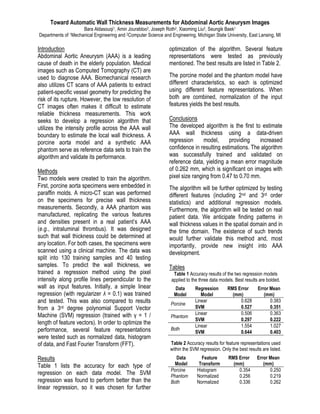
1. Toward Automatic Wall Thickness Measurements for Abdominal Aortic Aneurysm Images
Bara Aldasouqi1, Amin Jourabloo2, Joseph Roth2, Xiaoming Liu2, Seungik Baek1
Departments of 1Mechanical Engineering and 2Computer Science and Engineering, Michigan State University, East Lansing, MI
Introduction
Abdominal Aortic Aneurysm (AAA) is a leading
cause of death in the elderly population. Medical
images such as Computed Tomography (CT) are
used to diagnose AAA. Biomechanical research
also utilizes CT scans of AAA patients to extract
patient-specific vessel geometry for predicting the
risk of its rupture. However, the low resolution of
CT images often makes it difficult to estimate
reliable thickness measurements. This work
seeks to develop a regression algorithm that
utilizes the intensity profile across the AAA wall
boundary to estimate the local wall thickness. A
porcine aorta model and a synthetic AAA
phantom serve as reference data sets to train the
algorithm and validate its performance.
Methods
Two models were created to train the algorithm.
First, porcine aorta specimens were embedded in
paraffin molds. A micro-CT scan was performed
on the specimens for precise wall thickness
measurements. Secondly, a AAA phantom was
manufactured, replicating the various features
and densities present in a real patient’s AAA
(e.g., intraluminal thrombus). It was designed
such that wall thickness could be determined at
any location. For both cases, the specimens were
scanned using a clinical machine. The data was
split into 130 training samples and 40 testing
samples. To predict the wall thickness, we
trained a regression method using the pixel
intensity along profile lines perpendicular to the
wall as input features. Initially, a simple linear
regression (with regularizer λ = 0.1) was trained
and tested. This was also compared to results
from a 3rd degree polynomial Support Vector
Machine (SVM) regression (trained with γ = 1 /
length of feature vectors). In order to optimize the
performance, several feature representations
were tested such as normalized data, histogram
of data, and Fast Fourier Transform (FFT).
Results
Table 1 lists the accuracy for each type of
regression on each data model. The SVM
regression was found to perform better than the
linear regression, so it was chosen for further
optimization of the algorithm. Several feature
representations were tested as previously
mentioned. The best results are listed in Table 2.
The porcine model and the phantom model have
different characteristics, so each is optimized
using different feature representations. When
both are combined, normalization of the input
features yields the best results.
Conclusions
The developed algorithm is the first to estimate
AAA wall thickness using a data-driven
regression model, providing increased
confidence in resulting estimations. The algorithm
was successfully trained and validated on
reference data, yielding a mean error magnitude
of 0.262 mm, which is significant on images with
pixel size ranging from 0.47 to 0.70 mm.
The algorithm will be further optimized by testing
different features (including 2nd and 3rd order
statistics) and additional regression models.
Furthermore, the algorithm will be tested on real
patient data. We anticipate finding patterns in
wall thickness values in the spatial domain and in
the time domain. The existence of such trends
would further validate this method and, most
importantly, provide new insight into AAA
development.
Tables
Table 1 Accuracy results of the two regression models
applied to the three data models. Best results are bolded.
Data
Model
Regression
Model
RMS Error
(mm)
Error Mean
(mm)
Porcine
Linear 0.628 0.383
SVM 0.527 0.351
Phantom
Linear 0.506 0.363
SVM 0.297 0.222
Both
Linear 1.554 1.027
SVM 0.644 0.403
Table 2 Accuracy results for feature representations used
within the SVM regression. Only the best results are listed.
Data
Model
Feature
Transform
RMS Error
(mm)
Error Mean
(mm)
Porcine Histogram 0.354 0.250
Phantom Normalized 0.256 0.219
Both Normalized 0.336 0.262