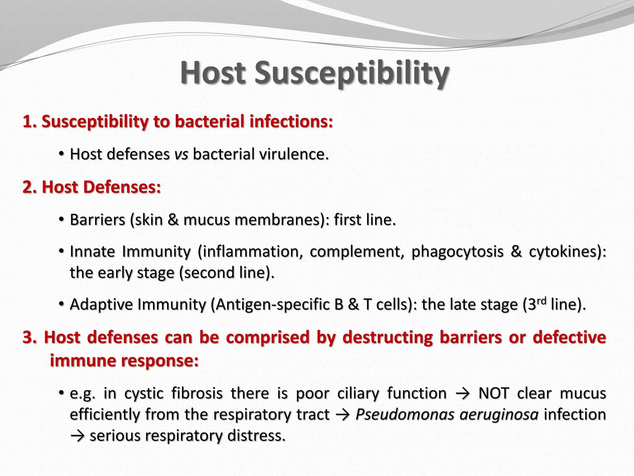
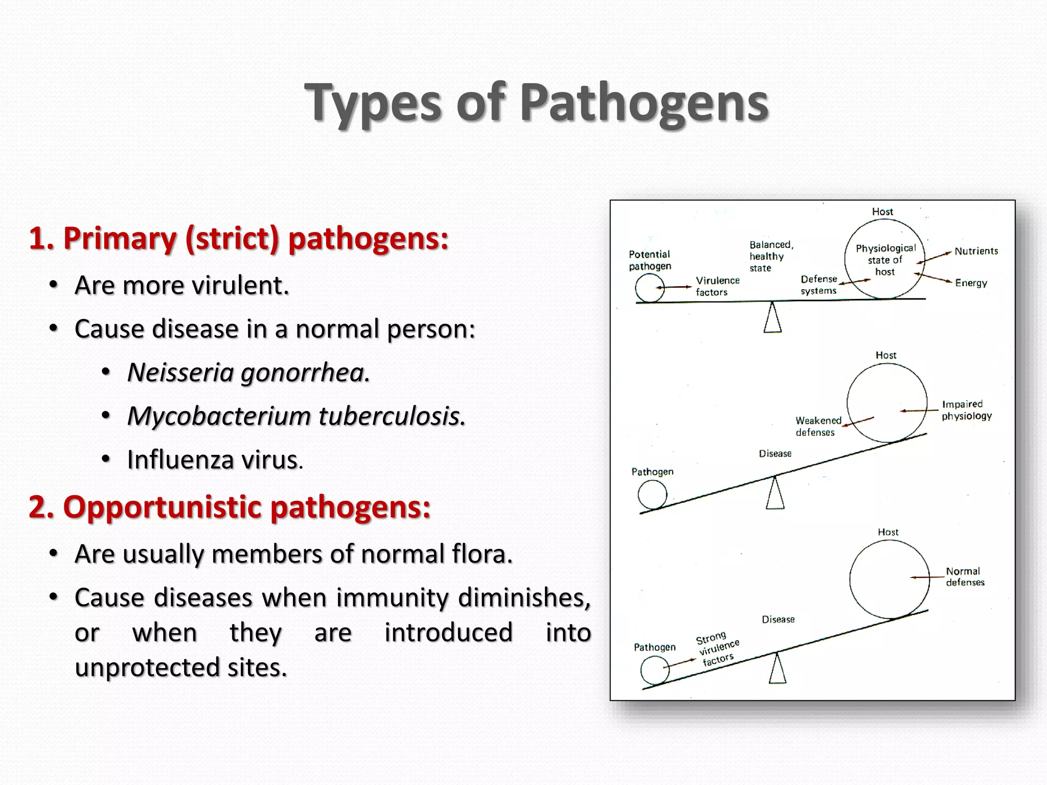
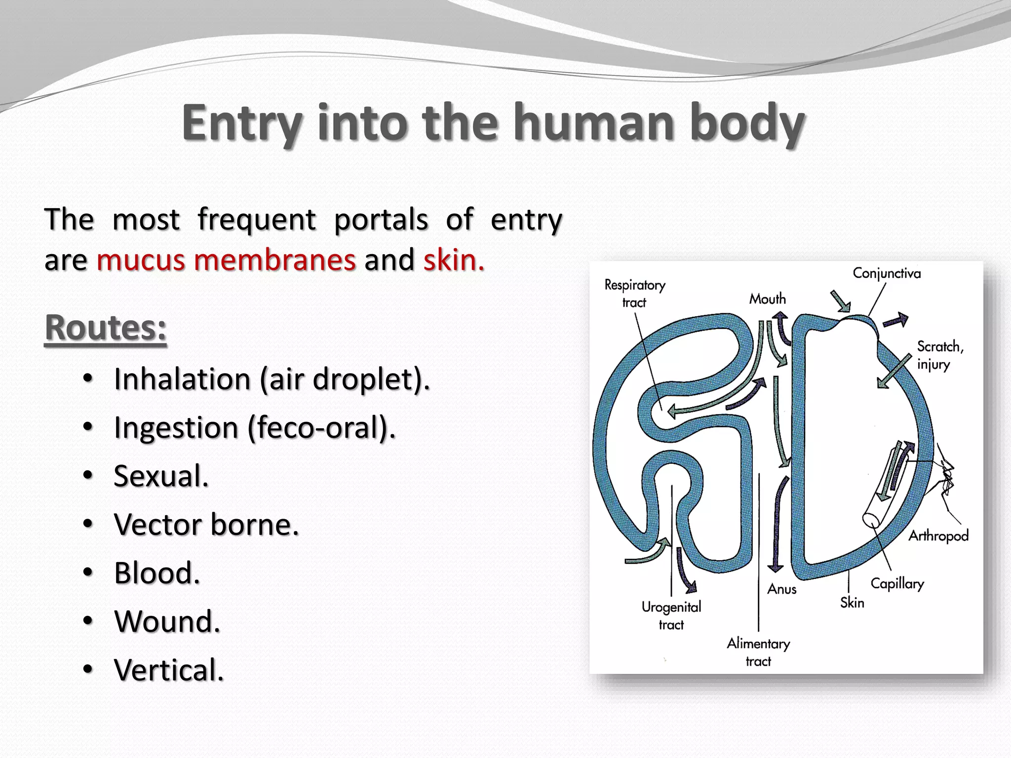
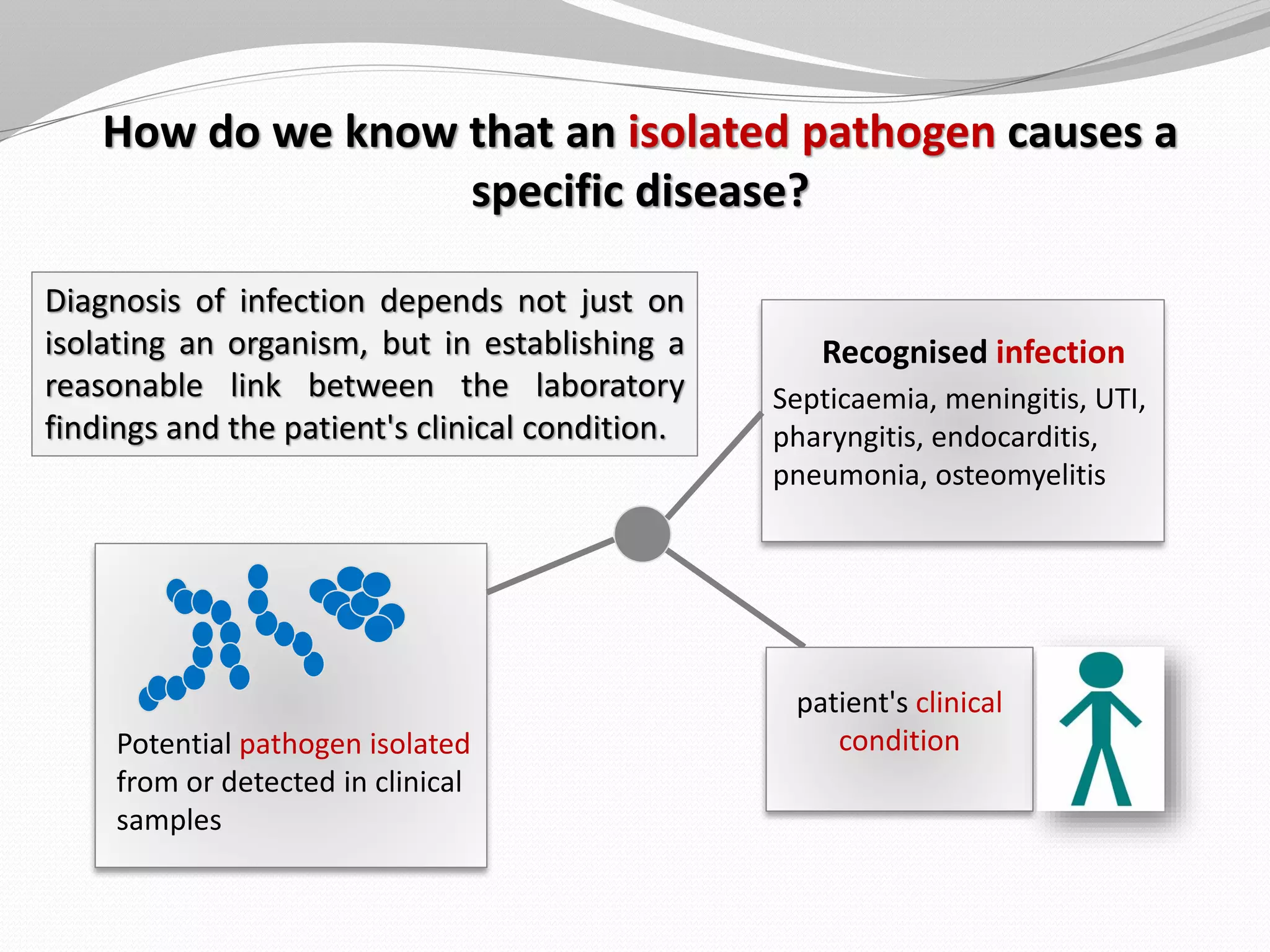
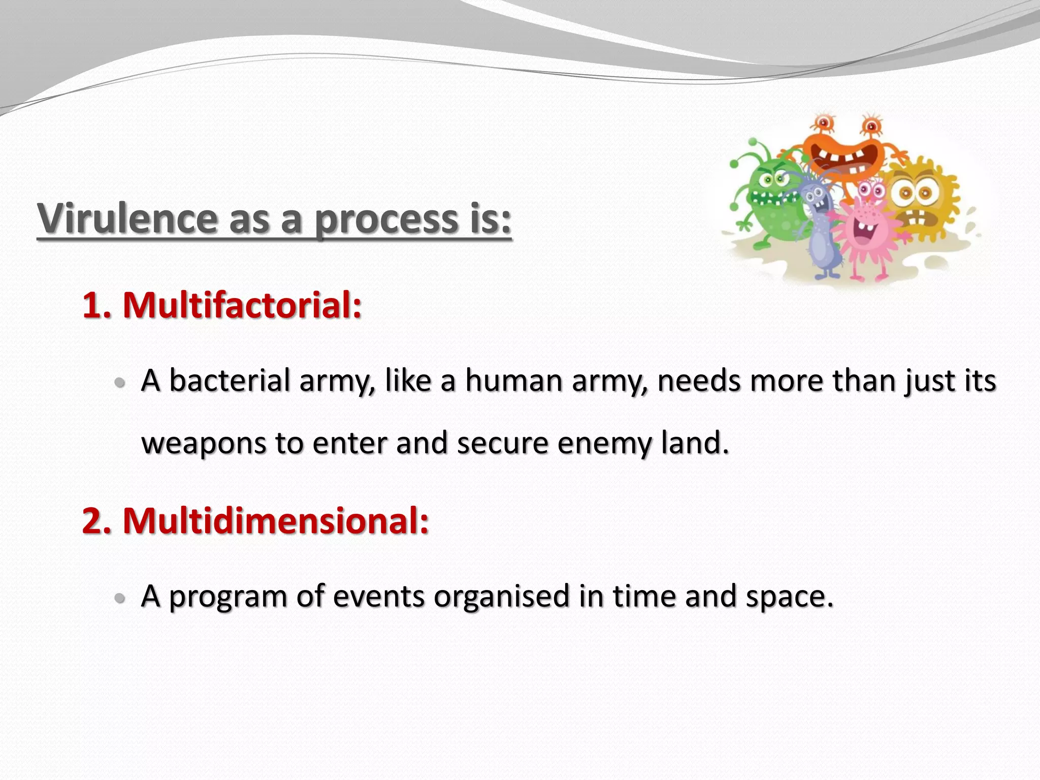
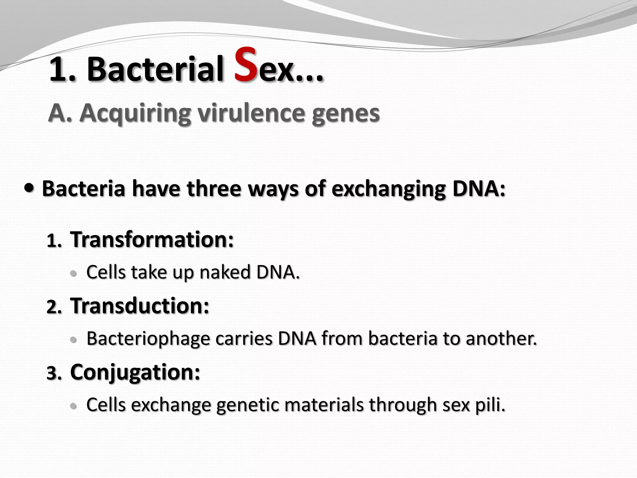
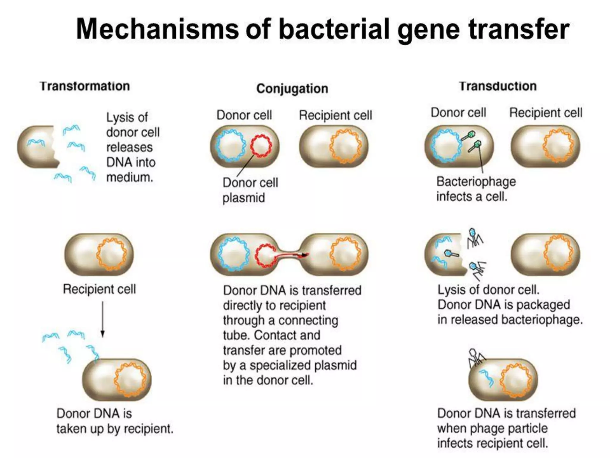
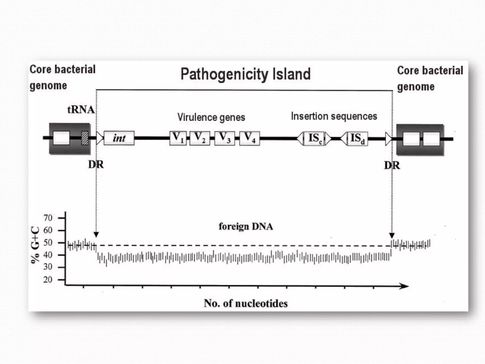
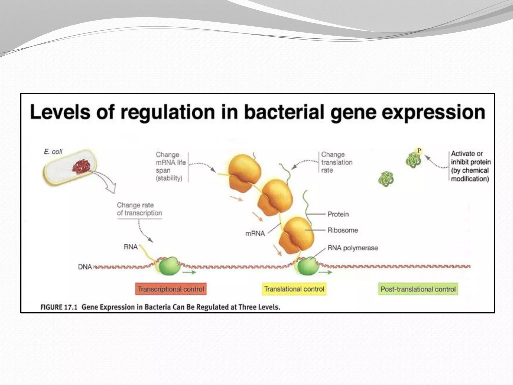
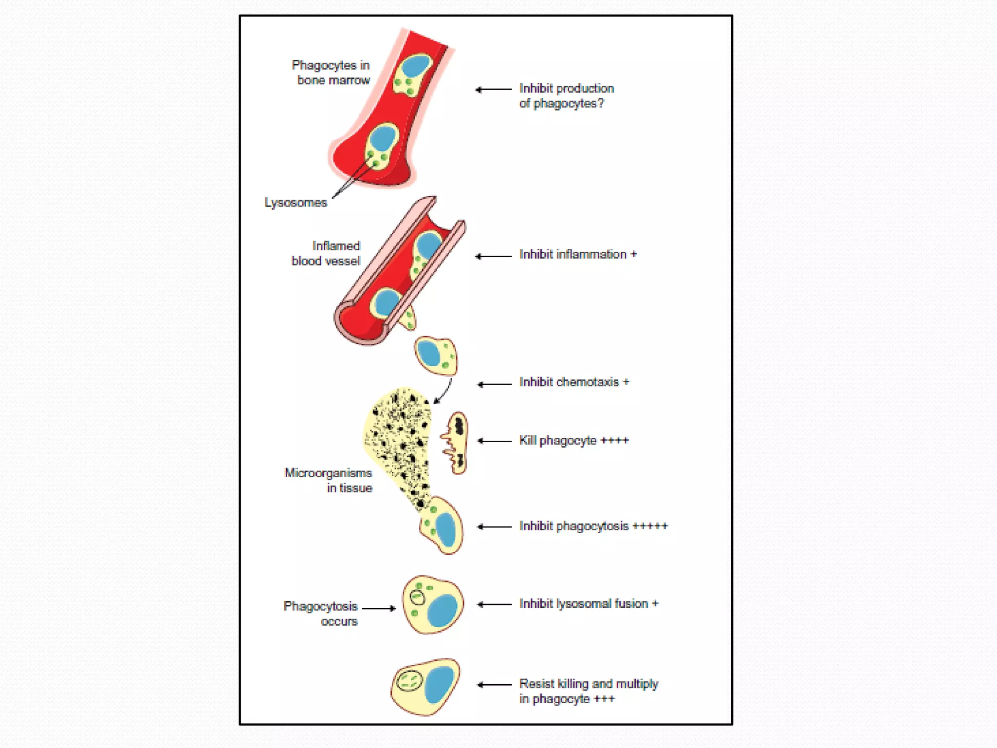
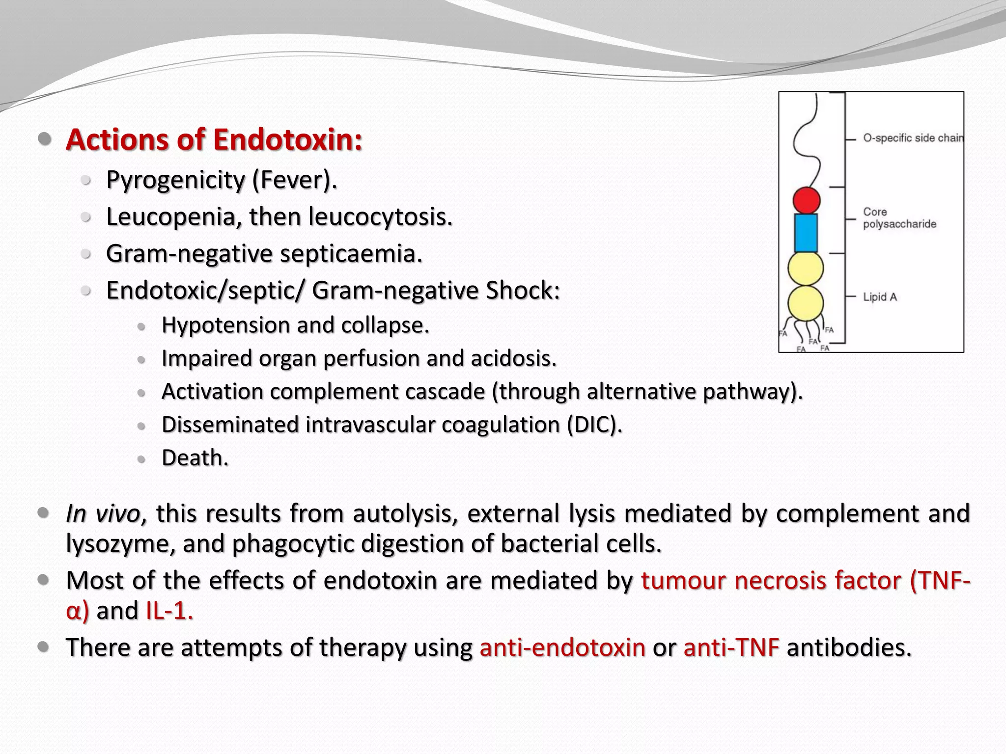
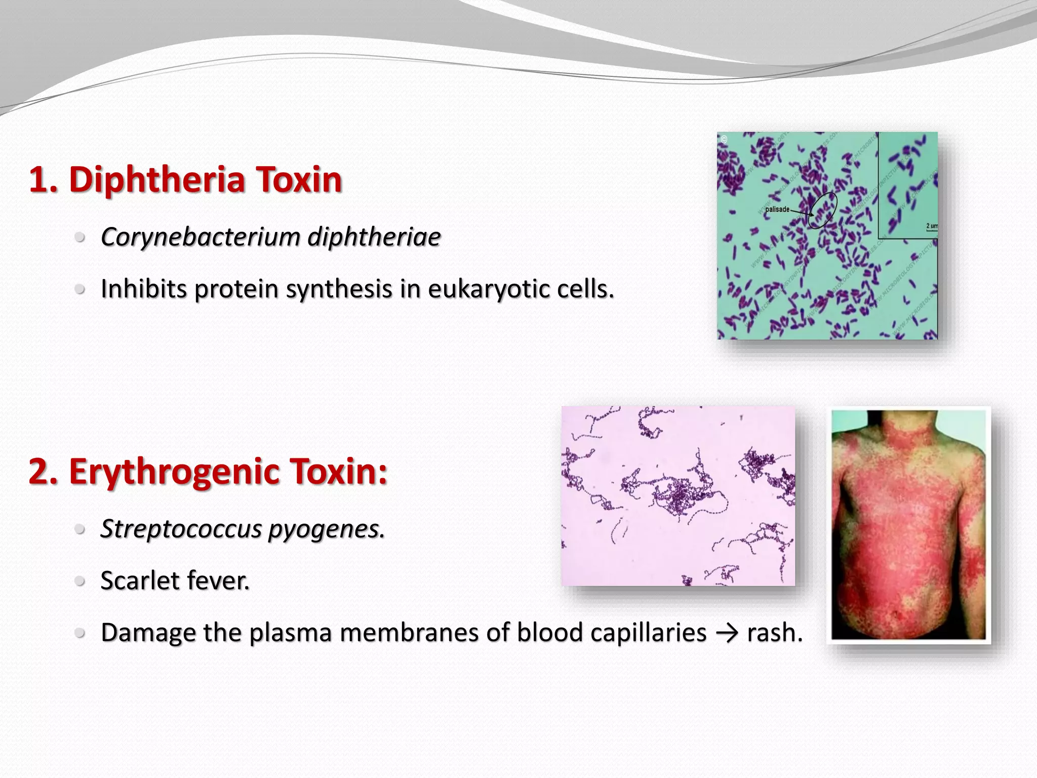
Bangalore Call Girl Whatsapp Number 100% Complete Your Sexual Needs
Bacterial Pathogenesis and Virulence Factors
1. Dr. Hany Lotfy
Assistant Professor of Medical Microbiology & Immunology
Faculty of Medicine, Sulaiman Al Rajhi University
2. Definitions
1. Infection: growth and multiplication of a microbe in or on
the body with/without the production of disease.
2. Pathogenicity: The capacity of a bacterium to cause disease.
3. Virulence: is the degree of the pathogenicity of a
microorganism.
4. Pathogenesis: the mechanism of infection, and the
mechanism by which disease develops.
3. Host Susceptibility
1. Susceptibility to bacterial infections:
• Host defenses vs bacterial virulence.
2. Host Defenses:
• Barriers (skin & mucus membranes): first line.
• Innate Immunity (inflammation, complement, phagocytosis & cytokines):
the early stage (second line).
• Adaptive Immunity (Antigen-specific B & T cells): the late stage (3rd line).
3. Host defenses can be comprised by destructing barriers or defective
immune response:
• e.g. in cystic fibrosis there is poor ciliary function → NOT clear mucus
efficiently from the respiratory tract → Pseudomonas aeruginosa infection
→ serious respiratory distress.
4. 1. Primary (strict) pathogens:
• Are more virulent.
• Cause disease in a normal person:
• Neisseria gonorrhea.
• Mycobacterium tuberculosis.
• Influenza virus.
2. Opportunistic pathogens:
• Are usually members of normal flora.
• Cause diseases when immunity diminishes,
or when they are introduced into
unprotected sites.
Types of Pathogens
5. Entry into the human body
The most frequent portals of entry
are mucus membranes and skin.
Routes:
• Inhalation (air droplet).
• Ingestion (feco-oral).
• Sexual.
• Vector borne.
• Blood.
• Wound.
• Vertical.
9. A bacterium may cause diseases by:
1. Destroying tissue (Invasiveness).
2. Producing toxins (Toxigenicity).
3. Stimulating overwhelming host immune responses.
4. Combination.
10.
11. Microbes and humans
Disease can come about in several overlapping ways:
1. Some bacteria from the normal flora can cause disease if they gain
access to deep tissues by trauma, surgery, or catheters; especially if
associated with a foreign body, e.g. Staphylococcus epidermidis.
2. In immunocompromized patients, many free-living bacteria and
components of the normal flora can cause disease, e.g. Acinetobacter.
3. Some bacteria which are part of the normal flora acquire extra
virulence factors making them pathogenic, e.g. Escherichia coli.
4. Some bacteria are never part of the normal flora and can cause
infection, e.g. Mycobacterium tuberculosis.
12. How do we know that a given pathogen
causes a specific disease?
Koch's postulates:
In 1884, Robert Koch proposed a series of postulates.
These postulates have been applied to link many specific bacterial species
with particular diseases.
1. The pathogen must be present in every case of the disease.
2. The pathogen must be isolated from the diseased host and grown in
pure culture.
3. The specific disease must be reproduced when a pure culture of the
pathogen is inoculated into a healthy susceptible host.
4. The pathogen must be recoverable from the experimentally infected
host.
13. Koch’s postulates are NOT feasible in all cases:
Some pathogens can’t be cultured in the laboratory.
Some diseases are caused by a combination of pathogens.
Ethical considerations prevent applying Koch’s postulates to
pathogens that require a human host.
14. Potential pathogen isolated
from or detected in clinical
samples
Recognised infection
Septicaemia, meningitis, UTI,
pharyngitis, endocarditis,
pneumonia, osteomyelitis
patient's clinical
condition
How do we know that an isolated pathogen causes a
specific disease?
Diagnosis of infection depends not just on
isolating an organism, but in establishing a
reasonable link between the laboratory
findings and the patient's clinical condition.
15. Evidence for a potential pathogen being of
clinical significance:
1. Isolated in abundance.
2. Isolated in pure culture.
3. Isolated on more than one occasion.
4. Isolated from deeper tissues.
5. Evidence of local inflammation.
6. Evidence of immune response to pathogen.
7. Fits with clinical picture.
16.
17. Infection:
Invasion by and multiplication of a microorganism or infectious agent in an
organ or tissue + tissue injury and progress to disease.
Colonization:
Colonization describes when microorganisms in or on a host with growth and
multiplication but without tissue invasion or damage.
These bacteria may be part of the normal flora of the individual, or NOT
necessarily normal flora.
Contamination:
Presence of infectious agents on various surfaces e.g. objects, instruments,
skin, mucous membranes.
Contamination of a sample from an external source (e.g. poor aseptic
technique when taking the sample, a swab touching a surface before being
used… etc).
18.
19. Types of Infections
Communicable (contagious) Infection:
An infection that can be transmitted from one individual to another
either directly (by contact) or indirectly (by fomites and vectors).
Symptomatic Infection:
A disease is considered symptomatic if a patient expressed
symptoms e.g. fever, erythema, pain … etc
Asymptomatic Infection:
A disease is considered asymptomatic if a patient is a carrier for a
disease or infection but experiences no symptoms.
Also called subclinical infections.
20. Endemic infection:
An infection prevalent in or restricted to a particular region,
community, or group of people (e.g. Cholera in Yemen, Malaria in
Nigeria).
Epidemic infection:
An epidemic occurs when new high number of cases of a certain
disease, in a given human population, and during a given period (e.g.
Dengue, Ebola).
Pandemic infection:
Pandemic is an epidemic of infectious disease that is spreading
through human populations across a large region; for instance
multiple countries, or even worldwide (e.g. COVID-19, Influenza).
21.
22.
23. Virulence as a process is:
1. Multifactorial:
A bacterial army, like a human army, needs more than just its
weapons to enter and secure enemy land.
2. Multidimensional:
A program of events organised in time and space.
24. Steps for Successful Bacterial Infection
Sex comes before disease.
Sense of environment.
Switch virulence genes on/off.
Swim to site of infection.
Stick to site of infection.
Scavenge nutrients (iron).
Secrete enzymes.
Survive stress.
Stealth: Evade immune system
Survival Intracellularly.
Strike-back.
Spread.
25. 1. Bacterial Sex...
A. Acquiring virulence genes
Bacteria have three ways of exchanging DNA:
1. Transformation:
Cells take up naked DNA.
2. Transduction:
Bacteriophage carries DNA from bacteria to another.
3. Conjugation:
Cells exchange genetic materials through sex pili.
26.
27. Bacterial Sex...
B. Mobile genetic elements
Transposons:
Class of genetic elements that can “jump” among different locations
within a genome.
Although these elements are frequently called “jumping genes,” they are
always maintained in an integrated site in the genome.
e.g. genes for heat-stable enterotoxin.
Plasmids:
Carry genes for exotoxins of Salmonella and E. coli.
Carry F-Factor essential for conjugation.
Phage-encoded toxins:
Botulism toxin.
Diphtheria toxin.
Shiga-like toxin.
Staphylococcal toxins.
28. Bacterial Sex...
C. Pathogenicity Islands (PAIs)
Pathogenicity islands (PAIs) are a distinct class of genomic islands which are acquired
by horizontal gene transfer.
Defining Features:
Carriage of one or many virulence genes.
Occupy large chromosomal regions (10-200 Kbp).
Present only in genomes of pathogenic strains.
Unstable, prone to be transferred, or deleted.
Different G+C content (lower) from host chromosome.
Compact distinct genetic units, often flanked by DRs.
Are associated with tRNA genes, which act as sites for recombination into the DNA.
They carry functional genes (e.g. integrases), to enable insertion into host DNA.
29.
30. Adherence factors: PAIs encode bacterial structures that enable them
to attach to host surfaces and, hence facilitate the infection process.
P‐pili of Escherichia coli.
Type IV pili of Vibrio cholerae.
Intimin of EPEC.
Siderophores: (used to deliver the essential iron)
Yersinia pestis chelates iron by Yersiniabactin.
Escherichia coli chelate iron by Aerobactin.
Exotoxins:
α‐haemolysin.
Pertussis toxin.
Invasion genes which mediate bacterial entry into eukaryotic cells
e.g. inv genes of Salmonella.
Pathogenicity Islands, encode..
31. 2. Sense environment… Quorum Sensing
Quorum sensing is the regulation of gene expression in response to
fluctuations in cell-population density.
Gram-positive Bacteria:
Gram-positive bacteria use autoinducing peptide (AIP) (= autoinducer).
When Gram-positive bacteria detect high concentration of AIP in their
environment, AIP binds to a receptor to activate a kinase
that phosphorylates a transcription factor, which regulates expression of
virulence genes.
Gram-negative Bacteria:
Gram-negative bacteria produce N-acyl homoserine lactones (AHL) as their
signaling molecule.
AHL binds directly to the transcription factor (LuxR) to regulate expression of
virulence genes.
33. 3. Switch virulence factors on / off
Changes in gene sequences:
Virulence gene amplification.
Genetic rearrangements: e.g. flagellar phase variation.
Regulation of transcription:
Switching on / off of activators or repressors.
mRNA folding → stops transcription of a gene.
Regulation of translation.
Regulation of post-translational modification.
34.
35. 4. Swim.... Motility
Many pathogenic bacteria are motile (e.g. Salmonella, Campylobacter,
Helicobacter, Listeria, Proteus, Spirochetes…etc).
Motility is crucial for virulence.
Sensor activation results in signals that regulate rotational movement.
36. 5. Stick.... adhesion
Common adherence mechanisms:
Capsule.
Biofilm formation.
Gram-positive adhesins:
MSCRAMMs (microbial surface components recognizing adhesive matrix
molecules), e.g. protein A in Staphylococci.
Lipoteichoic acid.
Gram-negative adhesins:
Fimbriae.
Afimbrial adhesin (Pertactin).
Outer membrane protein (OMP).
37. Bacterial biofilm:
Biofilms are bacterial populations that are enclosed in a matrix of
extracellular polymeric substances.
Bacteria form micro-colonies with cone-like and mushroom-shaped
morphologies by adhering to each other and to a surface.
Water-filled channels surround the micro-colonies and function like a
circulatory system, allowing access to nutrients, elimination of wastes,
and inter-bacterial communication.
38. Consequences of biofilm formation
In industrial settings, biofilms are responsible for fouling storage tanks
and clogging water pipes.
In the medical field, biofilms can form on virtually all synthetic medical
implants, including intravascular catheters, artificial valves,
pacemakers, orthopedic devices, and contact lenses.
Bacteria in biofilms have been reported to be up to 500 times more
resistant to antibiotics than planktonic cells.
The consequences of biofilm formation can include:
Chronic inflammation.
Impaired wound healing.
Increased antibiotic resistance.
Spread of infectious emboli.
39.
40. Stages of biofilm formation:
Biofilms are proposed to have five developmental phases:
(1) Initial reversible attachment.
(2) Irreversible attachment.
(3) Growth and micro-colony formation.
(4) Maturation and formation of water channels.
(5) Dispersion of cells from the biofilm to new sites.
43. Bacteria can adhere to:
Cell surfaces and extracellular matrix (in respiratory, gastrointestinal
and genitourinary tracts).
Solid surfaces: e.g. teeth, heart valves, prosthetic materials…etc
Other bacteria.
Some bacteria interact through molecular bridging:
Staphylococcus aureus binding via fibronectin bridging.
44.
45. 6. Scavenge nutrients....Iron
Free iron level is very low in body fluids:
Acute phase response causes further drop in levels of circulating iron.
Iron overload increases susceptibility to infection.
Bacterial systems for scavenging iron:
Siderophores chelate available iron & transport it into bacteria.
Iron can be scavenged directly from host iron-binding proteins, e.g.
lactoferrin-binding proteins (LBPs).
46. Key players in the host-pathogen “tug-of-war” for iron. Bacteria secrete siderophores to scavenge
iron. In response, the host cell defensively sequesters labile and complexes siderophores, via
lactoferrin and siderocalin (Lcn2), respectively. Stealth siderophores have features that render them
incompatible to siderocalin binding. Image from Wilson et al. (2016).
47. 7. Secrete Enzymes
Many pathogens secrete enzymes that enable them to:
1. Dissolve structural chemicals in the body.
2. Maintain an infection.
3. Invade further.
4. Avoid body defenses.
5. Inactivate antibiotic.
48. 1. Hyaluronidase:
“Spreading Factor”.
Digests hyaluronic acid (cement
substance between cells).
2. Collagenase:
Breaks down collagen.
49. 3. Coagulase:
Coagulates blood proteins.
Providing a “hiding place” for bacteria within a clot.
4. Kinases:
Such as staphylokinase and streptokinase.
Kinase enzymes convert inactive plasminogen to plasmin which
digests fibrin and prevents clotting of the blood.
50. 5. Ig A Proteases:
Split IgA at specific bonds.
Inactivates Ab activity.
6. Neuraminidase:
Produced by intestinal pathogens (Vibrio cholerae and Shigella
dysentriae).
Degrades neuraminic (sialic) acid; an intercellular cement of the
epithelial cells of the intestinal mucosa.
51. 7. Lecithinase:
Produced by Clostridium perferingens.
Destroys lecithin (phosphatidylcholine) in cell membranes.
8. Phospholipases (alpha toxin).
Produced by Clostridium perferingens.
Hydrolyzes phospholipids in cell membranes by removal of
polar head groups.
52. 9. Hemolysins
Produced by staphylococci (alpha toxin), streptococci
(streptolysins) and various Clostridia.
May be phospholipases or lecithinases that destroy red
blood cells and other cells (phagocytes) by lysis.
53. 8. Survive Stress
Pathogens face many stress conditions:
Heat shock during fever.
Oxidative stress within phagocytes.
pH stress.
Nutrient-limitation stress.
Stress response proteins, such as protein folding chaperones (heat shock
proteins) enable bacteria to tolerate fever.
Detoxification proteins play a role inside phagocytes (e.g. superoxide
dismutase to resist oxidative stress).
Infectious dose for enteric pathogens is much lower in cases of
achlorhydria (no need to overcome acid stress).
54. 9. Intracellular Survival
Some bacteria grow inside PMNL, macrophages or in monocytes
They survive by several mechanisms:
Avoid entry into phagosome.
Prevent phago-lysososme fusion (Mycobacterium tuberculosis).
Acquire resistance to lysosomal activity.
Examples: Mycobacterium tuberculosis, Brucella, Listeria.
55. 10. Stealth.... Overcome immune system
1. IgA protease (e.g. Neisseria gonorrheae).
2. Immunoglobulin-binding proteins:
e.g. protein A of Staphylococcus aureus.
3. Antiphagocytic factors:
Capsule: prevents adherence of phagocytic cells.
M-protein of Group A Streptococci.
Protein A of Staphylococcus aureus.
Leukocidin: destroys neutrophils, macrophages.
56. 4. Antigenic mimicry:
e.g. Sialic acid capsule of group B meningococcus.
5. Antigenic variation (phase variation):
Involves surface antigens such as proteins, LPS, capsules.
6. Adopt cryptic position:
Inside phagocytes.
Within biofilm.
57.
58. 11. Spread of bacteria
Through cells and organs.
Within macrophages, e.g. Salmonella in typhoid fever.
Through blood.
Within cells.
60. Actions of Endotoxin:
Pyrogenicity (Fever).
Leucopenia, then leucocytosis.
Gram-negative septicaemia.
Endotoxic/septic/ Gram-negative Shock:
Hypotension and collapse.
Impaired organ perfusion and acidosis.
Activation complement cascade (through alternative pathway).
Disseminated intravascular coagulation (DIC).
Death.
In vivo, this results from autolysis, external lysis mediated by complement and
lysozyme, and phagocytic digestion of bacterial cells.
Most of the effects of endotoxin are mediated by tumour necrosis factor (TNF-
α) and IL-1.
There are attempts of therapy using anti-endotoxin or anti-TNF antibodies.
61.
62. Exotoxins:
Bacterial cytotoxins form pores in eukaryotic cell membranes:
Streptolysin O of Streptococcus pyogenes.
Listeriolysin of Listeria monocytogenes.
Other toxins degrade components of the membrane:
e.g. Clostridium perfringens alpha toxin.
64. 1. Diphtheria Toxin
Corynebacterium diphtheriae
Inhibits protein synthesis in eukaryotic cells.
2. Erythrogenic Toxin:
Streptococcus pyogenes.
Scarlet fever.
Damage the plasma membranes of blood capillaries → rash.
65. 3. Botulinum Toxin:
Released by Clostridium botulinum.
Neurotoxin: It inhibits the release of acetylcholine (Ach) in
neuromuscular junctions → paralysis.