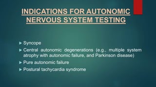
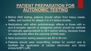
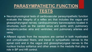
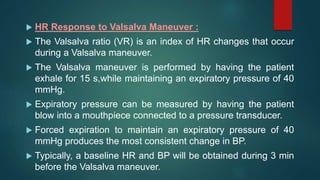
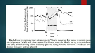
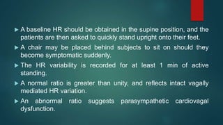
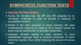
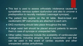
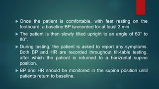
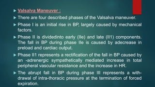
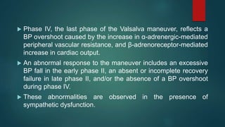
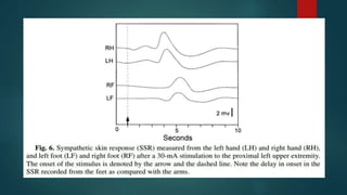
AUTONOMIC
NERVOUS
SYSTEM TESTING
PRESENTED BY
ARFA SULTANA MOHAMMED,
MPT 1ST YEAR,NEURO,
VAPMS COLLEGE OF PHYSIOTHERAPY.
INTRODUCTION
 Disorders of the autonomic nervous system (ANS) can be
found in many conditions thatspan the disciplinary
boundaries of medicine.
 The neurophysiological evaluation of the humanautonomic
nervous system function is challenging because most
autonomic structures arelocated at a distance from the skin,
and, thus, less amenable to direct observation or study.
 In addition, most neurophysiological measures of the ANS
reflect end-organ function (smoothmuscle, cardiac muscle,
and glandular organs), rather than direct neural activity in the
sympathetic and parasympathetic nerves.
 Despite these methodological difficulties, there are a number of
clinically useful techniques that test the functional integrity of
the sympathetic and parasympathetic nervous system.
 Neurophysiological tests of autonomic function are extensions
of the clinical exam, and their appropriate use requires a
thorough history and physical examination.
 In addition, a careful review of autonomic symptoms should be
taken, with attention to systems commonly affected by
autonomic dysfunction.
 As in all clinical tests, the pretest clinical probability affects the
power of ANS testing, and a clear, well-formulated question
should precede ANS testing.
COMMON AUTONOMIC
SYMPTOMS
 Orthostatic intolerance
 Dizziness
 Lightheadedness
 Fatigue
 “Coathanger” headache
 Nausea
 Palpitations
 Near-syncope and syncope
 Genitourinary
 Bladder urgency or frequency, Incontinence
 Nocturia
 Erectile dysfunction
 Ejaculatory disturbances
 Gastrointestinal
 Diarrhea
 Constipation
 Fecal incontinence
 Postprandial fullness, cramping, or bloating
 Sudomotor
 Hyperhidrosis
 Hypohidrosis and anhidrosis
NEUROANATOMIC AND
NEUROPHYSIOLOGICAL
PRINCIPLES
 A systematic approach to ANS testing is important given its
neuroanatomical complexity.
 A useful conceptual approach for understanding
sympathetic and parasympathetic function is to think in
terms of neuronal circuits and autonomic reflex arcs.
 Afferent information arises from end-organ receptors, such
as baroreceptors in vessel walls, and mechanoreceptors
and metaboreceptors in skeletal muscle.
 Signals arising from these structures are directed tocentral
autonomic structures via unmyelinated and myelinated autonomic
nerves.
 The nucleus tractus solitarius, located in the dorsal medulla,
receives much of the afferent peripheral autonomic input.
 This structure has reciprocal connections to central regions
regulating both sympathetic and parasympathetic function and
plays an important role in feedback regulatory circuits.
 Central regions with input to sympathetic preganglionic neurons
centers include the paraventricular nuclei of the hypothalamus,
the amygdala, and the insular and medial prefrontal cortices.
 Preganglionic sympathetic neurons are also located in the
rostral ventrolateral medulla, the caudal ventrolateral medulla,
the caudal raphe nuclei, and the A5 region of the pons.
 Efferent parasympathetic fibers originate in the Edinger–
Westphal nucleus, ciliary ganglion, superior and inferior
salivary nuclei, otic ganglion, and the nucleus ambiguous and
the dorsal motor nucleus of the vagus.
 These fibers are carried in cranial nerves III, VII, IX, and X, and
synapse on both end organs and parasympathetic ganglia
located within or in close proximity to their target organs.
 Preganglionic parasympathetic fibers related to genitourinary
function originate in the spinal cord at sacral cord levels 2, 3,
and 4.
 Efferent preganglionic sympathetic fibers originate in the
intermediolateral cell columns at levels T1–L2.
 These fibers synapse on postganglionic fibers in paravertebral,
prevertebral, and previsceral ganglia.
INDICATIONS FOR AUTONOMIC
NERVOUS SYSTEM TESTING
 Syncope
 Central autonomic degenerations (e.g., multiple system
atrophy with autonomic failure, and Parkinson disease)
 Pure autonomic failure
 Postural tachycardia syndrome
 Autonomic and small-fiber peripheral neuropathies (e.g.,
diabetic peripheral neuropathy, amyloid peripheral
neuropathy, and hereditary peripheral neuropathies)
 Sympathetically mediated pain
 Evaluating response to therapy
 Differentiating benign symptoms (e.g., dizziness) from life-
threatening autonomic disorders
PATIENT PREPARATION FOR
AUTONOMIC TESTING
 Before ANS testing, patients should refrain from heavy meals,
coffee, and nicotine for atleast 3 to 4 h before studies.
 Medications with either anticholinergic effects or those with α-
and β-receptor agonist or antagonist activity should be withheld
(if medically appropriate)24 to 48 h before testing, because these
can significantly affect the outcome of ANS tests.
 Most autonomic testing sessions are 60 to 90 min.
 Patients should wear loose-fitting clothes for comfort and to
facilitate the application of cardiac electrodes and blood
pressure(BP) cuffs
ELECTROPHYSIOLOGICAL
METHODS
 Heart Rate Recording and the RR Interval :
 Heart rate (HR) is routinely recorded with a three-lead
electrocardiograph (ECG) machine.
 Tests evaluating HR variability can only be performed in the
setting of sinus rhythm.
 The ECG leads are placed at anterior chest locations, which
minimize movement artifact, and the reference electrode is
usually placed at the mid-axillary line at approximately the
T4 level.
 Autonomic tests of HR variability may be based on HR
measure in beats per minute or its inverse, the RR interval,
measured in milliseconds
 BP Recording :
 BP during autonomic testing can be measured intermittently or
continuously.
 Some methods rely on brachial artery cuffs, whereas others
use devices that are applied to the wrist or the finger.
 The intermittent recording techniques rely on occlusive cuff
methods.
 Techniques for detecting arterial lumen opening include
auscultatory and oscillometric methods.
 Auscultation for Korotkoff sounds is a widely available, and
invaluable, bedside tool.
 The oscillometric technique is based on the observation that
during cuff deflation, the point of maximal oscillation between
systolic and diastolic pressure corresponds to the mean
arterial pressure.
 Although both the auscultatory and oscillometric techniques
are relatively easy to perform, they provide only periodic
sampling of BP.
 Plethysmography and arterial tonometry are methods used
for continuous, beat-to-beat,noninvasive BP recording.
 Plethysmography is an indirect measure of BP, whereby the
pulse pressure is derived from the blood volume.
 The volume clamp method of Penaz, also called arterial
counterpulsation, uses a servo-loop circuit that continuously
counter balances arterial pressure, thereby clamping the
volume of blood flow to maintain a transmural pressure of zero.
 The counter-pulsation pressure provides a real-time measure
of arterial pulse pressure.
 The volume clamp technique of Penaz has been used with
digital pressure cuffs and fingerphotoplethysmography.
 A potential drawback to this method is its sensitivity to
decreases indistal limb temperature, which may significantly
alter the measured BP.
 Arterial tonometry is a method in which arrays of pressure
sensors compress and partiallyflatten (applanation) an artery
at a constant pressure.
 The intra-arterial forces measured by the sensor array are
then translated into arterial pressure waveforms displayed in
real time.
 The tonometric sensors, which are placed over the radial
artery, may be affected by both improper sensor placement as
well as tremors.
 Both methods may be inaccurate in subjects with peripheral
vascular occlusive disease, and both are dependent on arm
position.
 Recordings should be made with the arm abducted or elbow
flexed so that the recording site is at the level of the heart
throughout autonomic testing.
 During formal tests of autonomic function, both intermittent
and continuous BP recordings are usually obtained.
 Oscillometric BP recordings may be used to verify the
accuracy of the continuous BP recordings, and may serve as
a more reliable means of BP recording in the event of
marked decreases in BP.
PARASYMPATHETIC FUNCTION
TESTS
 Neurophysiological tests of cardiovascular parasympathetic function
evaluate the integrity of a reflex arc that includes the vagus and
glossopharyngeal nerves; cardiopulmonary baroreceptors located in
structures, such as the carotid sinus and aortic arch; pulmonary
receptors;cardiac atria and ventricles; and pulmonary arteries and
veins.
 Afferent signals from the receptors are carried in both myelinated
and unmyelinated fibers, and travel in sympathetic nerves to the
spinal cord and in the vagus and glossopharyngeal nerves to the
nucleus tractus solitarius and other areas in the medulla that play a
role in BP and HR control.
 HR Response to Valsalva Maneuver :
 The Valsalva ratio (VR) is an index of HR changes that occur
during a Valsalva maneuver.
 The Valsalva maneuver is performed by having the patient
exhale for 15 s,while maintaining an expiratory pressure of 40
mmHg.
 Expiratory pressure can be measured by having the patient
blow into a mouthpiece connected to a pressure transducer.
 Forced expiration to maintain an expiratory pressure of 40
mmHg produces the most consistent change in BP.
 Typically, a baseline HR and BP will be obtained during 3 min
before the Valsalva maneuver.
 The patient is then instructed to inhale deeply, exhale completely,
then take another deep breath in and blow into the mouth piece.
 Both HR and BP are recorded during the maneuver and up to 60 s
after the Valsalva maneuver is terminated.
 A mild degree of light-headedness is often reported during this
procedure.
 Because of the need for prolonged expiratory effort, the elderly
and those with any underlying pulmonary disorders may find the
Valsalva maneuver difficult to perform.
 Typically, one to two trials are obtained for analysis,with an
adequate time between trials (3–5 min) to allow subjects to return
to a baseline state.
 Because intraocular pressure increases during Valsalva
maneuver, recent retinal surgery orhemorrhage is a relative
contraindication to performing this test.
 The VR is a measure of the HR response to BP changes resulting
from the mechanical and cardiovascular effects of the Valsalva
maneuver.
 There are four phases of BP, designated I to IV, which delineate
the cardiovascular changes during the Valsalva maneuver.
 Phase I consistsof a transient rise in BP that results from the
increased intrathoracic and intra-abdominal pressures.
 During phase II, there is a fall and recovery of BP.
 During phase III, there is a decrease in BP caused by the release
of intrathoracic pressure.
 During phase IV, there is an “overshoot” caused by the increase
cardiac output into a vasoconstricted peripheral circulation.
 As the BP falls in phase II, there is an increase in HR caused by
parasympathetic with-drawal that is then followed by sympathetic
activation.
 This results in an increase in HR during and shortly after the
Valsalva maneuver.
 In response to the BP overshoot during phase IV,approx 15 to 30 s
after the end of the maneuver, the HR falls and produces a
transient bradycardia that persists, in the normal state, until after
 This results in the minimum HR.
 The VR is calculated as the maximum HR, which occurs
during or shortly after the Valsalva maneuver, divided by the
minimum HR, which occurs after the cessation of the
Valsalva maneuver (VR = max HR/min HR).
 A normal VR reflects an intact baroreceptor-mediated rise
and fall in HR.
 The VR values are age related, and decline with increasing
age. A reduced VR reflects baroreceptor and cardiovagal
dysfunction.
 However, it is important that an adequate and steady
expiratory pressure is maintained during the maneuver for
these results to be meaningful.
 HR Variability With Respiration (Respiratory Sinus
Arrhythmia) :
 The expiratory/inspiratory (E/I) ratio, similar to the VR, is a
measure of parasympathetic function.
 Timed breathing potentiates the normal sinus arrhythmia that
occurs normally during respiratory cycles.
 The physiological mechanism of respiratory sinus arrhythmia is
complex and includes pulmonary stretch and cardiac
mechanoreceptor activation, baroreceptor activation, local
cardiac reflexes and central factors.
 During inspiration, there is a reflex withdrawal of parasympathetic
activity, increased excitability of cardiac pacemaker cells in the
sinoatrial node, and an increase in HR.
 Respiratory sinus arrhythmia provides a measure of
parasympathetic control of HR variation throughout the respiratory
cycle.
 The respiratory sinus arrhythmia is recorded with the patient
supine and breathing at a fixed rate.
 A respiratory frequency of six breaths per minute, with slow
inhalation and exhalation (5 s each) provides close to maximum
HR variability.
 The timed breathing is performed with the aid of either verbal
coaching, or an electronic visual cue.
 This is a safe,easily performed test, without contraindications.
 Typically, six to eight cycles are recorded,and one to two trials
are performed. The variation in HR across this cycle of six to
eight breaths is often expressed as the mean difference
between the maximum and minimum HR.
 Alternatively, one can calculate the E/I ratio, which is the sum
of the longest RR intervals divided by sum of the shortest RR
intervals (E/I = Σ longest RR/Σ shortest RR).
 HR variability with respiration decreases with increasing age.
 Reduced HR variability with respiration can be observed in
cases of autonomic failure caused by autonomic peripheral
neuropathies and central autonomic degenerations.
 Other factors known to influence the HR response to deep
breathing include poor respiratory effort, hypocapnia,
salicylates, positioning, and obesity.
 The 30:15 Ratio :
 The 30:15 ratio, similar to the VR and E/I ratio, is a measure of
parasympathetic cardiovagal function.
 Immediately after standing, there is an increase in HR that
reflects an exercise reflex and withdrawal of parasympathetic
tone.
 There is a further HR increase at approx 15 s that reflects a
compensatory response to decreased venous return and
decreased cardiac output and a fall in BP.
 This is followed by a second period of relative bradycardia at
approx 30 s.
 The 30:15 ratio is the RR interval at approx 30 s, divided by
theRR interval at approx 15 s.
 A baseline HR should be obtained in the supine position, and the
patients are then asked to quickly stand upright onto their feet.
 A chair may be placed behind subjects to sit on should they
become symptomatic suddenly.
 The HR variability is recorded for at least 1 min of active
standing.
 A normal ratio is greater than unity, and reflects intact vagally
mediated HR variation.
 An abnormal ratio suggests parasympathetic cardiovagal
dysfunction.
 A number of general medical conditions, such as
hypovolemia, medical deconditioning, and hypothyroidism
can lead to misinterpretation of the 30:15 ratio and, thus,
these factors should be taken into account when interpreting
the test results.
SYMPATHETIC FUNCTION TESTS
 Head-Up Tilt-Table Testing :
 During the tilt-table test, the BP and HR response to an
orthostatic challenge is used to provide a measure of
sympathetic function.
 Similar to the early response to active standing, the early
cardiovascular response to head-up tilt is largely caused by
blood volume redistribution to the lower extremities.
 However, tilt-table testing is more sensitive to such
redistribution because there is minimal contraction of lower
extremity muscles, thus, further reducing the amount of venous
return.
 This test is used to assess orthostatic intolerance caused by
sympathetic nervous system dysfunction and also to uncover a
predisposition to neurally mediated (vasovagal syncope).
 The patient lies supine on the tilt table. Beat-to-beat and
oscillometric BP instruments are attached to each arm.
 EKG monitoring should take place throughout the test.
 A large waist belt should be placed around patients to secure
them in case of syncope or unexpected falls.
 Other safety measures include the availability of cardiovascular
medications, including atropine, and a resuscitation cart that
can be used in the event of cardiac asystole and other
unexpected cardiac arrhythmias.
 Once the patient is comfortable, with feet resting on the
footboard, a baseline BP isrecorded for at least 3 min.
 The patient is then slowly tilted upright to an angle of 60° to
80°.
 During testing, the patient is asked to report any symptoms.
Both BP and HR are recorded throughout tilt-table testing,
after which the patient is returned to a horizontal supine
position.
 BP and HR should be monitored in the supine position until
patients return to baseline.
 Tilt-table test results are influenced by a number of conditions,
such as hypovolemia, cardio-vascular deconditioning,
medications, hypothyroidism, infection and sepsis, and
reduced myocardial function.
 These conditions must be accounted for during the
interpretation of abnormal tilt-table findings.
 Some laboratories use pharmacological measures to
potentiate the orthostatic challenge of head-up tilt.
 Intravenous administration of isoproterenol, a β-adrenergic
agonist that increases ventricular contractility and HR, is
frequently used.
 This intervention may increase the sensitivity of the test but is
associated with reduced specificity.
 In addition, the sight of needles andblood, as well as pain may
further reduce the specificity of the test.
 The use of sublingual nitro-glycerin may increase the sensitivity
of the test while avoiding these precipitants.
 A normal tilt-table test is one in which there are no symptoms,
and only a modest fall in systolic BP.
 Orthostatic hypotension is defined as a decrease in systolic BP of
greater than 20 mmHg, diastolic BP greater than 10 mmHg
accompanied by symptoms of orthostatic intolerance.
 The pattern, the temporal characteristics, and the degree of
changes that occur in both the BP and HR define the test
abnormality. The use of the term “positive tilt-table test”is
incomplete, uninformative, and should be avoided.
 Three well-described patterns of neurally mediated syncope can
occur during head-up tilt-table testing:
 1. Vasodepression resulting in hypotension without bradycardia.
 2. Cardioinhibition with a marked bradycardia (fewer than 40 beats
per minute) with or without significant hypotension.
 3. Mixed, with both bradycardia and hypotension.
 Valsalva Maneuver :
 There are four described phases of the Valsalva maneuver.
 Phase I is an initial rise in BP, largely caused by mechanical
factors.
 Phase II is dividedinto early (IIe) and late (II1) components.
The fall in BP during phase IIe is caused by adecrease in
preload and cardiac output.
 Phase II1 represents a rectification of the fall in BP caused by
an -adrenergic sympathetically mediated increase in total
peripheral vascular resistance and the increase in HR.
 The abrupt fall in BP during phase III represents a with-
drawal of intra-thoracic pressure at the termination of forced
expiration.
 Phase IV, the last phase of the Valsalva maneuver, reflects a
BP overshoot caused by the increase in α-adrenergic-mediated
peripheral vascular resistance, and β-adrenoreceptor-mediated
increase in cardiac output.
 An abnormal response to the maneuver includes an excessive
BP fall in the early phase II, an absent or incomplete recovery
failure in late phase II, and/or the absence of a BP overshoot
during phase IV.
 These abnormalities are observed in the presence of
sympathetic dysfunction.
 Sympathetic Cholinergic Sweat Function :
 The best-known neurophysiological test of sympathetic
sudomotor function is the sympathetic skin response (SSR), or
galvanic skin response.
 Increased sympathetic nerve fiber sudomotor activity causes
release of sweat from glands in the skin, which can be detected
as a change in surface conductivity.
 A standard EMG instrument can be used to measure this
response.
 A small recording electrode is attached to the palmar and dorsal
surface of both hands and feet, with a long sweep speed (10 s)
setting, and a bandpass filter of 1 Hz to 2000 Hz.
 After a stable baseline is obtained, a stimulus is administered
and the SSR is recorded.
 Any stimulus that activates the sympathetic system can be
administered, such as an electrical stimulus, a loud noise, or a
bright light.
 Simultaneous recording from both hand and foot can be
performed, which demonstrates the length-dependent latency
difference between these sites.
 Typically, three to four trials are obtained at each site.
 A normal SSR is a monophasic orbiphasic deflection that
habituates with time and repeated trials.
 An abnormal response is an absent SSR, and reflects
sympathetic sudomotor dysfunction.
 This test does not differentiate between preganglionic or
postganglionic sympathetic dysfunction.
 In the silastic imprint test, a plastic or silicone film is applied to
the area of sweat gland activity provoked by the iontophoresis
of a cholinergic agonist.
 The film is then removed, and the number of active sweat
glands, their density, and the volume of sweat secreted can
be determined by digital image analysis.
 The thermoregulatory sweat test is performed by the
application of an indicator, such as alizarin red, which
changes color in the presence of moisture.
 The indicator is applied to the body of the patient, who is
then placed in a thermally controlled environment, and the
core temperature is raised above baseline.
 The pattern of color changes, which can be expressed as
percentage of body surface, is used to quantify
hypohypohidrosis and anhidrosis.
 Iontophoresis of cholinergic agonists can also be used to
evoke an axon reflex-mediated response from a more distal
population of sweat glands.
 The response is recorded using a sudorometer, which
measures changes in humidity resulting from sweat
production.
 This quantitative sudomotor axon reflex test (Q-SART)
can be used to measure both sweat volumeand sweat
pattern.
 It is largely a measure of postganglionic sympathetic
function, but may be reduced with severe preganglionic
disorders.
MICRONEUROGRAPHY
 Intraneural microneurography enables the direct
measurement of muscle sympathetic nerve activity.
 A tungsten microelectrode of approx 5 µm in size is
inserted into a fascicle of a distal sympathetic nerve to the
skin or muscles.
 Using this technique, sympathetic outflow to skin and
muscle can be measured at rest and in response to
various physiological perturbations.
 Muscle sympathetic nerve activity, consisting predominantly of
efferent muscle blood vessel vasoconstrictor impulses, are
grouped in relationship to the cardiac rhythm.
 Skin sympathetic activity consists of vasoconstrictor impulses
to skin capillaries,arteriovenous shunts, and sudomotor and
pilomotor impulses.
 They appear as irregular burst of varying duration, often
occurring in relationship to the respiratory rhythm.
 This technique is primarily used in research, rather than in
clinical laboratories.
THANKYOU

Autonomic nervous system testing arfa sulthana

  • 1.
    AUTONOMIC NERVOUS SYSTEM TESTING PRESENTED BY ARFASULTANA MOHAMMED, MPT 1ST YEAR,NEURO, VAPMS COLLEGE OF PHYSIOTHERAPY.
  • 2.
    INTRODUCTION  Disorders ofthe autonomic nervous system (ANS) can be found in many conditions thatspan the disciplinary boundaries of medicine.  The neurophysiological evaluation of the humanautonomic nervous system function is challenging because most autonomic structures arelocated at a distance from the skin, and, thus, less amenable to direct observation or study.  In addition, most neurophysiological measures of the ANS reflect end-organ function (smoothmuscle, cardiac muscle, and glandular organs), rather than direct neural activity in the sympathetic and parasympathetic nerves.
  • 3.
     Despite thesemethodological difficulties, there are a number of clinically useful techniques that test the functional integrity of the sympathetic and parasympathetic nervous system.  Neurophysiological tests of autonomic function are extensions of the clinical exam, and their appropriate use requires a thorough history and physical examination.  In addition, a careful review of autonomic symptoms should be taken, with attention to systems commonly affected by autonomic dysfunction.  As in all clinical tests, the pretest clinical probability affects the power of ANS testing, and a clear, well-formulated question should precede ANS testing.
  • 5.
    COMMON AUTONOMIC SYMPTOMS  Orthostaticintolerance  Dizziness  Lightheadedness  Fatigue  “Coathanger” headache  Nausea  Palpitations  Near-syncope and syncope  Genitourinary
  • 6.
     Bladder urgencyor frequency, Incontinence  Nocturia  Erectile dysfunction  Ejaculatory disturbances  Gastrointestinal  Diarrhea  Constipation  Fecal incontinence  Postprandial fullness, cramping, or bloating  Sudomotor  Hyperhidrosis  Hypohidrosis and anhidrosis
  • 7.
    NEUROANATOMIC AND NEUROPHYSIOLOGICAL PRINCIPLES  Asystematic approach to ANS testing is important given its neuroanatomical complexity.  A useful conceptual approach for understanding sympathetic and parasympathetic function is to think in terms of neuronal circuits and autonomic reflex arcs.  Afferent information arises from end-organ receptors, such as baroreceptors in vessel walls, and mechanoreceptors and metaboreceptors in skeletal muscle.
  • 8.
     Signals arisingfrom these structures are directed tocentral autonomic structures via unmyelinated and myelinated autonomic nerves.  The nucleus tractus solitarius, located in the dorsal medulla, receives much of the afferent peripheral autonomic input.  This structure has reciprocal connections to central regions regulating both sympathetic and parasympathetic function and plays an important role in feedback regulatory circuits.  Central regions with input to sympathetic preganglionic neurons centers include the paraventricular nuclei of the hypothalamus, the amygdala, and the insular and medial prefrontal cortices.
  • 10.
     Preganglionic sympatheticneurons are also located in the rostral ventrolateral medulla, the caudal ventrolateral medulla, the caudal raphe nuclei, and the A5 region of the pons.  Efferent parasympathetic fibers originate in the Edinger– Westphal nucleus, ciliary ganglion, superior and inferior salivary nuclei, otic ganglion, and the nucleus ambiguous and the dorsal motor nucleus of the vagus.  These fibers are carried in cranial nerves III, VII, IX, and X, and synapse on both end organs and parasympathetic ganglia located within or in close proximity to their target organs.
  • 11.
     Preganglionic parasympatheticfibers related to genitourinary function originate in the spinal cord at sacral cord levels 2, 3, and 4.  Efferent preganglionic sympathetic fibers originate in the intermediolateral cell columns at levels T1–L2.  These fibers synapse on postganglionic fibers in paravertebral, prevertebral, and previsceral ganglia.
  • 12.
    INDICATIONS FOR AUTONOMIC NERVOUSSYSTEM TESTING  Syncope  Central autonomic degenerations (e.g., multiple system atrophy with autonomic failure, and Parkinson disease)  Pure autonomic failure  Postural tachycardia syndrome
  • 13.
     Autonomic andsmall-fiber peripheral neuropathies (e.g., diabetic peripheral neuropathy, amyloid peripheral neuropathy, and hereditary peripheral neuropathies)  Sympathetically mediated pain  Evaluating response to therapy  Differentiating benign symptoms (e.g., dizziness) from life- threatening autonomic disorders
  • 14.
    PATIENT PREPARATION FOR AUTONOMICTESTING  Before ANS testing, patients should refrain from heavy meals, coffee, and nicotine for atleast 3 to 4 h before studies.  Medications with either anticholinergic effects or those with α- and β-receptor agonist or antagonist activity should be withheld (if medically appropriate)24 to 48 h before testing, because these can significantly affect the outcome of ANS tests.  Most autonomic testing sessions are 60 to 90 min.  Patients should wear loose-fitting clothes for comfort and to facilitate the application of cardiac electrodes and blood pressure(BP) cuffs
  • 15.
    ELECTROPHYSIOLOGICAL METHODS  Heart RateRecording and the RR Interval :  Heart rate (HR) is routinely recorded with a three-lead electrocardiograph (ECG) machine.  Tests evaluating HR variability can only be performed in the setting of sinus rhythm.  The ECG leads are placed at anterior chest locations, which minimize movement artifact, and the reference electrode is usually placed at the mid-axillary line at approximately the T4 level.
  • 16.
     Autonomic testsof HR variability may be based on HR measure in beats per minute or its inverse, the RR interval, measured in milliseconds
  • 17.
     BP Recording:  BP during autonomic testing can be measured intermittently or continuously.  Some methods rely on brachial artery cuffs, whereas others use devices that are applied to the wrist or the finger.  The intermittent recording techniques rely on occlusive cuff methods.  Techniques for detecting arterial lumen opening include auscultatory and oscillometric methods.  Auscultation for Korotkoff sounds is a widely available, and invaluable, bedside tool.
  • 18.
     The oscillometrictechnique is based on the observation that during cuff deflation, the point of maximal oscillation between systolic and diastolic pressure corresponds to the mean arterial pressure.  Although both the auscultatory and oscillometric techniques are relatively easy to perform, they provide only periodic sampling of BP.  Plethysmography and arterial tonometry are methods used for continuous, beat-to-beat,noninvasive BP recording.  Plethysmography is an indirect measure of BP, whereby the pulse pressure is derived from the blood volume.
  • 19.
     The volumeclamp method of Penaz, also called arterial counterpulsation, uses a servo-loop circuit that continuously counter balances arterial pressure, thereby clamping the volume of blood flow to maintain a transmural pressure of zero.  The counter-pulsation pressure provides a real-time measure of arterial pulse pressure.  The volume clamp technique of Penaz has been used with digital pressure cuffs and fingerphotoplethysmography.  A potential drawback to this method is its sensitivity to decreases indistal limb temperature, which may significantly alter the measured BP.
  • 20.
     Arterial tonometryis a method in which arrays of pressure sensors compress and partiallyflatten (applanation) an artery at a constant pressure.  The intra-arterial forces measured by the sensor array are then translated into arterial pressure waveforms displayed in real time.  The tonometric sensors, which are placed over the radial artery, may be affected by both improper sensor placement as well as tremors.  Both methods may be inaccurate in subjects with peripheral vascular occlusive disease, and both are dependent on arm position.
  • 21.
     Recordings shouldbe made with the arm abducted or elbow flexed so that the recording site is at the level of the heart throughout autonomic testing.  During formal tests of autonomic function, both intermittent and continuous BP recordings are usually obtained.  Oscillometric BP recordings may be used to verify the accuracy of the continuous BP recordings, and may serve as a more reliable means of BP recording in the event of marked decreases in BP.
  • 22.
    PARASYMPATHETIC FUNCTION TESTS  Neurophysiologicaltests of cardiovascular parasympathetic function evaluate the integrity of a reflex arc that includes the vagus and glossopharyngeal nerves; cardiopulmonary baroreceptors located in structures, such as the carotid sinus and aortic arch; pulmonary receptors;cardiac atria and ventricles; and pulmonary arteries and veins.  Afferent signals from the receptors are carried in both myelinated and unmyelinated fibers, and travel in sympathetic nerves to the spinal cord and in the vagus and glossopharyngeal nerves to the nucleus tractus solitarius and other areas in the medulla that play a role in BP and HR control.
  • 23.
     HR Responseto Valsalva Maneuver :  The Valsalva ratio (VR) is an index of HR changes that occur during a Valsalva maneuver.  The Valsalva maneuver is performed by having the patient exhale for 15 s,while maintaining an expiratory pressure of 40 mmHg.  Expiratory pressure can be measured by having the patient blow into a mouthpiece connected to a pressure transducer.  Forced expiration to maintain an expiratory pressure of 40 mmHg produces the most consistent change in BP.  Typically, a baseline HR and BP will be obtained during 3 min before the Valsalva maneuver.
  • 24.
     The patientis then instructed to inhale deeply, exhale completely, then take another deep breath in and blow into the mouth piece.  Both HR and BP are recorded during the maneuver and up to 60 s after the Valsalva maneuver is terminated.  A mild degree of light-headedness is often reported during this procedure.  Because of the need for prolonged expiratory effort, the elderly and those with any underlying pulmonary disorders may find the Valsalva maneuver difficult to perform.  Typically, one to two trials are obtained for analysis,with an adequate time between trials (3–5 min) to allow subjects to return to a baseline state.
  • 25.
     Because intraocularpressure increases during Valsalva maneuver, recent retinal surgery orhemorrhage is a relative contraindication to performing this test.  The VR is a measure of the HR response to BP changes resulting from the mechanical and cardiovascular effects of the Valsalva maneuver.  There are four phases of BP, designated I to IV, which delineate the cardiovascular changes during the Valsalva maneuver.  Phase I consistsof a transient rise in BP that results from the increased intrathoracic and intra-abdominal pressures.  During phase II, there is a fall and recovery of BP.
  • 26.
     During phaseIII, there is a decrease in BP caused by the release of intrathoracic pressure.  During phase IV, there is an “overshoot” caused by the increase cardiac output into a vasoconstricted peripheral circulation.  As the BP falls in phase II, there is an increase in HR caused by parasympathetic with-drawal that is then followed by sympathetic activation.  This results in an increase in HR during and shortly after the Valsalva maneuver.  In response to the BP overshoot during phase IV,approx 15 to 30 s after the end of the maneuver, the HR falls and produces a transient bradycardia that persists, in the normal state, until after
  • 27.
     This resultsin the minimum HR.  The VR is calculated as the maximum HR, which occurs during or shortly after the Valsalva maneuver, divided by the minimum HR, which occurs after the cessation of the Valsalva maneuver (VR = max HR/min HR).  A normal VR reflects an intact baroreceptor-mediated rise and fall in HR.  The VR values are age related, and decline with increasing age. A reduced VR reflects baroreceptor and cardiovagal dysfunction.  However, it is important that an adequate and steady expiratory pressure is maintained during the maneuver for these results to be meaningful.
  • 29.
     HR VariabilityWith Respiration (Respiratory Sinus Arrhythmia) :  The expiratory/inspiratory (E/I) ratio, similar to the VR, is a measure of parasympathetic function.  Timed breathing potentiates the normal sinus arrhythmia that occurs normally during respiratory cycles.  The physiological mechanism of respiratory sinus arrhythmia is complex and includes pulmonary stretch and cardiac mechanoreceptor activation, baroreceptor activation, local cardiac reflexes and central factors.
  • 30.
     During inspiration,there is a reflex withdrawal of parasympathetic activity, increased excitability of cardiac pacemaker cells in the sinoatrial node, and an increase in HR.  Respiratory sinus arrhythmia provides a measure of parasympathetic control of HR variation throughout the respiratory cycle.  The respiratory sinus arrhythmia is recorded with the patient supine and breathing at a fixed rate.  A respiratory frequency of six breaths per minute, with slow inhalation and exhalation (5 s each) provides close to maximum HR variability.
  • 31.
     The timedbreathing is performed with the aid of either verbal coaching, or an electronic visual cue.  This is a safe,easily performed test, without contraindications.  Typically, six to eight cycles are recorded,and one to two trials are performed. The variation in HR across this cycle of six to eight breaths is often expressed as the mean difference between the maximum and minimum HR.  Alternatively, one can calculate the E/I ratio, which is the sum of the longest RR intervals divided by sum of the shortest RR intervals (E/I = Σ longest RR/Σ shortest RR).
  • 35.
     HR variabilitywith respiration decreases with increasing age.  Reduced HR variability with respiration can be observed in cases of autonomic failure caused by autonomic peripheral neuropathies and central autonomic degenerations.  Other factors known to influence the HR response to deep breathing include poor respiratory effort, hypocapnia, salicylates, positioning, and obesity.
  • 36.
     The 30:15Ratio :  The 30:15 ratio, similar to the VR and E/I ratio, is a measure of parasympathetic cardiovagal function.  Immediately after standing, there is an increase in HR that reflects an exercise reflex and withdrawal of parasympathetic tone.  There is a further HR increase at approx 15 s that reflects a compensatory response to decreased venous return and decreased cardiac output and a fall in BP.  This is followed by a second period of relative bradycardia at approx 30 s.  The 30:15 ratio is the RR interval at approx 30 s, divided by theRR interval at approx 15 s.
  • 38.
     A baselineHR should be obtained in the supine position, and the patients are then asked to quickly stand upright onto their feet.  A chair may be placed behind subjects to sit on should they become symptomatic suddenly.  The HR variability is recorded for at least 1 min of active standing.  A normal ratio is greater than unity, and reflects intact vagally mediated HR variation.  An abnormal ratio suggests parasympathetic cardiovagal dysfunction.
  • 39.
     A numberof general medical conditions, such as hypovolemia, medical deconditioning, and hypothyroidism can lead to misinterpretation of the 30:15 ratio and, thus, these factors should be taken into account when interpreting the test results.
  • 40.
    SYMPATHETIC FUNCTION TESTS Head-Up Tilt-Table Testing :  During the tilt-table test, the BP and HR response to an orthostatic challenge is used to provide a measure of sympathetic function.  Similar to the early response to active standing, the early cardiovascular response to head-up tilt is largely caused by blood volume redistribution to the lower extremities.  However, tilt-table testing is more sensitive to such redistribution because there is minimal contraction of lower extremity muscles, thus, further reducing the amount of venous return.
  • 41.
     This testis used to assess orthostatic intolerance caused by sympathetic nervous system dysfunction and also to uncover a predisposition to neurally mediated (vasovagal syncope).  The patient lies supine on the tilt table. Beat-to-beat and oscillometric BP instruments are attached to each arm.  EKG monitoring should take place throughout the test.  A large waist belt should be placed around patients to secure them in case of syncope or unexpected falls.  Other safety measures include the availability of cardiovascular medications, including atropine, and a resuscitation cart that can be used in the event of cardiac asystole and other unexpected cardiac arrhythmias.
  • 42.
     Once thepatient is comfortable, with feet resting on the footboard, a baseline BP isrecorded for at least 3 min.  The patient is then slowly tilted upright to an angle of 60° to 80°.  During testing, the patient is asked to report any symptoms. Both BP and HR are recorded throughout tilt-table testing, after which the patient is returned to a horizontal supine position.  BP and HR should be monitored in the supine position until patients return to baseline.
  • 43.
     Tilt-table testresults are influenced by a number of conditions, such as hypovolemia, cardio-vascular deconditioning, medications, hypothyroidism, infection and sepsis, and reduced myocardial function.  These conditions must be accounted for during the interpretation of abnormal tilt-table findings.  Some laboratories use pharmacological measures to potentiate the orthostatic challenge of head-up tilt.  Intravenous administration of isoproterenol, a β-adrenergic agonist that increases ventricular contractility and HR, is frequently used.
  • 44.
     This interventionmay increase the sensitivity of the test but is associated with reduced specificity.  In addition, the sight of needles andblood, as well as pain may further reduce the specificity of the test.  The use of sublingual nitro-glycerin may increase the sensitivity of the test while avoiding these precipitants.  A normal tilt-table test is one in which there are no symptoms, and only a modest fall in systolic BP.  Orthostatic hypotension is defined as a decrease in systolic BP of greater than 20 mmHg, diastolic BP greater than 10 mmHg accompanied by symptoms of orthostatic intolerance.
  • 45.
     The pattern,the temporal characteristics, and the degree of changes that occur in both the BP and HR define the test abnormality. The use of the term “positive tilt-table test”is incomplete, uninformative, and should be avoided.  Three well-described patterns of neurally mediated syncope can occur during head-up tilt-table testing:  1. Vasodepression resulting in hypotension without bradycardia.  2. Cardioinhibition with a marked bradycardia (fewer than 40 beats per minute) with or without significant hypotension.  3. Mixed, with both bradycardia and hypotension.
  • 48.
     Valsalva Maneuver:  There are four described phases of the Valsalva maneuver.  Phase I is an initial rise in BP, largely caused by mechanical factors.  Phase II is dividedinto early (IIe) and late (II1) components. The fall in BP during phase IIe is caused by adecrease in preload and cardiac output.  Phase II1 represents a rectification of the fall in BP caused by an -adrenergic sympathetically mediated increase in total peripheral vascular resistance and the increase in HR.  The abrupt fall in BP during phase III represents a with- drawal of intra-thoracic pressure at the termination of forced expiration.
  • 49.
     Phase IV,the last phase of the Valsalva maneuver, reflects a BP overshoot caused by the increase in α-adrenergic-mediated peripheral vascular resistance, and β-adrenoreceptor-mediated increase in cardiac output.  An abnormal response to the maneuver includes an excessive BP fall in the early phase II, an absent or incomplete recovery failure in late phase II, and/or the absence of a BP overshoot during phase IV.  These abnormalities are observed in the presence of sympathetic dysfunction.
  • 50.
     Sympathetic CholinergicSweat Function :  The best-known neurophysiological test of sympathetic sudomotor function is the sympathetic skin response (SSR), or galvanic skin response.  Increased sympathetic nerve fiber sudomotor activity causes release of sweat from glands in the skin, which can be detected as a change in surface conductivity.  A standard EMG instrument can be used to measure this response.  A small recording electrode is attached to the palmar and dorsal surface of both hands and feet, with a long sweep speed (10 s) setting, and a bandpass filter of 1 Hz to 2000 Hz.
  • 51.
     After astable baseline is obtained, a stimulus is administered and the SSR is recorded.  Any stimulus that activates the sympathetic system can be administered, such as an electrical stimulus, a loud noise, or a bright light.  Simultaneous recording from both hand and foot can be performed, which demonstrates the length-dependent latency difference between these sites.  Typically, three to four trials are obtained at each site.  A normal SSR is a monophasic orbiphasic deflection that habituates with time and repeated trials.
  • 53.
     An abnormalresponse is an absent SSR, and reflects sympathetic sudomotor dysfunction.  This test does not differentiate between preganglionic or postganglionic sympathetic dysfunction.  In the silastic imprint test, a plastic or silicone film is applied to the area of sweat gland activity provoked by the iontophoresis of a cholinergic agonist.  The film is then removed, and the number of active sweat glands, their density, and the volume of sweat secreted can be determined by digital image analysis.
  • 54.
     The thermoregulatorysweat test is performed by the application of an indicator, such as alizarin red, which changes color in the presence of moisture.  The indicator is applied to the body of the patient, who is then placed in a thermally controlled environment, and the core temperature is raised above baseline.  The pattern of color changes, which can be expressed as percentage of body surface, is used to quantify hypohypohidrosis and anhidrosis.  Iontophoresis of cholinergic agonists can also be used to evoke an axon reflex-mediated response from a more distal population of sweat glands.
  • 55.
     The responseis recorded using a sudorometer, which measures changes in humidity resulting from sweat production.  This quantitative sudomotor axon reflex test (Q-SART) can be used to measure both sweat volumeand sweat pattern.  It is largely a measure of postganglionic sympathetic function, but may be reduced with severe preganglionic disorders.
  • 58.
    MICRONEUROGRAPHY  Intraneural microneurographyenables the direct measurement of muscle sympathetic nerve activity.  A tungsten microelectrode of approx 5 µm in size is inserted into a fascicle of a distal sympathetic nerve to the skin or muscles.  Using this technique, sympathetic outflow to skin and muscle can be measured at rest and in response to various physiological perturbations.
  • 59.
     Muscle sympatheticnerve activity, consisting predominantly of efferent muscle blood vessel vasoconstrictor impulses, are grouped in relationship to the cardiac rhythm.  Skin sympathetic activity consists of vasoconstrictor impulses to skin capillaries,arteriovenous shunts, and sudomotor and pilomotor impulses.  They appear as irregular burst of varying duration, often occurring in relationship to the respiratory rhythm.  This technique is primarily used in research, rather than in clinical laboratories.
  • 61.