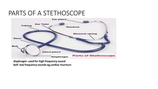
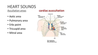
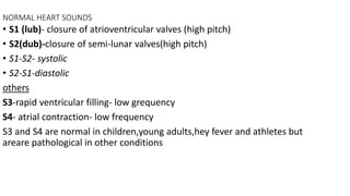
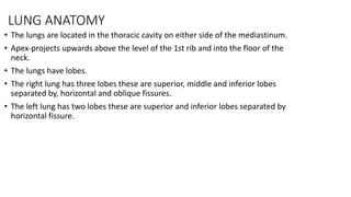
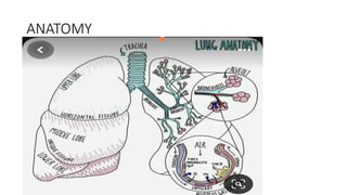
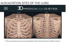
AUSCULTATION
PARTS OF A STETHOSCOPE
diaphragm- used for high frequency sound
bell- low frequency sounds eg,cardiac murmurs
HEART SOUNDS
Ascultation areas
• Aotic area
• Pulmonary area
• Erbs point
• Tricuspid area
• Mitral area
NORMAL HEART SOUNDS
• S1 (lub)- closure of atrioventricular valves (high pitch)
• S2(dub)-closure of semi-lunar valves(high pitch)
• S1-S2- systolic
• S2-S1-diastolic
others
S3-rapid ventricular filling- low grequency
S4- atrial contraction- low frequency
S3 and S4 are normal in children,young adults,hey fever and athletes but
areare pathological in other conditions
CARDIAC MURMURS/CARDIAC BRUIT
• Murmurs are unsual or abnormal heart sounds
• Most common presentation of CHD
• CAUSES
1. Valvular disease
2. septal defect
3. vascular defects
valvular diseases
1. stenosis- narrowing
2. incompetence/regurgitation/insuficiency -weakening of heart valvesleading to backflow
CLASSIFICATION OF MURMURS
I. systolic eg a.v valve regurgitation,semilunar valves stenosis,murmur due to anaemia,septal
defect,coactation of aorta
II. diastolic eg stenosis of a.v valves,regurgitation of semilunar valves
III. continous eg potent ductus arteriosus
Assesment of murmurs
AORTIC
STENOSIS
MITRAL
STENOSIS
PULMONARY
STENOSIS
MITRAL
REGURGITATION
PULMONARY
REGURGITATION
AORTIC
REGURGITATION
SITE aortic area apex/mitral
area
pulmonary
area
apex/mitral pulmonary
area
2th-4th left i.c.s
CHARACTER crescendo/dec
resendo
rumbling harsh blowing decresendo blowing
RADIATION carotid artery none left shoulder axilla right sternal
edge
none
INTENSITY variable variabe variable variable variable variable
PITCH high low high high high
TIMING systolic mid diastole pancytolic/thr
ougt systole
early diastole early diastoles
ABDOMEN
• Ausclulation of the abdomen is generally to asses bowel sounds or bruits
• -we use the diaphragm of the stethoscope
• 1.BOWEL SOUNDS
• -listen for bowel sounds in the abdomen to the right of the umbilicus, this is where the mid portion of the small bowel is
located
• -proceed to listen to all four quadrants
• Normal sound
• Low pitch, gurgling sound that occurs every 5-10seconds with peristalsis or bowel movements
• Frequency varies from person to person, therefore listen for 2 minutes before concluding that the bowel sounds are absent.
• Absence of sound
• For more than 2 minutes indicate no peristalsis which implies an alias
• Alias- intolerance of oral intake due to inhibition of GI propulsion with no sign of mechanical obstruction. Caused by surgery
or some medication.
• High pitch sound
• Can be as associated with a mechanical obstruction eg. Small bowel obstruction which increases volume and the frequency
of bowel sounds
BRUITS
• Is an abnormal swishing or blowing sound from blood flowing through
a narrow or partially occluded artery (turbulent blood flow)
• PLACEMENT OF STETHOSCOPE
• AORTA- about ⅔ down from the xiphoid process between umbilicus
• BILATERAL RENAL ARTERY - about 3cm superior and lateral to
umbilicus (along midclavicular line)
• BILATERAL ILIAC ARTERY- about 3cm inferior and lateral to umbilicus
• HEPATIC ARTERY -right costal margin, approximately mid clavicular
line
• SPLENIC ARTERY - left costal margin, approximately 2cm inferior and
lateral moving posteriorly to the mid clavicular line
AUSCALTATION OF THE LUNG
• When listening to the sounds of the lungs four features to look out for:
I. Pitch -High or Low
II. Amplitude -loud or soft
III. Quality-is there swishling or gargling
NB:a normal breath sound should not have any additional sounds.
The patient is asked to take deep breaths in and out and as air flows in and out check for abnormal breath sounds.
Listen to the full cycle of breathing.
Breath sounds
Classified into normal and and abnormal
Normal
1.Bronchial -Heard anteriorly over the tracheal area ,high pitched and loud, inspiration is longer than expiration.
NB:IT IS ABNORMAL IF HEARD OVER THE PERIPHERAL LUNG AREA. It could mean lung consolidation and pneuomonia
2.Bronchovesicular- Heard anteriorly and posteriorly, medium pitched and Inspiration and expiration is equal in
duration.
3.Vesicular- Heard Both anteriorly and posteriorly throughout the peripheral lung,it is low pitched and soft. Inspiration
is greater than expiration.
Abnormal
Divided into discontinuous and continuous
Continous
Extra sound lasting more than 0.2 seconds in a full respiration.
1.High pitched polyphonic wheeze-Mainly heard in expiration but also in inspiration
High pitch with several sound qualities hence polyphonic.
2.Low pitched monophonic wheeze- Mainly heard in expiration but also in inspiration.
Low pitched whistle with one sound quality hence monophonic.
3.Stridor -Heard in ispiration.Cused by obstruction of airways maybe by inflammation or foreign object. High
pitch whistling or gasping sound
Discontinous
Extra sound lasting less than 0.2 seconds in afull respiration.
1)Coarse crackles-mainly heard on inspiration and can extend into expiration. It is a low pitched bubbling sound
or shovelling of rocks.
2)Fine crackles-Mainly heard on inspiration .It is a high pitched fire crackling sound.
3)Pleural friction rub-Heard in both expiration and inspiration.
Low pitched harsh grating sound. Caused by rubbing or friction between the two layers of pleura.
Question :How to differentiate Between a pleural friction rub and a pericardial friction rub?
LUNG ANATOMY
• The lungs are located in the thoracic cavity on either side of the mediastinum.
• Apex-projects upwards above the level of the 1st rib and into the floor of the
neck.
• The lungs have lobes.
• The right lung has three lobes these are superior, middle and inferior lobes
separated by, horizontal and oblique fissures.
• The left lung has two lobes these are superior and inferior lobes separated by
horizontal fissure.
ANATOMY
AUSCALTATION SITES OF THE LUNG
• These sites are found both on the chest wall and the back, anteriorly and posteriorly respectively.
• While listening compare both left and right lung.
• Keep moving an intercostal space downwards.
Anteriorly
• Apex- slightly above the clavicle to listen to the apex of the lungs.
• Superior lobes-2nd and 3rd intercostal space mid clavicular.
• Middle lobe (right lung) -4th and 5th intercostal space mid clavicular.
• Inferior lobes-6th and 7th intercostal space mid axillary.
Posteriorly
• Apex -Above the scapula.
• Superior lobes- C7 to T3 vertebrae.
• Inferior Lobes- T3 to T10 vertebrae.

AUSCULTATION POINTS FOR PHARMACY STUDENTS.pptx

  • 1.
  • 2.
    PARTS OF ASTETHOSCOPE diaphragm- used for high frequency sound bell- low frequency sounds eg,cardiac murmurs
  • 3.
    HEART SOUNDS Ascultation areas •Aotic area • Pulmonary area • Erbs point • Tricuspid area • Mitral area
  • 4.
    NORMAL HEART SOUNDS •S1 (lub)- closure of atrioventricular valves (high pitch) • S2(dub)-closure of semi-lunar valves(high pitch) • S1-S2- systolic • S2-S1-diastolic others S3-rapid ventricular filling- low grequency S4- atrial contraction- low frequency S3 and S4 are normal in children,young adults,hey fever and athletes but areare pathological in other conditions
  • 5.
    CARDIAC MURMURS/CARDIAC BRUIT •Murmurs are unsual or abnormal heart sounds • Most common presentation of CHD • CAUSES 1. Valvular disease 2. septal defect 3. vascular defects valvular diseases 1. stenosis- narrowing 2. incompetence/regurgitation/insuficiency -weakening of heart valvesleading to backflow CLASSIFICATION OF MURMURS I. systolic eg a.v valve regurgitation,semilunar valves stenosis,murmur due to anaemia,septal defect,coactation of aorta II. diastolic eg stenosis of a.v valves,regurgitation of semilunar valves III. continous eg potent ductus arteriosus
  • 6.
    Assesment of murmurs AORTIC STENOSIS MITRAL STENOSIS PULMONARY STENOSIS MITRAL REGURGITATION PULMONARY REGURGITATION AORTIC REGURGITATION SITEaortic area apex/mitral area pulmonary area apex/mitral pulmonary area 2th-4th left i.c.s CHARACTER crescendo/dec resendo rumbling harsh blowing decresendo blowing RADIATION carotid artery none left shoulder axilla right sternal edge none INTENSITY variable variabe variable variable variable variable PITCH high low high high high TIMING systolic mid diastole pancytolic/thr ougt systole early diastole early diastoles
  • 7.
    ABDOMEN • Ausclulation ofthe abdomen is generally to asses bowel sounds or bruits • -we use the diaphragm of the stethoscope • 1.BOWEL SOUNDS • -listen for bowel sounds in the abdomen to the right of the umbilicus, this is where the mid portion of the small bowel is located • -proceed to listen to all four quadrants • Normal sound • Low pitch, gurgling sound that occurs every 5-10seconds with peristalsis or bowel movements • Frequency varies from person to person, therefore listen for 2 minutes before concluding that the bowel sounds are absent. • Absence of sound • For more than 2 minutes indicate no peristalsis which implies an alias • Alias- intolerance of oral intake due to inhibition of GI propulsion with no sign of mechanical obstruction. Caused by surgery or some medication. • High pitch sound • Can be as associated with a mechanical obstruction eg. Small bowel obstruction which increases volume and the frequency of bowel sounds
  • 8.
    BRUITS • Is anabnormal swishing or blowing sound from blood flowing through a narrow or partially occluded artery (turbulent blood flow) • PLACEMENT OF STETHOSCOPE • AORTA- about ⅔ down from the xiphoid process between umbilicus • BILATERAL RENAL ARTERY - about 3cm superior and lateral to umbilicus (along midclavicular line) • BILATERAL ILIAC ARTERY- about 3cm inferior and lateral to umbilicus • HEPATIC ARTERY -right costal margin, approximately mid clavicular line • SPLENIC ARTERY - left costal margin, approximately 2cm inferior and lateral moving posteriorly to the mid clavicular line
  • 9.
    AUSCALTATION OF THELUNG • When listening to the sounds of the lungs four features to look out for: I. Pitch -High or Low II. Amplitude -loud or soft III. Quality-is there swishling or gargling NB:a normal breath sound should not have any additional sounds. The patient is asked to take deep breaths in and out and as air flows in and out check for abnormal breath sounds. Listen to the full cycle of breathing. Breath sounds Classified into normal and and abnormal Normal 1.Bronchial -Heard anteriorly over the tracheal area ,high pitched and loud, inspiration is longer than expiration. NB:IT IS ABNORMAL IF HEARD OVER THE PERIPHERAL LUNG AREA. It could mean lung consolidation and pneuomonia 2.Bronchovesicular- Heard anteriorly and posteriorly, medium pitched and Inspiration and expiration is equal in duration. 3.Vesicular- Heard Both anteriorly and posteriorly throughout the peripheral lung,it is low pitched and soft. Inspiration is greater than expiration.
  • 10.
    Abnormal Divided into discontinuousand continuous Continous Extra sound lasting more than 0.2 seconds in a full respiration. 1.High pitched polyphonic wheeze-Mainly heard in expiration but also in inspiration High pitch with several sound qualities hence polyphonic. 2.Low pitched monophonic wheeze- Mainly heard in expiration but also in inspiration. Low pitched whistle with one sound quality hence monophonic. 3.Stridor -Heard in ispiration.Cused by obstruction of airways maybe by inflammation or foreign object. High pitch whistling or gasping sound Discontinous Extra sound lasting less than 0.2 seconds in afull respiration. 1)Coarse crackles-mainly heard on inspiration and can extend into expiration. It is a low pitched bubbling sound or shovelling of rocks. 2)Fine crackles-Mainly heard on inspiration .It is a high pitched fire crackling sound. 3)Pleural friction rub-Heard in both expiration and inspiration. Low pitched harsh grating sound. Caused by rubbing or friction between the two layers of pleura. Question :How to differentiate Between a pleural friction rub and a pericardial friction rub?
  • 11.
    LUNG ANATOMY • Thelungs are located in the thoracic cavity on either side of the mediastinum. • Apex-projects upwards above the level of the 1st rib and into the floor of the neck. • The lungs have lobes. • The right lung has three lobes these are superior, middle and inferior lobes separated by, horizontal and oblique fissures. • The left lung has two lobes these are superior and inferior lobes separated by horizontal fissure.
  • 12.
  • 13.
  • 14.
    • These sitesare found both on the chest wall and the back, anteriorly and posteriorly respectively. • While listening compare both left and right lung. • Keep moving an intercostal space downwards. Anteriorly • Apex- slightly above the clavicle to listen to the apex of the lungs. • Superior lobes-2nd and 3rd intercostal space mid clavicular. • Middle lobe (right lung) -4th and 5th intercostal space mid clavicular. • Inferior lobes-6th and 7th intercostal space mid axillary. Posteriorly • Apex -Above the scapula. • Superior lobes- C7 to T3 vertebrae. • Inferior Lobes- T3 to T10 vertebrae.