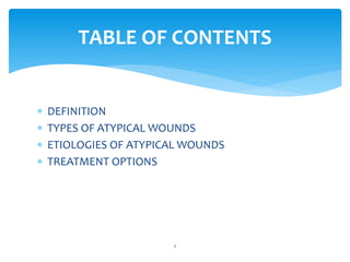
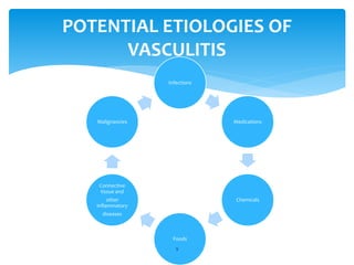
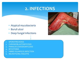
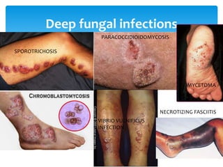
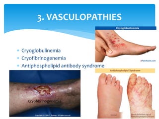
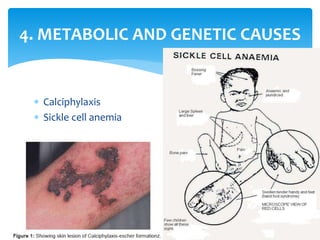
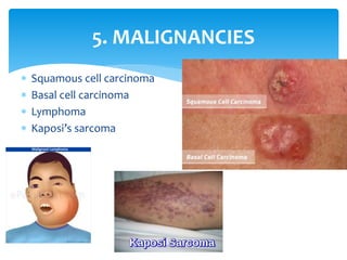
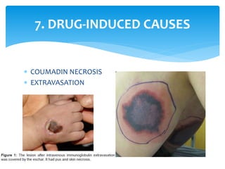
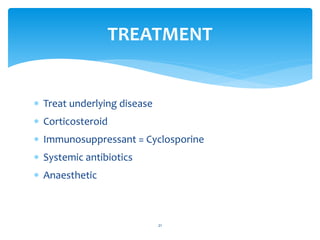
ATYPICAL WOUNDS
QURATULAIN MUGHAL
DOCTOR OF PHYSICAL THERAPY
IIRS
BATCH IV
1
 DEFINITION
 TYPES OF ATYPICAL WOUNDS
 ETIOLOGIES OF ATYPICAL WOUNDS
 TREATMENT OPTIONS
TABLE OF CONTENTS
2
 “Wounds resulting from uncommon etiologies called
atypical wounds.”
OR
 “Any wound that is not healing after 3 to 6 months of
appropriate treatment should raise the consideration
of an atypical wounds.”
3
ATYPICAL WOUNDS
 Pressure ulcers due to prolonged pressure.
 Venous leg ulcers due to venous insufficiency.
 Diabetic foot ulcers due to complications of
longstanding diabetes mellitus.
 Arterial ulcers due to poor vascular supply.
4
TYPES OF ATYPICAL WOUNDS
 They are less frequently encountered and less well
understood.
 Their prevalence has not been studied extensively.
 But it is estimated that at least 10% of the more than
500,000 leg ulcers in the United States may be due to
unusual causes.
5
EPIDEMIOLOGY
 It is present in a location different from that of a
common chronic wound.
 Its appearance varies from that of a common chronic
wound.
 It does not respond to conventional therapy.
6
A WOUND SHOULD BE EVALUATED
FOR AN ATYPICAL ETIOLOGY IF:
1. Inflammatory causes
2. Infections
3. Vasculopathies
4. Metabolic and genetic causes
5. Malignancies
6. External causes
7. Drug-induced causes
7
ETIOLOGIES OF ATYPICAL WOUNDS
Although not all-inclusive, this list presents some of the most commonly
encountered etiologies for an atypical wound.
Tissue samples are
mandatory for
atypical wounds
 Vasculitis
 Pyoderma gangrenosum (diagnostic test to confirm &
Curative treatment does not exist).
8
1. INFLAMMATORY CAUSES
Infections
Medications
Chemicals
Foods
Connective
tissue and
other
inflammatory
diseases
Malignancies
9
POTENTIAL ETIOLOGIES OF
VASCULITIS
DIAGNOSTIC TESTS FOR
VASCULITIS
To
determine
the etiology
of vasculitis.
To
determine
the extent
of disease.
10
•Leg elevation
•Compression dressingsMild
•Systemic steroids
•Plasmapheresis
Extensive
or systemic
11
VASCULITIS TREATMENT OPTIONS
 Atypical mycobacteria
 Buruli ulcer
 Deep fungal infections
12
2. INFECTIONS
1. SPOROTRICHOSIS
2. CHROMOBLASTOMYCOSIS
3. PARACOCCIDIOIDOMYCOSIS
4. MYCETOMA
5. VIBRIO VULNIFICUS INFECTION
6. NECROTIZING FASCIITIS
13
Deep fungal infections
SPOROTRICHOSIS
PARACOCCIDIOIDOMYCOSIS
MYCETOMA
VIBRIO VULNIFICUS
INFECTION
NECROTIZING FASCIITIS
 Age 50 and older
 Alcoholism
 Malignancy
 Malnutrition
 Obesity
 Renal failure
 Smoking
 Diabetes mellitus
 HypertensionSurgery
14
PATIENTS AT RISK FOR
NECROTIZING FASCIITIS
 Cryoglobulinemia
 Cryofibrinogenemia
 Antiphospholipid antibody syndrome
15
3. VASCULOPATHIES
Cryofibrinogenemia
 Calciphylaxis
 Sickle cell anemia
16
4. METABOLIC AND GENETIC CAUSES
CALCIPHYLAXIS TREATMENT
MEDICAL TREATMENT
 Decreased calcium in dialysate
 Antibiotics
 Low phosphate diet
 Bisphosphonates
 Sodium thiosulfate
 Avoidance of challenging agents
 Avoidance of systemic steroids
 Anticoagulation
SURGICAL TREATMENT
 Parathyroidectomy
 Wound care and debridement
 Amputation
 Renal transplantation
 Skin grafting using either
autologous or tissue
engineered skin
17
 Squamous cell carcinoma
 Basal cell carcinoma
 Lymphoma
 Kaposi’s sarcoma
18
5. MALIGNANCIES
 Burns
 Bites
 Stings
 Radiation
 Factitial dermatitis
19
6. EXTERNAL CAUSES
 COUMADIN NECROSIS
 EXTRAVASATION
20
7. DRUG-INDUCED CAUSES
 Treat underlying disease
 Corticosteroid
 Immunosuppressant = Cyclosporine
 Systemic antibiotics
 Anaesthetic
21
TREATMENT
 Control pain
 Necrotic tissue
 surgical debridement is contraindicated as it may
result in even worse ulceration
 Avoidance of trauma at dressing removal
 disturbance can generate an even greater
inflammatory response and stimulate deterioration.
22
WOUND MANAGEMENT
 Negative pressure therapy
 may be used to assist debridement when the
disease is stable
 Debridement and skin grafting
 can be considered when condition is under
controlled
 surgery may reactive the disease
23
WOUND MANAGEMENT
24
REFERENCES
25

Atypical wounds

  • 1.
    ATYPICAL WOUNDS QURATULAIN MUGHAL DOCTOROF PHYSICAL THERAPY IIRS BATCH IV 1
  • 2.
     DEFINITION  TYPESOF ATYPICAL WOUNDS  ETIOLOGIES OF ATYPICAL WOUNDS  TREATMENT OPTIONS TABLE OF CONTENTS 2
  • 3.
     “Wounds resultingfrom uncommon etiologies called atypical wounds.” OR  “Any wound that is not healing after 3 to 6 months of appropriate treatment should raise the consideration of an atypical wounds.” 3 ATYPICAL WOUNDS
  • 4.
     Pressure ulcersdue to prolonged pressure.  Venous leg ulcers due to venous insufficiency.  Diabetic foot ulcers due to complications of longstanding diabetes mellitus.  Arterial ulcers due to poor vascular supply. 4 TYPES OF ATYPICAL WOUNDS
  • 5.
     They areless frequently encountered and less well understood.  Their prevalence has not been studied extensively.  But it is estimated that at least 10% of the more than 500,000 leg ulcers in the United States may be due to unusual causes. 5 EPIDEMIOLOGY
  • 6.
     It ispresent in a location different from that of a common chronic wound.  Its appearance varies from that of a common chronic wound.  It does not respond to conventional therapy. 6 A WOUND SHOULD BE EVALUATED FOR AN ATYPICAL ETIOLOGY IF:
  • 7.
    1. Inflammatory causes 2.Infections 3. Vasculopathies 4. Metabolic and genetic causes 5. Malignancies 6. External causes 7. Drug-induced causes 7 ETIOLOGIES OF ATYPICAL WOUNDS Although not all-inclusive, this list presents some of the most commonly encountered etiologies for an atypical wound. Tissue samples are mandatory for atypical wounds
  • 8.
     Vasculitis  Pyodermagangrenosum (diagnostic test to confirm & Curative treatment does not exist). 8 1. INFLAMMATORY CAUSES
  • 9.
  • 10.
    DIAGNOSTIC TESTS FOR VASCULITIS To determine theetiology of vasculitis. To determine the extent of disease. 10
  • 11.
    •Leg elevation •Compression dressingsMild •Systemicsteroids •Plasmapheresis Extensive or systemic 11 VASCULITIS TREATMENT OPTIONS
  • 12.
     Atypical mycobacteria Buruli ulcer  Deep fungal infections 12 2. INFECTIONS 1. SPOROTRICHOSIS 2. CHROMOBLASTOMYCOSIS 3. PARACOCCIDIOIDOMYCOSIS 4. MYCETOMA 5. VIBRIO VULNIFICUS INFECTION 6. NECROTIZING FASCIITIS
  • 13.
  • 14.
     Age 50and older  Alcoholism  Malignancy  Malnutrition  Obesity  Renal failure  Smoking  Diabetes mellitus  HypertensionSurgery 14 PATIENTS AT RISK FOR NECROTIZING FASCIITIS
  • 15.
     Cryoglobulinemia  Cryofibrinogenemia Antiphospholipid antibody syndrome 15 3. VASCULOPATHIES Cryofibrinogenemia
  • 16.
     Calciphylaxis  Sicklecell anemia 16 4. METABOLIC AND GENETIC CAUSES
  • 17.
    CALCIPHYLAXIS TREATMENT MEDICAL TREATMENT Decreased calcium in dialysate  Antibiotics  Low phosphate diet  Bisphosphonates  Sodium thiosulfate  Avoidance of challenging agents  Avoidance of systemic steroids  Anticoagulation SURGICAL TREATMENT  Parathyroidectomy  Wound care and debridement  Amputation  Renal transplantation  Skin grafting using either autologous or tissue engineered skin 17
  • 18.
     Squamous cellcarcinoma  Basal cell carcinoma  Lymphoma  Kaposi’s sarcoma 18 5. MALIGNANCIES
  • 19.
     Burns  Bites Stings  Radiation  Factitial dermatitis 19 6. EXTERNAL CAUSES
  • 20.
     COUMADIN NECROSIS EXTRAVASATION 20 7. DRUG-INDUCED CAUSES
  • 21.
     Treat underlyingdisease  Corticosteroid  Immunosuppressant = Cyclosporine  Systemic antibiotics  Anaesthetic 21 TREATMENT
  • 22.
     Control pain Necrotic tissue  surgical debridement is contraindicated as it may result in even worse ulceration  Avoidance of trauma at dressing removal  disturbance can generate an even greater inflammatory response and stimulate deterioration. 22 WOUND MANAGEMENT
  • 23.
     Negative pressuretherapy  may be used to assist debridement when the disease is stable  Debridement and skin grafting  can be considered when condition is under controlled  surgery may reactive the disease 23 WOUND MANAGEMENT
  • 24.
  • 25.