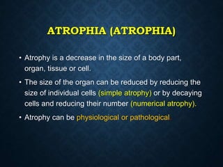
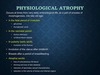
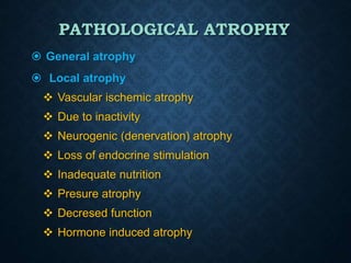
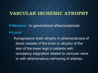
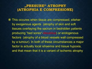
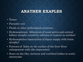
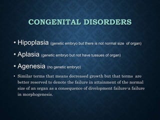
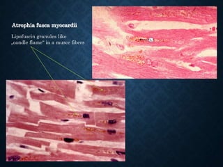
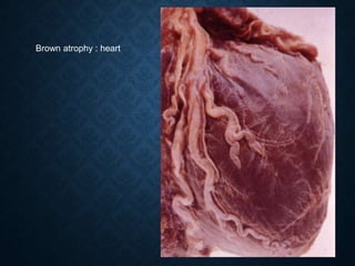
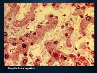
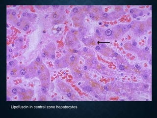
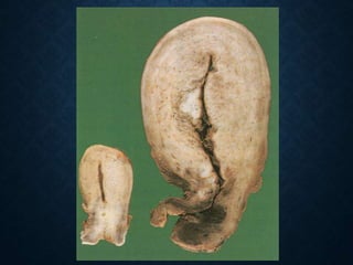
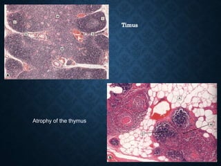
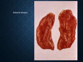
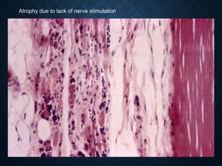
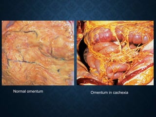
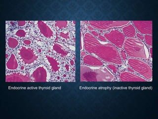
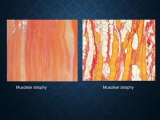
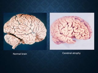
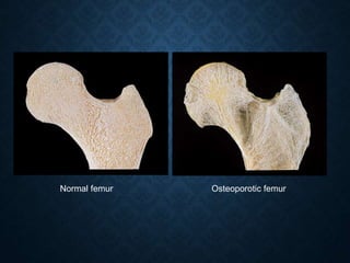
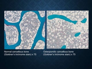
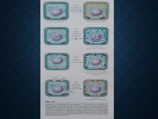
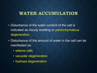
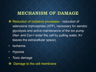
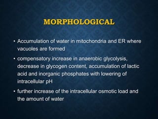
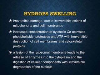
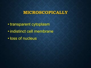
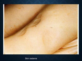
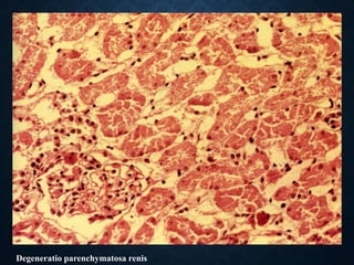
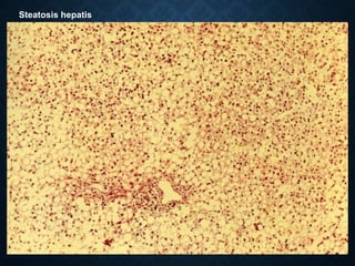
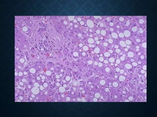
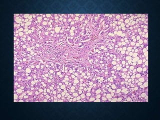
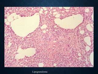
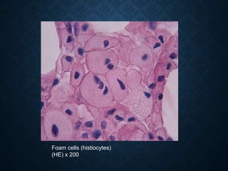
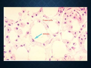
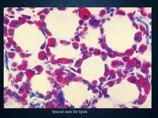
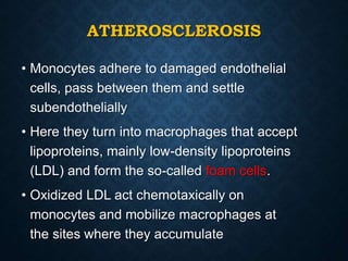
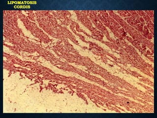
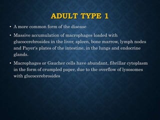
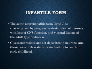
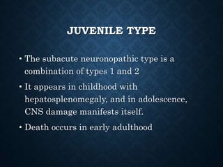
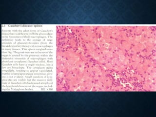
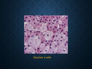
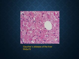
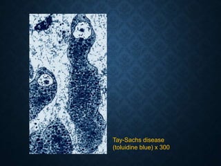
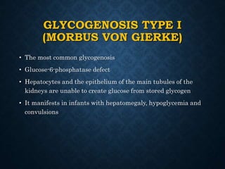
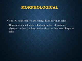
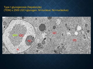
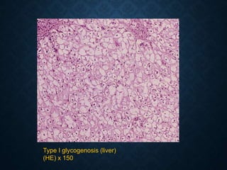
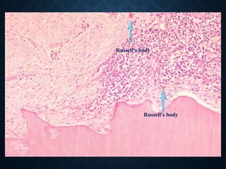
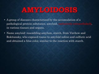
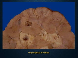
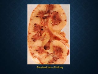
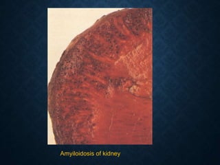
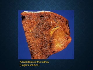
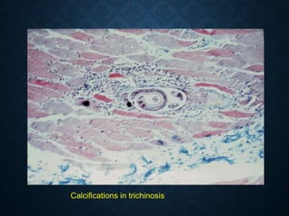
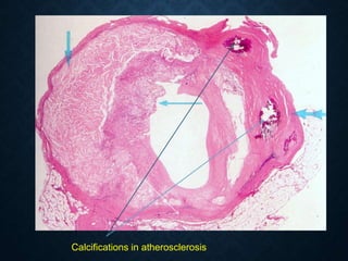
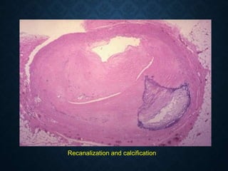
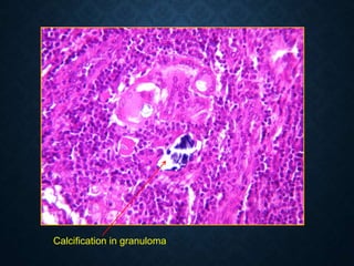
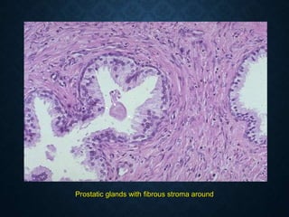
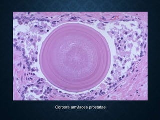
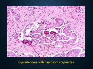
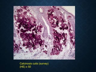
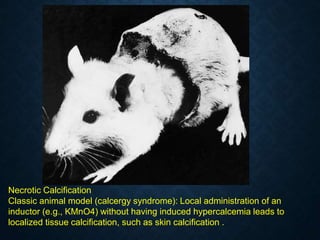
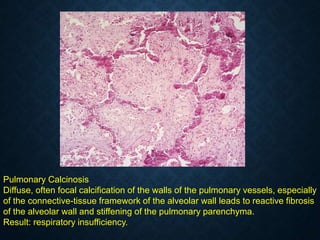
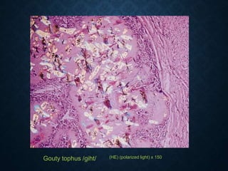
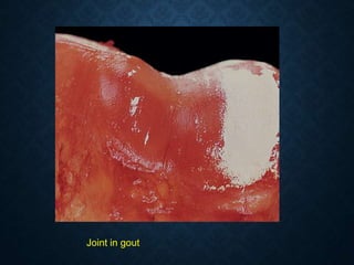
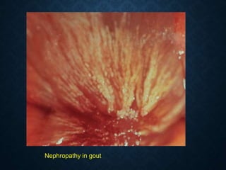
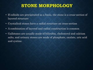
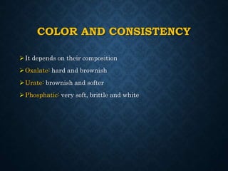
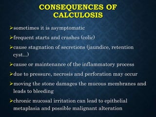
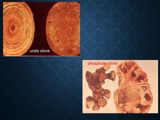
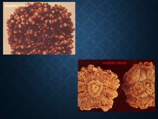
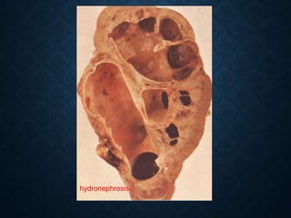
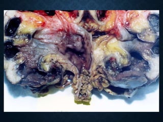
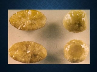
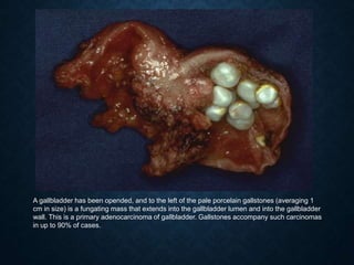
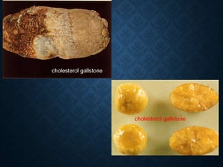
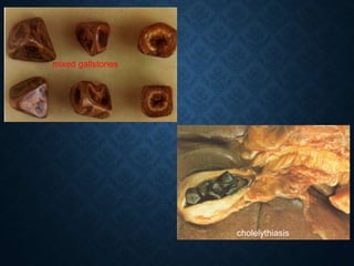
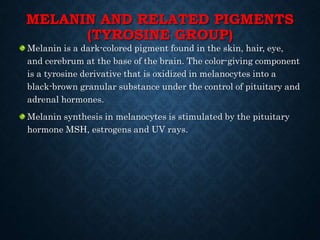
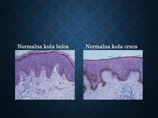
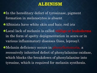
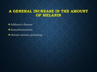
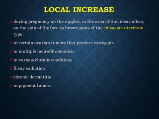
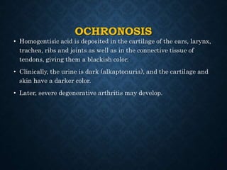
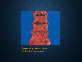
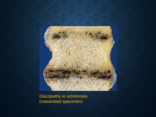
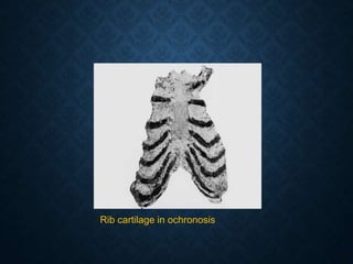
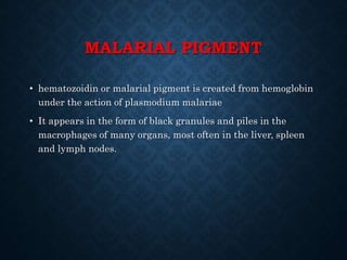
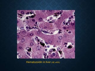
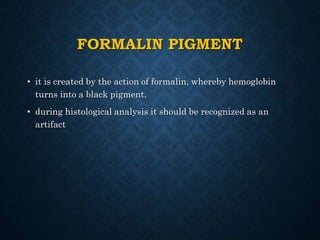
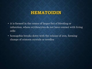
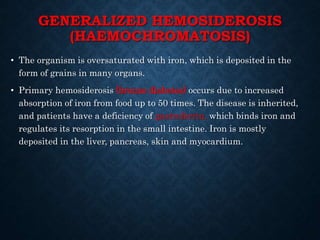
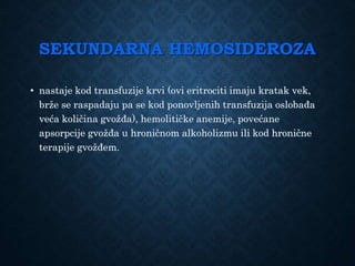
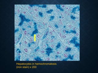
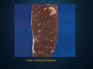
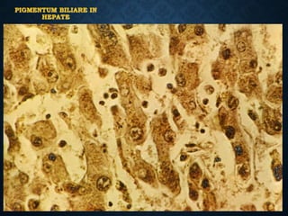
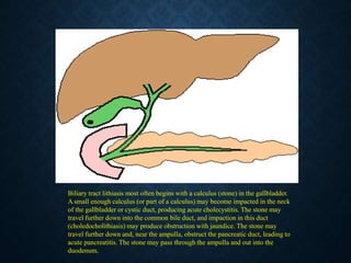
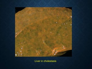
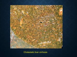
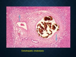
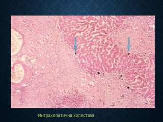
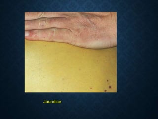
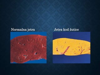
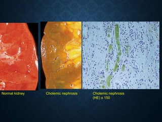
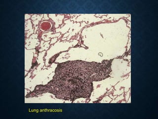
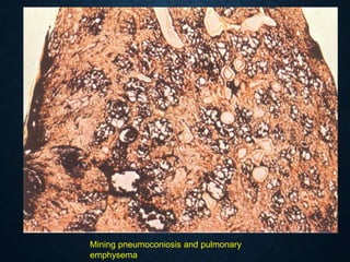
ATROPHY AND
DYSTROPHY
(accumulation)
Prof. dr Nebojša Mitić
FACULTY OF MEDICINE IN PRIŠTINA
Institute of pathology
ATROPHIA (ATROPHIA)
• Atrophy is a decrease in the size of a body part,
organ, tissue or cell.
• The size of the organ can be reduced by reducing the
size of individual cells (simple atrophy) or by decaying
cells and reducing their number (numerical atrophy).
• Atrophy can be physiological or pathological.
PHYSIOLOGICAL ATROPHY
Occurs at times from very early embryological life, as a part of process of
morphogenesis, into late old age:
• in the fetal period of involution
• gill arches
• thyroglossal canal
• In the neonatal period
• ductus arteriosus
• umbilical blood vessels
• In puberty (early adult)
• involution of the thymus
• Involution of the uterus after childbirth
• Breasts after a period of breastfeeding
• Atrophia senilis
• loss of subcutaneous fat tissue
• thinning and loss of skin elasticity
• reduction of secondary sexual characteristics
• reduction in the volume of bones and internal organs
PATHOLOGICAL ATROPHY
 General atrophy
 Local atrophy
 Vascular ischemic atrophy
 Due to inactivity
 Neurogenic (denervation) atrophy
 Loss of endocrine stimulation
 Inadequate nutrition
 Presure atrophy
 Decresed function
 Hormone induced atrophy
VASCULAR ISCHEMIC ATROPHY
General : in generalized atherosclerosis
Local
progressive brain atrophy in atherosclerosis of
blood vessels of the brain or atrophy of the
skin of the lower legs in patients with
circulatory stagnation related to varicose veins
or with atheromatous narrowing of arteries.
„PRESURE“ ATROPHY
(ATROPHIA E COMPRESIONE)
 This occures when tissue are compressed, eiteher
by exogenous agents (atrophy of skin and soft
tissues overlaying the sacrum in bedridden patients
producing “bed sores“-decubitus) or endogenous
factors (atrophy of a blood vessels wall compressed
by a tumour). In both of these circumstances a major
factor is actually local ishaemia and tissue hypoxia,
and that mean that it is a variant of ischemic atrophy
ANATHER EXAPLES
• Tumor
• Parasitic cyst
• Fluids or other pathological processes
• Hydronephrosis (dilatation of renal pelvis and cortical
kidney atrophy caused by calculus or tumour in urether)
• Hydrocephalus (opstruction of liquor supply with brain
atrophy)
• Furrows of Zahn on the surface of the liver (liver
enlargement with ribs impression)
• Scars on the ribs, sternum and vertebral bodies in aortic
aneurysms
ATROPHIA EX INACTIVITATE
• General
• Local
• Most often as a result of decreased function as, for
example, in a limb immobilised as a consequence of a
fracture, there may be marked muscle atrophy, due to
decrease in muscle fibre size. In extreme case bone
atrophy may lead to osteoporosis end bone weakening.
NEUROGENIC ATROPHY
• Loss of innervation of muscle causes muscle atrophy
as is seen in:
• Nerve transection
• Neuropathy
• Motor neuron diseases (poliomyelitis, amyotrophic
lateral sclerosis)
• In paraplegics, loss of innervation may also precipitate
atrophy of bone which become osteoporotic
LOSS OF ENDOCRINE STIMULATION
• Atrophy of the „target“ organ of a hormone
may occure if endocrine stimulation is
inadequate:
• Adrenal gland may become atrophic as a
consequence of decreased ACTH secretion
by the anterior pituitary (destruction by a
tumour or infarction-bleeding or infarction
in the pituitary gland - Sheehan's
syndrome
HORMONE –INDUCED ATROPHY
• This form of atrophy may be seen in the skin,as a
result of the growth –inhibiting actions of
corticosteroids. When corticosteroids are applied
topically in high concetration to the skin, they may
cause dermal and epidermal atophy.
ATROPHY DUE TO INADEQUATE
NUTRITION
• Inanition or marantic atrophy (an extreme form of
systemic atrophy similar to that seen in severe
starvation is teremed „cachexia“; this may be seen in
patients in the late stage of severe illnesses such as
cancer. Cytokines such as tumour necrosis factor
(TNF) are postulated to influence the development of
cachexia.
• insufficient intake of food
• diseases of the digestive tract
CONGENITAL DISORDERS
• Hipoplasia (genetic embryo but there is not normal size of organ)
• Aplasia (genetic embryo but not have tussues of organ)
• Agenesia (no genetic embryo)
• Similar terms that means decreased growth but that terms are
better reserved to denote the failure in attainment of the normal
size of an organ as a consequence of divelopment failure-a failure
in morphogenesis.
Atrophia fusca myocardii
Lipofuscin granules like
„candle flame“ in a musce fibers
Brown atrophy : heart
Atrophia fusca hepatitis
Lipofuscin in central zone hepatocytes
Timus
Atrophy of the thymus
Adrenal atrophy
Atrophy due to lack of nerve stimulation
Normal omentum Omentum in cachexia
Endocrine active thyroid gland Endocrine atrophy (inactive thyroid gland)
Musclear atrophy Musclear atrophy
Normal brain Cerebral atrophy
Normal femur Osteoporotic femur
Normal cancellous bone
(Goldner’s trichrome stain) x 75
Osteoporotic cancellous bone
(Goldner’s trichrome stain) x 75
DYSTROPHIA
• Dystrophies or degenerations are old terms
that include reversible or irreversible
morphological-functional damage of cellular
structures that are manifested by intracellular
or extracellular accumulation of various
substances in abnormal amounts
WATER ACCUMULATION
• Disturbance of the water content of the cell is
indicated as cloudy swelling or parenchymatous
degeneration.
• Disturbance of the amount of water in the cell can be
manifested as:
• edema cells
• vacuolar degeneration
• hydrops degeneration
MECHANISM OF DAMAGE
 Reduction of oxidative processes - reduction of
adenosine triphosphate (ATP), necessary for aerobic
glycolysis and active maintenance of the ion pump
(Na+ and Ca++ enter the cell by pulling water, K+
leaves the extracellular space)
• Ischemia
• Hypoxia
• Toxic damage
 Damage to the cell membrane
MORPHOLOGICAL
• Accumulation of water in mitochondria and ER where
vacuoles are formed
• compensatory increase in anaerobic glycolysis,
decrease in glycogen content, accumulation of lactic
acid and inorganic phosphates with lowering of
intracellular pH
• further increase of the intracellular osmotic load and
the amount of water
HYDROPS SWELLING
 irreversible damage, due to irreversible lesions of
mitochondria and cell membranes
 increased concentration of cytosolic Ca activates
phospholipids, proteases and ATP with irreversible
destruction of cell membranes and cytoskeletal
proteins
 a lesion of the lysosomal membrane leads to the
release of enzymes into the cytoplasm and the
digestion of cellular components with irreversible
degradation of the nucleus
MICROSCOPICALLY
• transparent cytoplasm
• indistinct cell membrane
• loss of nucleus
Skin oedema
Degeneratio parenchymatosa renis
ACCUMULATION OF LIPIDS
• neutral fat metabolism disorder (fat
change and lipomatosis)
• genetically determined deficiencies of
enzymes that break down fat
macromolecules (lipoidosis)
FAT CHANGE
fat metamorphosis or fat
transformation or steatosis
• Abnormal accumulation of neutral fats,
most often triglycerides, in the cytoplasm of
parenchymal cells that microscopically do
not contain fats (liver, heart muscle,
kidneys, skeletal muscles).
• Most often a reversible change
STEATOSIS HEPATIS
 Alcohol
 Marked hypoxia
 Protein malnutrition
 Hepatotoxins
 Obesity
 Diabetes mellitus
 Starvation
 Pregnancy
 Total parenteral nutrition
 Rey's syndrome
PATHOGENESIS
• Increased supply of free fatty acids‚ (obesity,
starvation, adrenaline, somatotropic and
adenocorticotropic hormone, corticosteroid therapy
• Increased synthesis of fatty acids in the liver with
increased esterification into triglycerides (hypoxia)
• Reduced oxidation of fatty acids with increased
esterification into triglycerides (hypoxia)
• Increased retention of triglycerides due to the lack of
apoprotein, necessary for their excretion from
hepatocytes into the circulation (hepatotoxins,
starvation, malabsorption, kwashiorkor).
MORPHOLOGICAL
• enlarged liver
• soft consistency
• yellow color
Steatosis hepatis
HISTOLOGICALLY
• macrovesicular-macrovesicular fatty change
• small droplet-microvesicular
• diffuse
• zonal
Steatosis hepatis
FATTY CHANGE OF THE
MYOCARDIUM
• Tiger heart
• chronic severe hypoxia
• anemia
• hypoxemia
• chronic protein malnutrition
• Diffuse form
• diphtheria myocarditis
Fatty change of the myocardium
Tiger-stripped pattern of fatty myocardial
deposits
FATTY CHANGE OF THE
KIDNEYS
• Fat deposition in renal tubular cells, with increased
reabsorption, and due to the enormous passage of
lipids through damaged glomeruli, usually in
glomerulonephritis or severe hypoxia
• Macroscopic: enlarged kidneys, soft consistency and
yellow color.
• Histological: fat droplets in the tubular epithelium
Steatosis of the renal tubules
(oil-red stain) x 150 (G=glomerulus)
RESORPTIVE FATTY CHANGE
DUE TO PHAGOCYTOSIS
• Deposition of lipids, cholesterol and
cholesterol esters in macrophages after
phagocytosis
• Fat-changed macrophages: lipophages,
pseudoxanthoma cells, xanthoma or foam
cells
NECROSIS AND ABSCESS
• Lipids are released from the membranes of
necrotic cells and are phagocytosed by
macrophages-lipophages
• In the focus of the abscess, fats are
released from necrotic neutrophils
• In encephalomalacia, lipids from necrotic
brain tissue, primarily myelin, are
phagocytosed by microglia-foam cells.
• Fat tissue necrosis - lipogranulomas
Encephalomalata alba - foam cells
Lipogranuloma
Foam cells (histiocytes)
(HE) x 200
Foam cells
mitosis
Special stain for lipids
ATHEROSCLEROSIS
• Monocytes adhere to damaged endothelial
cells, pass between them and settle
subendothelially
• Here they turn into macrophages that accept
lipoproteins, mainly low-density lipoproteins
(LDL) and form the so-called foam cells.
• Oxidized LDL act chemotaxically on
monocytes and mobilize macrophages at
the sites where they accumulate
Chylomicron-micelles
(TEM) x 300 000
VLDL
(TEM) x 300 000
LDL
(TEM) x 300 000
HDL
(TEM) x 300 000
Foam cells
Cholesterol crystals
Foam cells
CHOLESTEROLOSIS OF THE
GALLBLADDER
• Accumulation of macrophages that have
phagocytosed cholesterol, within the
mucosa of the gallbladder.
• Change of unknown origin
Cholesterolosis vesicae feleae
Foam cells
XANTHOMA AND XANTHELASMA
• Xanthomas are clusters of macrophages that have
phagocytosed cholesterol esters.
• They are found in the skin and tendons in
hyperlipoproteinemia
• Localized usually on the extensor sides of the
extremities, elbows, back, fingers, palms and
shoulders
• In the area of the corner of the eye-xanthelasma
Eruptive xanthomas
Tuberous xanthomas
xanthelasma
Xanthoma
(HE) x 200
Touton giant cell
(EvG) x 200
LIPOMATOSIS
• Accumulation of already existing fat tissue, or fat
transformation of connective tissue cells in the
interstitium of organs.
• It can be:
• generalized (overweight or obesity)
• localized
OBESITY
• excessive deposition of neutral fats
and quantitative increase of adipose
tissue in existing fat depots, with an
increase in body weight.
LOCALIZED FATTY
INFILTRATION
• Converting undifferentiated interstitium
mesenchyme cells into fat cells
• After physiological atrophy of the parenchyma
• thymus
• bone marrow of tubular bones
• Lipomatosis of salivary glands, lymph nodes and
pancreas
• Myocardial lipomatosis is a common finding in
general obesity
LIPOMATOSIS
CORDIS
LIPIDOSES
(DYSLIPIDOSES, SPHINGOLIPIDOSES)
• Congenital metabolic diseases due to
enzymatic defects in the breakdown of fat
macromolecules that are progressively
deposited in cells, primarily by lysosomes,
damage the cell and eventually lead to its
death.
• They are inherited autosomally recessively
GAUCHER DISEASE (MORBUS
GAUCHER)
• Glucosylceramide lipidosis
• Mutation of the gene that regulates the synthesis of
glucocerebrosidase enzyme
• Reduced enzyme values ​​lead to type 1, the adult form
• Complete enzyme deficiency - type 2, infantile acute
cerebral form
ADULT TYPE 1
• A more common form of the disease
• Massive accumulation of macrophages loaded with
glucocerebrosides in the liver, spleen, bone marrow, lymph nodes
and Payer's plates of the intestine, in the lungs and endocrine
glands.
• Macrophages or Gaucher cells have abundant, fibrillar cytoplasm
in the form of crumpled paper, due to the overflow of lysosomes
with glucocerebrosides
INFANTILE FORM
• The acute neuronopathic form (type 2) is
characterized by progressive destruction of neurons
with loss of CNS function, and visceral lesions of
the adult type of disease.
• Glycocerebrosides are not deposited in neurons, and
these nevertheless deteriorate leading to death in
early childhood.
JUVENILE TYPE
• The subacute neuronopathic type is a
combination of types 1 and 2
• It appears in childhood with
hepatosplenomegaly, and in adolescence,
CNS damage manifests itself.
• Death occurs in early adulthood
Gaucher`s cells
Gaucher`s disease of the liver
PASx75
NIEMANN-PICK DISEASE
(MORBUS NIEMANN-PICK) SPHINGOMYELIN
• Accumulation of sphingomyelin in the mononuclear-phagocytic
system and parenchymal cells of many organs due to the
deficiency of the enzyme sphingomyelinase.
• Type A-neuronopathic form is more common, with deposition of
sphingomyelin in neurons and glial cells of the brain and visceral
organs. Death in early childhood.
• Type B-visceral lesion, without brain accumulation. Live longer.
TAY-SACHS DISEASE (MORBUS
TAY-SACHS)
• GM2-gangliosidosis type I
• Accumulation of GM2-ganglioside
predominantly in the CNS and
peripheral nervous system
• (familial amaurotic idiocy)
Tay-Sachs disease
(freeze-etching, TEM) x 40 000
Tay-Sachs disease
(toluidine blue) x 300
ACCUMULATION OF
CARBOHYDRATES
• The most important sources of energy in the body
• If they are not taken in as monosaccharides, they must
be broken down in the GIT by enterocyte enzymes
• Enzymatic cleavage and resorption of the resulting
monosaccharides take place simultaneously
• Glucose is a circulating sugar
• Energy is obtained by breaking down glucose
• By incorporating glucose, macromolecules containing
carbohydrates are created, so the glucose is deposited
as glycogen
DISORDERS
•glycogenosis
•glucose malabsorption
•disorders of glyconeogenesis,
regulation and use of
carbohydrates
GLYCOGENOSIS
• Genetic diseases with abnormal deposition of glycogen in tissues
due to deficiency of enzymes involved in its metabolism
• The most pronounced deposition in the heart, skeletal muscles,
liver and kidneys
• Only in type II lysosomal deposition, in all others the deposition is
diffuse in the cytoplasm
GLYCOGENOSIS TYPE I
(MORBUS VON GIERKE)
• The most common glycogenosis
• Glucose-6-phosphatase defect
• Hepatocytes and the epithelium of the main tubules of the
kidneys are unable to create glucose from stored glycogen
• It manifests in infants with hepatomegaly, hypoglycemia and
convulsions
MORPHOLOGICAL
• The liver and kidneys are enlarged and brown in color
• Hepatocytes and kidney tubule epithelial cells contain
glycogen in the cytoplasm and nucleus, so they look like plant
cells
Type I glycogenosis (hepatocyte)
(TEM) x 2500 (GC=glycogen; N=nucleus; Nc=nucleolus)
GC
GC
GC
N
NC
Type I glycogenosis (liver)
(HE) x 150
Type I glycogenosis (liver)
x 150
GLYCOGENOSIS TYPE II
(MORBUS POMPE)
• Deficiency of the lysosomal enzyme acid alpha-(1,4)-
glucosidase (acid maltase)
• Infantile form: deposition in lysosomes of hepatocytes, heart,
skeletal muscles, CNS, lymphocytes and other cells
• In children and adults: only skeletal muscles are affected
Type II glycogenosis (hepatocyte)
(TEM) x 5000 (M=mitochondrion; GC: lysosomal glycogen)
M
GC
GLYCOGENOSIS TYPE III
(MORBUS CORI)
• Amylo-1,6-glucosidase deficiency in hepatocytes, enterocytes,
cardiac muscle and skeletal muscle
• Morphologically, cardiohepatomegaly
• Clinically, muscle weakness, heart failure, liver fibrosis and
susceptibility to infections
• Many sufferers live to adulthood
GLYCOGENOSIS TYPE IV
(MORBUS ANDERSON)
• A rare guy
• Branching enzyme defect with generalized accumulation of
glycogen, mostly in the liver, heart, skeletal muscles and brain
• Cirrhosis and portal hypertension at an early age
GLYCOGENOSIS TYPE V
(MORBUS MCARDLE)
• Muscle phosphorylase defect
• Weakness and pain in the muscles
• Rhabdomyolysis with myoglobinuria and subsequent acute
renal failure can occur during heavy exertion.
HYALINE DEGENERATION
• Deposition of hyaline in cells and extracellular space
• Hyaline is a homogeneous, structureless substance that stains
with acid dyes (eosin).
• It consists mainly of proteins but can also contain fats and
carbohydrates
• Conventional classification
• intracellular hyaline
• extracellular hyaline
INTRACELLULAR HYALINE
• "Epithelial hyaline"
• Mallory's hyaline, coral-like appearance in hepatocytes in
alcoholic liver disease and Wilson's disease
KAUNSILMAN’S BODY
• Round, red, hyaline globules in the liver
• Apoptotic hepatocytes in acute viral
hepatitis
• Pronounced eosinophilia and
condensation of fragmented cytoplasm
creates the impression of hyaline
changes
HYALINE DROPS
• in the epithelial cells of the proximal kidney tubules, proteins
are deposited in a state of increased reabsorption, mainly in
nephrotic syndrome.
RUSSELL'S BODY
•are spherical deposits of
immunoglobulins, in the rough
endoplasmic reticulum of plasma
cells.
Russell's body
Russell's body
VIRAL INCLUSIONS
•nuclear and/or
intracytoplasmic inclusions
ZENKER'S WAXY DEGENERATION
• Limited necrosis of the striated muscle sarcoplasm
• The hyaline appearance originates from the
condensation of disintegrated contractile fibrils of
skeletal muscle
• Most often in the diaphragm and m. rectus abdominis
under the influence of bacterial toxins and the
consequent accumulation of lactic acid in the cell.
EXTRACELLULAR HYALINE
A structureless, eosinophilic substance that is never degraded by
immunopathological processes.
in preformed spaces (hyaline thrombi-hematogenous hyaline,
pulmonary hyaline membranes, hyaline renal cylinders)
in blood vessels (vascular hyaline)
in connective tissue (connective tissue hyaline)
in glomeruli
HYALINE THROMBI
• they are caused by the drying of blood clots in blood vessels
when their disintegration or connective tissue organization is
absent.
• parietal hyaline macrothrombi in larger blood vessels
• hyaline microthrombi in capillaries, arterioles and venules in
shock or DIC syndrome (Disseminated Intravasscular
Coagulation).
Pulmonary shock. DIK, microthrombi in a dilated small blood vessel
PULMONARY HYALINE
MEMBRANES
• homogeneous, eosinophilic, ribbon-like deposits on the walls of
respiratory bronchioles, alveolar ducts and alveoli, consisting of
fibrin and damaged alveolar epithelium
• They occur as part of hypoxic damage to the capillaries of the
lungs, due to the lack of surfactant in RDS of newborns, in adults
in shock, ARDS, toxic damage.
Hyaline membranes in RDS adults. Microthrombus (DIC)
Lung shock, hyaline membrane
Hyaline membranes and blood aspiration
Aspiration of amniotic fluid and meconium, hyaline membrane, partial fetal
atelectasis
Hyaline membranes
Aspiration of amniotic fluid, meconium and blood. Hyaline membrane
HYALINE CYLINDERS
• They are formed in the loop of Henle and the distal tubules of
the kidney
• They consist of precipitated proteins of primary urine, and can
be proven in urinary sediment.
VASCULAR HYALINE
• it is located in the wall of small arteries
• thickening of the wall and narrowing of the blood vessel
lumen consists of precipitated plasma proteins that infiltrated
the wall under elevated hydrostatic pressure, in hypertension,
as well as elements of the endothelial basement membrane
(collagen type IV)
VASCULAR HYALINE
• Physiological hyalinosis in old age, primarily in the arteries of
the white pulp of the spleen
• in systemic hypertension
• diabetes mellitus (diabetic microangiopathy)
DEGENERATIO HYALINEA VASORUM
LIENIS
CONNECTIVE TISSUE HYALINE
• Collagen connective tissue in scars is prone to hyalinization,
especially after burns, in silicosis or keloid.
• It is found on serous membranes (asbestosis), in the capsule of
the spleen ("perisplenitis cartilaginea") and tumors.
Keloid
GLOMERULAR HYALINOSIS
• a consequence of chronic damage and leads to the
transformation of glomeruli into an amorphous, eosinophilic
mass
• hyaline consists of plasma proteins, basement membrane
material, and mesangial matrix.
AMYLOIDOSIS
• A group of diseases characterized by the accumulation of a
pathological protein substance, amyloid, exclusively extracellularly,
in various tissues and organs.
• Name amyloid: resembling amylum, starch, from Virchow and
Rokitansky, who exposed tissue to amyloid iodine and sulfuric acid
and obtained a blue color, similar to the reaction with starch.
Amyloidosis of the spleen
Amyloidosis of the tongue
(Lugol’s solution)
AMYLOID COMPOSITION
•90% fibrillar proteins ("Amyloid
Light Chain"-AL; "Amyloid
Associated Protein" - AA,
2microglobulin, transferrin,
procalcitonin)
•10% glycoproteins
HISTOLOGICALLY
• In standard tissue staining with hematoxylin-eosin, amyloid has the
appearance of an amorphous pink mass.
• The deposition is progressive and irreversible, its pressure leads to
atrophy of the surrounding cells, it is chemically inert and there is no
inflammatory reaction.
• To distinguish from hyaline complementary staining, Congo red for
polarizing microscope, thioflavin T for fluorescent microscope.
CLASSIFICATION OF AMYLOIDOSIS
• Based on the chemical composition of amyloid
(division based on the different structure of the
fibrillar protein)
• AL amyloid consists of an immunoglobulin light chain,
usually  or less often , and is synthesized by B
lymphocytes in lymphocytic dyscrasia and malignancy
(multiple myeloma).
• AA amyloid is synthesized by the liver in chronic
inflammatory conditions, osteomyelitis,
bronchiectasis, rheumatoid arthritis, systemic
connective tissue diseases.
CLASSIFICATION OF
AMYLOIDOSIS
• 2 microglobulin is a component of HLA class I
molecules and is deposited in tissues due to long-term
hemodialysis
• Transferrin, a protein whose fragments are deposited in
familial polyneuropathy
• -amyloid protein is deposited in cerebral blood vessels
and plaques in Alzheimer's disease
• Procalcitonin is found in medullary thyroid carcinoma
PRIMARY AMYLOIDOSIS
• rare and mostly characterized by deposition of AL in the body
• B cell dyscrasia and malignant processes are usually not
found
SECUNDARY AMYLOIDOSIS
• Chronic inflammatory processes such as: tuberculosis,
osteomyelitis, bronchiectasis, lues
• Chronic systemic diseases: rheumatoid arthritis,
sarcoidosis, ulcerative colitis, Morbus Crohn
• Malignant diseases: Hodgkin's lymphoma, multiple
myeloma.
• Amyloid is deposited in the kidneys (renal failure), liver,
spleen, digestive tract (malabsorption syndrome), heart,
adrenal glands, skin, lymph nodes.
LOCALIZED AMYLOIDOSIS
 Usually one organ or tissue is affected
 Amyloidosis of the tongue, of unknown cause
 Age-related amyloid build-up in the heart, seminal vesicles, or
brain
 In Alzheimer's disease in the brain
 In medullary carcinoma of the thyroid gland
 In the pancreas in diabetes mellitus
HEREDOFAMILIAL
AMYLOIDOSIS
• They are usually of the isolated type
• familial amyloid polyneuropathy
• familial cardiac amyloidosis
• familial occurrence of medullary thyroid cancer.
• Systemic familial amyloidosis:
• Familial Mediterranean fever (febrile attacks, polyserositis and
renal amyloidosis)
MORPHOLOGY
• An organ affected by amyloid is firmer or stiffer, usually
slightly enlarged, heavier, has a waxy appearance and shine.
• As a result of atrophy of the parenchyma, function is
impaired.
KIDNEYS
• the most commonly affected organs
• slightly enlarged, firmer, pale, waxy
• amyloid can be deposited in glomeruli, interstitium, along the
tubular basement membrane, in the walls of blood vessels
Amiloidoza bubrega
Amyiloidosis of kidney
Amyloidosis of the kidney
(Lugol’s solution)
DEGENERATIO AMYLOIDEA
RENIS
Amyloidosis of the kidney
(Congo red) x 200
Amyloidosis - hystology
SPLEEN
• significantly increased up to 800 g
• "Sago spleen" - amyloid is deposited along the dense reticulin
weft of lymphatic follicles with their transformation into
amyloid corpuscles.
• "Bacon spleen" - amyloid is deposited in the walls of venous
sinusoids, connective tissue of the red pulp and arterial blood
vessels.
Spleen amyloidosis
Sago spleen
(Congo red) x 75
Lardaceous spleen
(Congo red) x 75
DEGENERATIO AMYLOIDEA
LIENIS
LIVER
• enlarged, firmer, pale with a glassy sheen
• amyloid is deposited in the walls of blood vessels and spaces of
Dise, penetrating to the sinusoids, exerting pressure on
hepatocytes, which atrophy.
Amyloidosis of the liver
(Congo red) x 150
Amyloidosis of the liver
(Congo red) x 150
DIGESTIVE TRACT
• Amyloid is deposited in blood vessels, submucosa, lamina
muscularis mucosae, muscle layer and subserosa.
• Dyspepsia and diarrhea
• Biopsy of the rectal mucosa in order to diagnose systemic
amyloidosis.
HEART
 Within systemic or isolated amyloidosis
 The heart is easily enlarged, firm and on the endocardium
grayish-white changes in the size of the pin head are
observed.
 Amyloid is deposited in the walls of blood vessels, in the
interstitium and subendocardium
 Damage to the conduction system is common
Congo red-amyloidosis of the heart, polarizing microscope
PATHOLOGICAL CALCIFICATION
• Under normal conditions, calcium salts are deposited
exclusively during the formation of bones and teeth
• Pathological calcification is the abnormal deposition of
calcium salts (in the form of calcium phosphate and calcium
carbonate) together with small amounts of iron, magnesium
and other mineral salts.
DYSTROPHIC CALCIFICATION
• Deposition takes place in dead or dying tissues, or
dystrophically altered tissues that have a reduced
metabolism.
• The level of serum calcium is normal, and the deposition is
conditioned by a special property of the tissue.
DYSTROPHIC CALCIFICATION
• in areas of necrosis
• dead fetus-lithopedion
• extinct parasites (trichinella or echinococcus)
• hyalinized connective tissue or scar
• in altered heart valves (rheumatic endocarditis)
• atheroma
• thrombi (phleboliths)
• Mönckeberg's medial calcifying sclerosis
• in thickened inflammatory exudate ("heart in armor")
• in glands and excretory ducts (calculi)
• in aging tissues (plexus chorioides, pineal gland, prostate-corpora
aranacea)
• in some tumors (meningoma, papillary carcinoma of the thyroid gland
and ovary, epithelioma calcificans and others)
DYSTROPHIC MITRAL VALVE
CALCIFICATION
Calcifications in trichinosis
Calcifications in atherosclerosis
Recanalization and calcification
Calcification in granuloma
Prostatic glands with fibrous stroma around
Corpora amylacea prostatae
Cystadenoma with psamsom corpuscles
Calcinosis cutis (survey)
(HE) x 50
Calcinosis cutis (detail)
HE x 200
Epithelioma calcificans-Malherbe
Arteriosclerosis Typ Mönckeberg
(HE) x 75
METASTATIC CALCIFICATION
• It can occur in normal tissues when hypercalcemia is present.
• Hypercalcemia also increases dystrophic calcification if it
exists.
CAUSES OF HYPERCALCEMIA
 hyperparathyroidism
 vitamin D intoxication
 systemic sarcoidosis
 idiopathic hypercalcemia in children
 hyperthyroidism
 Addison's disease
 increased bone destruction due to bone tumors
(multiple myeloma, leukemia, bone metastases)
 chronic renal failure with phosphate retention
causing secondary hyperparathyroidism
LOCALIZATION
 It can occur anywhere, but most often in the interstitium of the
kidneys, lungs and stomach lining, as well as in the walls of blood
vessels
 Excretion of uric acid, carbonic acid and hydrochloric acid creates
local alkalinity, which favors calcification.
 In blood vessels, the change of polysaccharides in the basic
substance creates a predisposition for the binding of calcium ions.
METASTATIC CALCIFICATION
IN THE KIDNEY
• Nephrocalcinosis: fine-spotted calcifications of the basal
membrane of medullary canals
• Calcareous infarction: calcium is deposited in the interstitium
of the pyramids
Renal calcinosis:
Calcium accumulates in the epithelium of the renal tubules, forming strips of
calcification. This calcification causes the necrotic tubular epithelium to be sloughed
off. This cast-off tissue forms cylindrical plugs of calcium in the tubules, creating strips
of calcification (1) in the medullary interstitium between the tubules (2). Exacerbating
this is the calcification of the vascular walls.
Result: progressive kidney failure.
1
2
MORPHOLOGICAL
• On the surface of the cross-section of the organ, whitish islets,
with a hard consistency
• More pronounced calcification makes it impossible to cut
organs, so decalcification is necessary (conc. HCL).
HISTOLOGICALLY
 Precipitated calcium salts are seen as basophilic, bluish-
purple granular or lumpy deposits, either intracellularly or
extracellularly.
 Over time, ossification can occur in the calcifications.
Necrotic Calcification
Classic animal model (calcergy syndrome): Local administration of an
inductor (e.g., KMnO4) without having induced hypercalcemia leads to
localized tissue calcification, such as skin calcification .
Pulmonary Calcinosis
Diffuse, often focal calcification of the walls of the pulmonary vessels, especially
of the connective-tissue framework of the alveolar wall leads to reactive fibrosis
of the alveolar wall and stiffening of the pulmonary parenchyma.
Result: respiratory insufficiency.
Muscle calcification
(HE) x 50
Muscle calcification
HE x 50
Myocardial calcinosis: Strips of calcification in individual groups of myocardial cells
lead to reactive myocardial fibrosis. This damages the structure of the myocardium.
The muscular pump responds with compensatory myocardial hypertrophy, i.e.,
secondary metabolic cardiomyopathy.
Result: heart failure.
SIGNIFICANCE OF
CALCIFICATIONS
• depends on localization
• organ failure (kidney or lung)
• weaker mobility of the valves and narrowing of the openings
• difficult heart contractions in case of calcified pericardium
• loss of elasticity of blood vessels and their easier rupture
DISORDER OF NUCLEOPROTEID
METABOLISM
Deposition of uric acid salts
Uric acid and its salts are products of
nitrogen metabolism.
Pathological deposition of uric acid is
in tissues with weaker circulation
(articular cartilage and joint capsule).
This causes a disease known as gout.
SYMPTOMATIC GOUT
(SECONDARY)
It occurs as a result of damage to the tubular secretory
mechanism of the kidney with a disorder of uric acid
excretion.
It can also be a matter of hyperproduction of uric acid in
hematological diseases in which there is an increased
breakdown of cells (leukemia).
In case of decay of cancer after radiation.
PRIMARNI GIHT
• Nasledna anomalija metabolizma sa autozomno dominantnim
nasleđem.
• Javlja se kod muškaraca posle četvrte decenije života.
• Renalna teorija enzimski uslovljeno kočenje sekrecije u
tubulima bubrega.
• Teorija hiperprodukcije enzimski defekt vezan na X-
hromozomu.
Granulomas in gout
Gouty tophus /giht/ (HE) (polarized light) x 150
Joint in gout
Nephropathy in gout
FORMATION OF STONES
(CALCULOSIS)
• Stones or concretions (calculus, concrementum, lithus) are
solid formations that lie freely in various hollow organs or
their drainage channels.
• The disease that develops is called calculosis or lithiasis.
LOCALIZATION
• Gallbladder and ducts (cholelithiasis)
• Kidneys and urinary tract (nephrolithiasis, urolithiasis)
• Salivary glands (sialolithiasis)
• Pancreas
• Intestines (coprolithiasis)
• Bronchi and alveoli (broncholithiasis)
• Prostate
• Tonsil crypts
• Calcified thrombi (phlebolithiasis)
CAUSES
changes in local solubility, concentration and pH ratios
local stagnation, disorder in secretion flow, as well as local
inflammatory processes
metabolic disorder (hypercalcemia, hypercholesterolemia,
hyperuricemia, cystinosis, oxalosis)
constitutional features of the organism and nutrition
MECHANISM OF ORIGIN
 Local stagnation of secretions as well as inflammation in a
hollow organ or outlet channel favors stone formation by
favoring the formation of a crystallization nucleus in which
fibrin, pigments, desquamated epithelial cells and
microorganisms are found.
 This is the base in which crystalloids (salts) are deposited,
resulting in mixed colloid-crystalloid stones
MACROSCOPICALLY
The appearance of calculus depends on the way it is
formed
The size is very variable
Number and shape variable (solitary, usually larger,
multiple usually smaller). Sometimes they are
round, sometimes they take the shape of the cavity
in which they are located
If there are more of them in the cavity, their contact
surfaces are usually smooth and rounded (faceted
calculi, characteristic of the gallbladder)
STONE MORPHOLOGY
• If colloids are precipitated as a basis, the stone is a cross-section of
layered structure
• Crystalloid stones have a radial structure on cross-section
• A combination of layered and radial construction is common
• Gallstones are usually made of bilirubin, cholesterol and calcium
salts, and urinary stones are made of phosphate, oxalate, uric acid
and cystine.
COLOR AND CONSISTENCY
It depends on their composition
Oxalate: hard and brownish
Urate: brownish and softer
Phosphatic: very soft, brittle and white
CONSEQUENCES OF
CALCULOSIS
sometimes it is asymptomatic
frequent starts and crashes (colic)
cause stagnation of secretions (jaundice, retention
cyst...)
cause or maintenance of the inflammatory process
due to pressure, necrosis and perforation may occur
moving the stone damages the mucous membranes and
leads to bleeding
chronic mucosal irritation can lead to epithelial
metaplasia and possible malignant alteration
urate stone
phosphate stone
oxalate stone
oxalate stone
hydronephrosis
Multiple yellow-tan faceted gallstones are seen in
the opened gallbladder pictured here. It is
possible for a stone to exit the gallbladder via the
cystic duct. It may then produce obstruction of
cystic duct, or it may get into the common bile
duct and obstruct that. It may obstruct at the
ampulla of Vater and produce a pancreatitis.
Biliary tract obstruction leads to jaundice with
increased total and direct bilirubin in serum.
A gallbladder has been opended, and to the left of the pale porcelain gallstones (averaging 1
cm in size) is a fungating mass that extends into the gallbladder lumen and into the gallbladder
wall. This is a primary adenocarcinoma of gallbladder. Gallstones accompany such carcinomas
in up to 90% of cases.
cholesterol gallstone
mixed gallstones
cholelythiasis
PATHOLOGICAL PIGMENTATION AND
DEPOSITATION OF PIGMENTS
Pigments are substances that are mainly deposited
in granular form in cells and tissues and have their
own color. They are mostly harmless.
Pigments can enter the organism from the external
environment (exogenous pigments)
Other pigments are produced in the body as
products of metabolism (endogenous pigments).
According to the chemical composition of the
colored component, the following groups of
pigments are distinguished:
MELANIN AND RELATED PIGMENTS
(TYROSINE GROUP)
Melanin is a dark-colored pigment found in the skin, hair, eye,
and cerebrum at the base of the brain. The color-giving component
is a tyrosine derivative that is oxidized in melanocytes into a
black-brown granular substance under the control of pituitary and
adrenal hormones.
Melanin synthesis in melanocytes is stimulated by the pituitary
hormone MSH, estrogens and UV rays.
Normalna koža belca Normalna koža crnca
ALBINISM
In the hereditary defect of tyrosinase, pigment
formation in melanocytes is absent
Albinists have white skin and hair, red iris
Local lack of melanin is called vitiligo or leukoderma
in the form of spotty depigmentation in scars or in
various inflammatory diseases (lues, leprosy).
Melanin deficiency occurs in phenylketonuria, a
recessively inherited defect of phenylalanine oxidase,
which blocks the breakdown of phenylalanine into
tyrosine, which is required for melanin synthesis.
Albino with parents
A GENERAL INCREASE IN THE AMOUNT
OF MELANIN
Addison's disease
hemochromatosis
chronic arsenic poisoning
LOCAL INCREASE
during pregnancy on the nipples, in the area of the lineae albae,
on the skin of the face as brown spots of the chloasma uterinum
type
in certain ovarian tumors that produce estrogens
in multiple neurofibromatosis
in various chronic conditions
X-ray radiation
chronic dermatitis
in pigment tumors
Melasma ("mask of pregnancy")
Melanoma malignum
Blue nevus
OCHRONOSIS
• A rare disease in which there is an accumulation of ochronosis
pigment, which is related to melanin.
• This occurs in alkaptonuria, an autosomal dominant inherited
deficiency of homogentisic acid oxidase, which is formed from
phenylalanine and tyrosine.
OCHRONOSIS
• Homogentisic acid is deposited in the cartilage of the ears, larynx,
trachea, ribs and joints as well as in the connective tissue of
tendons, giving them a blackish color.
• Clinically, the urine is dark (alkaptonuria), and the cartilage and
skin have a darker color.
• Later, severe degenerative arthritis may develop.
Discopathy in ochronosis
(untreated specimen)
Discopathy in ochronosis
(macerated specimen)
Rib cartilage in ochronosis
Ochronosis pigment
(TEM) x 1000
Ochronosis pigment
(safranin) x 300
Aorta in ochronosis
LIPOPIGMENTI
• Lipopigmenti su žuto-mrka fluorescentna zrnca koja sadrže proteine i
teško rastvorljive masti.
• lipofuscin
• ceroid
• lipohrom
LIPOFUSCIN
• “fuscus”-brown
• yellowish-white "wear pigment" or "aging pigment"
• the appearance of lipofuscin in the cell is a sign of cell damage by
free radicals and lipid peroxidation of organelle membranes
• atrophia fusca myocardi
• atrophia fusca hepatis
• adrenal glands, seminal vesicles, smooth and striated muscles
BROWN HEART
ATROPHY
HISTOLOGICALLY
•yellowish-brownish, fine-
grained pigment, with a
characteristic arrangement of
grains around the poles of the
nucleus
Lipofuscin in central zone hepatocytes
ATROPHIA FUSCA
MYOCARDII
CEROID
Yellowish to yellowish-brown pigment resulting from
phagocytosed unsaturated fatty substances.
It occurs in cirrhotic liver, especially in hemochromatosis
In the myocardium in severe hypovitaminosis
LIPOCHROME
•A yellowish pigment normally
found in the corpus luteum,
testis, adrenal cortex, and
adipose tissue
CORPUS LUTHEUM OVARII
HEMOGLOBINOGENIC PIGMENTS
• Hemoglobin can pass through the glomerular filter in case of
acute hemolysis, after transfusion of an inappropriate blood
group, under the action of hemolyzing immune complexes, in
some poisonings.
• In these cases, it appears in the form of dark red grains in the
kidney epithelium and in the lumen of the tubules in the form of
hyaline cylinders.
MYOGLOBIN
• It appears in acute myolysis, most often in Crush syndrome.
• It is found in the form of cylinders in the lumen of the tubules
and in the form of reabsorbed droplets in the epithelium of the
convoluted tubules of the kidney.
HEMATIN
• It can be formed from hemoglobin under the action of HCl in
the stomach.
• After massive hemolysis, it appears in the epithelium of the
renal tubules in the form of yellow-brown granules
Hematin in the stomach
MALARIAL PIGMENT
• hematozoidin or malarial pigment is created from hemoglobin
under the action of plasmodium malariae
• It appears in the form of black granules and piles in the
macrophages of many organs, most often in the liver, spleen
and lymph nodes.
Hematozoidin in liver (HE x400)
FORMALIN PIGMENT
• it is created by the action of formalin, whereby hemoglobin
turns into a black pigment.
• during histological analysis it should be recognized as an
artifact
HEMATOIDIN
• it is formed in the center of larger foci of bleeding or
infarction, where erythrocytes do not have contact with living
cells
• hemogobin breaks down with the release of iron, forming
clumps of crimson crystals or needles
Hematoidin
(HE) x 150
HEMOSIDERIN
• It is the most important iron-containing pigment.
• Iron pigment occurs in the form of yellow-brown grains in
parenchyma cells.
• Gives Berlin blue reaction (Perls reaction)
• Deposition of hemosiderin can occur locally or generalized.
LOCAL HEMOSIDEROSIS
• It represents the deposition of hemosiderin in macrophages in
areas of bleeding, hemorrhagic infarcts, in chronic lung
congestion.
HAEMOSIDEROSIS
PULMONIS
GENERALIZED HEMOSIDEROSIS
(HAEMOCHROMATOSIS)
• The organism is oversaturated with iron, which is deposited in the
form of grains in many organs.
• Primary hemosiderosis (bronze diabetes) occurs due to increased
absorption of iron from food up to 50 times. The disease is inherited,
and patients have a deficiency of gastroferrin, which binds iron and
regulates its resorption in the small intestine. Iron is mostly
deposited in the liver, pancreas, skin and myocardium.
SEKUNDARNA HEMOSIDEROZA
• nastaje kod transfuzije krvi (ovi eritrociti imaju kratak vek,
brže se raspadaju pa se kod ponovljenih transfuzija oslobađa
veća količina gvožđa), hemolitičke anemije, povećane
apsorpcije gvožđa u hroničnom alkoholizmu ili kod hronične
terapije gvožđem.
Hepatocytes in hemochromatosis
(iron stain) x 200
Liver in hemochromatosis
Pigment cirrhosis
(iron stain) x 100
Normal pancreas “Rusty” pancreas
Pancreas in hemochromatosis
(iron stain) x 100
BILIRUBIN
Bilirubin is a bile pigment, a normal breakdown product of
hemoglobin.
It is produced by the breakdown of erythrocytes in the spleen and
Kupffer cells. In the liver, it is conjugated and passes into the bile,
and together with the bile, passes to the intestines, where it is
changed into urobilin and biliverdin.
An increase in bilirubin in the serum leads to icterus, when the bile
color passes into the blood and reaches all the organs, turning them
yellow.
According to the localization of the basic disorders, icterus is divided
into:
prehepatic icterus
hepatic icterus
posthepatic icterus
JAUNDICE AND CHOLESTASIS
HEREDITARY HYPERBILIRUBINEMIA
• UNCONJUGATED
• Crigler-Najjar syndrome type I: autosomal
recessive; absent bilirubin UGT (uridine
diphosphate-glucuronosyl-transferase) activity;
canalicular cholestasis; fatal in the neonatal
period.
• Crigler-Najjar syndrome type II: autosomal
dominant; decreased bilirubin UGT activity;
normal-appearing liver; the clinical picture is
mostly mild, rare icterus can occur.
• Gilibert syndrome: autosomal dominant;
reduced bilirubin UGT activity with reduced
uptake of bilirubin in the liver; normal-looking
liver, clinically harmless (bilirubinemia during
physical exertion, exhaustion, malnutrition,
etc.).
CONJUGATED
• Dubin-Johnson syndrome: autosomal
recessive; impaired biliary excretion of
bilirubin glucuronidase, canalicular
membrane defect of transmission; in the
liver, pigment cytoplasmic globules,
epinephrine metabolites; clinically harmless.
• Rotor's syndrome: autosomal recessive;
decreased uptake and retention and
decreased biliary excretion; normal-
appearing liver; clinically harmless.
Dubin-Johnson sindrom
PIGMENTUM BILIARE IN
HEPATE
Biliary tract lithiasis most often begins with a calculus (stone) in the gallbladder.
A small enough calculus (or part of a calculus) may become impacted in the neck
of the gallbladder or cystic duct, producing acute cholecystitis. The stone may
travel further down into the common bile duct, and impaction in this duct
(choledocholithiasis) may produce obstruction with jaundice. The stone may
travel further down and, near the ampulla, obstruct the pancreatic duct, leading to
acute pancreatitis. The stone may pass through the ampulla and out into the
duodenum.
Liver in cholestasis
Cholestatic liver cirrhosis
Extrahepatic cholestasis
Интрахепатична холестаза
Јaundice
Normalna jetra Jetra kod žutice
Cholemic nephrosis
(HE) x 150
Normal kidney Cholemic nephrosis
INORGANIC EXOGENE
PIGMENTS
• These pigments enter the body in various ways:
 by inhalation
 injections
 through injuries
 orally
• These pigments are found in the lysosomes of parenchyma cells
and macrophages.
CHARCOAL PIGMENT
• Enter into the body through inhaled air in the form of coal
dust particles, causing black pigmentation of the lungs and
regional lymph nodes. This phenomenon is referred to as
anthracosis.
Lung anthracosis
Lung anthracosis
Mining pneumoconiosis and pulmonary
emphysema
PIGMENTS CONTAINING SILVER
(ARGYROSIS)
• They can appear with long-term use of preparations
containing silver.
• Their presence causes a dark gray color of the skin, kidneys,
and liver, but does not lead to cell damage.
LEAD PIGMENT (PLUMBISAM)
• It occurs among painters and painters, due to the presence of
lead in paints. On this occasion, a greenish-black pigment can
be seen in the cells of the gingival mesenchyme in the form of
a lead edge or edge.
Gingiva in amalgam carrier
(HE) x 150
COPPER DEPOSITION
• It occurs in the liver leading to cirrhosis (Morbus Wilson-
hepato-cerebro-lenticular degeneration).
• It is an inherited disorder of copper metabolism, with a defect
in the synthesis of the copper-binding serum protein
ceruloplasmin.
Liver cirrhosis in Wilson’s disease
Copper storage in the liver
(copper stain) x 300
Wilson’s hepatitis
HE x 100
Wilson’s hepatitis
HE x 100
Kayser-Fleischer corneal ring
SKIN TATTOOING
• Black ink, cinnabar (red mercuric sulphide, kaolin) are
deposited in dermal macrophages or absorbed on connective
tissue fibers.
Atrophy and dystrophy-accumulation.ppt

Atrophy and dystrophy-accumulation.ppt

  • 1.
    ATROPHY AND DYSTROPHY (accumulation) Prof. drNebojša Mitić FACULTY OF MEDICINE IN PRIŠTINA Institute of pathology
  • 2.
    ATROPHIA (ATROPHIA) • Atrophyis a decrease in the size of a body part, organ, tissue or cell. • The size of the organ can be reduced by reducing the size of individual cells (simple atrophy) or by decaying cells and reducing their number (numerical atrophy). • Atrophy can be physiological or pathological.
  • 3.
    PHYSIOLOGICAL ATROPHY Occurs attimes from very early embryological life, as a part of process of morphogenesis, into late old age: • in the fetal period of involution • gill arches • thyroglossal canal • In the neonatal period • ductus arteriosus • umbilical blood vessels • In puberty (early adult) • involution of the thymus • Involution of the uterus after childbirth • Breasts after a period of breastfeeding • Atrophia senilis • loss of subcutaneous fat tissue • thinning and loss of skin elasticity • reduction of secondary sexual characteristics • reduction in the volume of bones and internal organs
  • 4.
    PATHOLOGICAL ATROPHY  Generalatrophy  Local atrophy  Vascular ischemic atrophy  Due to inactivity  Neurogenic (denervation) atrophy  Loss of endocrine stimulation  Inadequate nutrition  Presure atrophy  Decresed function  Hormone induced atrophy
  • 5.
    VASCULAR ISCHEMIC ATROPHY General: in generalized atherosclerosis Local progressive brain atrophy in atherosclerosis of blood vessels of the brain or atrophy of the skin of the lower legs in patients with circulatory stagnation related to varicose veins or with atheromatous narrowing of arteries.
  • 6.
    „PRESURE“ ATROPHY (ATROPHIA ECOMPRESIONE)  This occures when tissue are compressed, eiteher by exogenous agents (atrophy of skin and soft tissues overlaying the sacrum in bedridden patients producing “bed sores“-decubitus) or endogenous factors (atrophy of a blood vessels wall compressed by a tumour). In both of these circumstances a major factor is actually local ishaemia and tissue hypoxia, and that mean that it is a variant of ischemic atrophy
  • 7.
    ANATHER EXAPLES • Tumor •Parasitic cyst • Fluids or other pathological processes • Hydronephrosis (dilatation of renal pelvis and cortical kidney atrophy caused by calculus or tumour in urether) • Hydrocephalus (opstruction of liquor supply with brain atrophy) • Furrows of Zahn on the surface of the liver (liver enlargement with ribs impression) • Scars on the ribs, sternum and vertebral bodies in aortic aneurysms
  • 8.
    ATROPHIA EX INACTIVITATE •General • Local • Most often as a result of decreased function as, for example, in a limb immobilised as a consequence of a fracture, there may be marked muscle atrophy, due to decrease in muscle fibre size. In extreme case bone atrophy may lead to osteoporosis end bone weakening.
  • 9.
    NEUROGENIC ATROPHY • Lossof innervation of muscle causes muscle atrophy as is seen in: • Nerve transection • Neuropathy • Motor neuron diseases (poliomyelitis, amyotrophic lateral sclerosis) • In paraplegics, loss of innervation may also precipitate atrophy of bone which become osteoporotic
  • 10.
    LOSS OF ENDOCRINESTIMULATION • Atrophy of the „target“ organ of a hormone may occure if endocrine stimulation is inadequate: • Adrenal gland may become atrophic as a consequence of decreased ACTH secretion by the anterior pituitary (destruction by a tumour or infarction-bleeding or infarction in the pituitary gland - Sheehan's syndrome
  • 11.
    HORMONE –INDUCED ATROPHY •This form of atrophy may be seen in the skin,as a result of the growth –inhibiting actions of corticosteroids. When corticosteroids are applied topically in high concetration to the skin, they may cause dermal and epidermal atophy.
  • 12.
    ATROPHY DUE TOINADEQUATE NUTRITION • Inanition or marantic atrophy (an extreme form of systemic atrophy similar to that seen in severe starvation is teremed „cachexia“; this may be seen in patients in the late stage of severe illnesses such as cancer. Cytokines such as tumour necrosis factor (TNF) are postulated to influence the development of cachexia. • insufficient intake of food • diseases of the digestive tract
  • 13.
    CONGENITAL DISORDERS • Hipoplasia(genetic embryo but there is not normal size of organ) • Aplasia (genetic embryo but not have tussues of organ) • Agenesia (no genetic embryo) • Similar terms that means decreased growth but that terms are better reserved to denote the failure in attainment of the normal size of an organ as a consequence of divelopment failure-a failure in morphogenesis.
  • 14.
    Atrophia fusca myocardii Lipofuscingranules like „candle flame“ in a musce fibers
  • 15.
  • 16.
  • 17.
    Lipofuscin in centralzone hepatocytes
  • 19.
  • 20.
  • 21.
    Atrophy due tolack of nerve stimulation
  • 22.
  • 23.
    Endocrine active thyroidgland Endocrine atrophy (inactive thyroid gland)
  • 24.
  • 25.
  • 26.
  • 27.
    Normal cancellous bone (Goldner’strichrome stain) x 75 Osteoporotic cancellous bone (Goldner’s trichrome stain) x 75
  • 28.
    DYSTROPHIA • Dystrophies ordegenerations are old terms that include reversible or irreversible morphological-functional damage of cellular structures that are manifested by intracellular or extracellular accumulation of various substances in abnormal amounts
  • 30.
    WATER ACCUMULATION • Disturbanceof the water content of the cell is indicated as cloudy swelling or parenchymatous degeneration. • Disturbance of the amount of water in the cell can be manifested as: • edema cells • vacuolar degeneration • hydrops degeneration
  • 31.
    MECHANISM OF DAMAGE Reduction of oxidative processes - reduction of adenosine triphosphate (ATP), necessary for aerobic glycolysis and active maintenance of the ion pump (Na+ and Ca++ enter the cell by pulling water, K+ leaves the extracellular space) • Ischemia • Hypoxia • Toxic damage  Damage to the cell membrane
  • 32.
    MORPHOLOGICAL • Accumulation ofwater in mitochondria and ER where vacuoles are formed • compensatory increase in anaerobic glycolysis, decrease in glycogen content, accumulation of lactic acid and inorganic phosphates with lowering of intracellular pH • further increase of the intracellular osmotic load and the amount of water
  • 33.
    HYDROPS SWELLING  irreversibledamage, due to irreversible lesions of mitochondria and cell membranes  increased concentration of cytosolic Ca activates phospholipids, proteases and ATP with irreversible destruction of cell membranes and cytoskeletal proteins  a lesion of the lysosomal membrane leads to the release of enzymes into the cytoplasm and the digestion of cellular components with irreversible degradation of the nucleus
  • 34.
    MICROSCOPICALLY • transparent cytoplasm •indistinct cell membrane • loss of nucleus
  • 35.
  • 36.
  • 37.
    ACCUMULATION OF LIPIDS •neutral fat metabolism disorder (fat change and lipomatosis) • genetically determined deficiencies of enzymes that break down fat macromolecules (lipoidosis)
  • 38.
    FAT CHANGE fat metamorphosisor fat transformation or steatosis • Abnormal accumulation of neutral fats, most often triglycerides, in the cytoplasm of parenchymal cells that microscopically do not contain fats (liver, heart muscle, kidneys, skeletal muscles). • Most often a reversible change
  • 39.
    STEATOSIS HEPATIS  Alcohol Marked hypoxia  Protein malnutrition  Hepatotoxins  Obesity  Diabetes mellitus  Starvation  Pregnancy  Total parenteral nutrition  Rey's syndrome
  • 40.
    PATHOGENESIS • Increased supplyof free fatty acids‚ (obesity, starvation, adrenaline, somatotropic and adenocorticotropic hormone, corticosteroid therapy • Increased synthesis of fatty acids in the liver with increased esterification into triglycerides (hypoxia) • Reduced oxidation of fatty acids with increased esterification into triglycerides (hypoxia) • Increased retention of triglycerides due to the lack of apoprotein, necessary for their excretion from hepatocytes into the circulation (hepatotoxins, starvation, malabsorption, kwashiorkor).
  • 41.
    MORPHOLOGICAL • enlarged liver •soft consistency • yellow color
  • 44.
  • 45.
    HISTOLOGICALLY • macrovesicular-macrovesicular fattychange • small droplet-microvesicular • diffuse • zonal
  • 46.
  • 49.
    FATTY CHANGE OFTHE MYOCARDIUM • Tiger heart • chronic severe hypoxia • anemia • hypoxemia • chronic protein malnutrition • Diffuse form • diphtheria myocarditis
  • 50.
    Fatty change ofthe myocardium
  • 51.
    Tiger-stripped pattern offatty myocardial deposits
  • 52.
    FATTY CHANGE OFTHE KIDNEYS • Fat deposition in renal tubular cells, with increased reabsorption, and due to the enormous passage of lipids through damaged glomeruli, usually in glomerulonephritis or severe hypoxia • Macroscopic: enlarged kidneys, soft consistency and yellow color. • Histological: fat droplets in the tubular epithelium
  • 53.
    Steatosis of therenal tubules (oil-red stain) x 150 (G=glomerulus)
  • 54.
    RESORPTIVE FATTY CHANGE DUETO PHAGOCYTOSIS • Deposition of lipids, cholesterol and cholesterol esters in macrophages after phagocytosis • Fat-changed macrophages: lipophages, pseudoxanthoma cells, xanthoma or foam cells
  • 55.
    NECROSIS AND ABSCESS •Lipids are released from the membranes of necrotic cells and are phagocytosed by macrophages-lipophages • In the focus of the abscess, fats are released from necrotic neutrophils • In encephalomalacia, lipids from necrotic brain tissue, primarily myelin, are phagocytosed by microglia-foam cells. • Fat tissue necrosis - lipogranulomas
  • 56.
  • 57.
  • 58.
  • 59.
  • 60.
  • 61.
    ATHEROSCLEROSIS • Monocytes adhereto damaged endothelial cells, pass between them and settle subendothelially • Here they turn into macrophages that accept lipoproteins, mainly low-density lipoproteins (LDL) and form the so-called foam cells. • Oxidized LDL act chemotaxically on monocytes and mobilize macrophages at the sites where they accumulate
  • 62.
    Chylomicron-micelles (TEM) x 300000 VLDL (TEM) x 300 000 LDL (TEM) x 300 000 HDL (TEM) x 300 000
  • 64.
  • 65.
  • 66.
    CHOLESTEROLOSIS OF THE GALLBLADDER •Accumulation of macrophages that have phagocytosed cholesterol, within the mucosa of the gallbladder. • Change of unknown origin
  • 67.
  • 68.
  • 69.
    XANTHOMA AND XANTHELASMA •Xanthomas are clusters of macrophages that have phagocytosed cholesterol esters. • They are found in the skin and tendons in hyperlipoproteinemia • Localized usually on the extensor sides of the extremities, elbows, back, fingers, palms and shoulders • In the area of the corner of the eye-xanthelasma
  • 70.
  • 71.
  • 72.
  • 74.
  • 75.
  • 76.
    LIPOMATOSIS • Accumulation ofalready existing fat tissue, or fat transformation of connective tissue cells in the interstitium of organs. • It can be: • generalized (overweight or obesity) • localized
  • 77.
    OBESITY • excessive depositionof neutral fats and quantitative increase of adipose tissue in existing fat depots, with an increase in body weight.
  • 78.
    LOCALIZED FATTY INFILTRATION • Convertingundifferentiated interstitium mesenchyme cells into fat cells • After physiological atrophy of the parenchyma • thymus • bone marrow of tubular bones • Lipomatosis of salivary glands, lymph nodes and pancreas • Myocardial lipomatosis is a common finding in general obesity
  • 79.
  • 80.
    LIPIDOSES (DYSLIPIDOSES, SPHINGOLIPIDOSES) • Congenitalmetabolic diseases due to enzymatic defects in the breakdown of fat macromolecules that are progressively deposited in cells, primarily by lysosomes, damage the cell and eventually lead to its death. • They are inherited autosomally recessively
  • 81.
    GAUCHER DISEASE (MORBUS GAUCHER) •Glucosylceramide lipidosis • Mutation of the gene that regulates the synthesis of glucocerebrosidase enzyme • Reduced enzyme values ​​lead to type 1, the adult form • Complete enzyme deficiency - type 2, infantile acute cerebral form
  • 82.
    ADULT TYPE 1 •A more common form of the disease • Massive accumulation of macrophages loaded with glucocerebrosides in the liver, spleen, bone marrow, lymph nodes and Payer's plates of the intestine, in the lungs and endocrine glands. • Macrophages or Gaucher cells have abundant, fibrillar cytoplasm in the form of crumpled paper, due to the overflow of lysosomes with glucocerebrosides
  • 83.
    INFANTILE FORM • Theacute neuronopathic form (type 2) is characterized by progressive destruction of neurons with loss of CNS function, and visceral lesions of the adult type of disease. • Glycocerebrosides are not deposited in neurons, and these nevertheless deteriorate leading to death in early childhood.
  • 84.
    JUVENILE TYPE • Thesubacute neuronopathic type is a combination of types 1 and 2 • It appears in childhood with hepatosplenomegaly, and in adolescence, CNS damage manifests itself. • Death occurs in early adulthood
  • 86.
  • 87.
    Gaucher`s disease ofthe liver PASx75
  • 88.
    NIEMANN-PICK DISEASE (MORBUS NIEMANN-PICK)SPHINGOMYELIN • Accumulation of sphingomyelin in the mononuclear-phagocytic system and parenchymal cells of many organs due to the deficiency of the enzyme sphingomyelinase. • Type A-neuronopathic form is more common, with deposition of sphingomyelin in neurons and glial cells of the brain and visceral organs. Death in early childhood. • Type B-visceral lesion, without brain accumulation. Live longer.
  • 89.
    TAY-SACHS DISEASE (MORBUS TAY-SACHS) •GM2-gangliosidosis type I • Accumulation of GM2-ganglioside predominantly in the CNS and peripheral nervous system • (familial amaurotic idiocy)
  • 90.
  • 91.
  • 92.
    ACCUMULATION OF CARBOHYDRATES • Themost important sources of energy in the body • If they are not taken in as monosaccharides, they must be broken down in the GIT by enterocyte enzymes • Enzymatic cleavage and resorption of the resulting monosaccharides take place simultaneously • Glucose is a circulating sugar • Energy is obtained by breaking down glucose • By incorporating glucose, macromolecules containing carbohydrates are created, so the glucose is deposited as glycogen
  • 93.
    DISORDERS •glycogenosis •glucose malabsorption •disorders ofglyconeogenesis, regulation and use of carbohydrates
  • 94.
    GLYCOGENOSIS • Genetic diseaseswith abnormal deposition of glycogen in tissues due to deficiency of enzymes involved in its metabolism • The most pronounced deposition in the heart, skeletal muscles, liver and kidneys • Only in type II lysosomal deposition, in all others the deposition is diffuse in the cytoplasm
  • 95.
    GLYCOGENOSIS TYPE I (MORBUSVON GIERKE) • The most common glycogenosis • Glucose-6-phosphatase defect • Hepatocytes and the epithelium of the main tubules of the kidneys are unable to create glucose from stored glycogen • It manifests in infants with hepatomegaly, hypoglycemia and convulsions
  • 96.
    MORPHOLOGICAL • The liverand kidneys are enlarged and brown in color • Hepatocytes and kidney tubule epithelial cells contain glycogen in the cytoplasm and nucleus, so they look like plant cells
  • 97.
    Type I glycogenosis(hepatocyte) (TEM) x 2500 (GC=glycogen; N=nucleus; Nc=nucleolus) GC GC GC N NC
  • 98.
    Type I glycogenosis(liver) (HE) x 150
  • 99.
    Type I glycogenosis(liver) x 150
  • 100.
    GLYCOGENOSIS TYPE II (MORBUSPOMPE) • Deficiency of the lysosomal enzyme acid alpha-(1,4)- glucosidase (acid maltase) • Infantile form: deposition in lysosomes of hepatocytes, heart, skeletal muscles, CNS, lymphocytes and other cells • In children and adults: only skeletal muscles are affected
  • 101.
    Type II glycogenosis(hepatocyte) (TEM) x 5000 (M=mitochondrion; GC: lysosomal glycogen) M GC
  • 102.
    GLYCOGENOSIS TYPE III (MORBUSCORI) • Amylo-1,6-glucosidase deficiency in hepatocytes, enterocytes, cardiac muscle and skeletal muscle • Morphologically, cardiohepatomegaly • Clinically, muscle weakness, heart failure, liver fibrosis and susceptibility to infections • Many sufferers live to adulthood
  • 103.
    GLYCOGENOSIS TYPE IV (MORBUSANDERSON) • A rare guy • Branching enzyme defect with generalized accumulation of glycogen, mostly in the liver, heart, skeletal muscles and brain • Cirrhosis and portal hypertension at an early age
  • 104.
    GLYCOGENOSIS TYPE V (MORBUSMCARDLE) • Muscle phosphorylase defect • Weakness and pain in the muscles • Rhabdomyolysis with myoglobinuria and subsequent acute renal failure can occur during heavy exertion.
  • 105.
    HYALINE DEGENERATION • Depositionof hyaline in cells and extracellular space • Hyaline is a homogeneous, structureless substance that stains with acid dyes (eosin). • It consists mainly of proteins but can also contain fats and carbohydrates • Conventional classification • intracellular hyaline • extracellular hyaline
  • 106.
    INTRACELLULAR HYALINE • "Epithelialhyaline" • Mallory's hyaline, coral-like appearance in hepatocytes in alcoholic liver disease and Wilson's disease
  • 108.
    KAUNSILMAN’S BODY • Round,red, hyaline globules in the liver • Apoptotic hepatocytes in acute viral hepatitis • Pronounced eosinophilia and condensation of fragmented cytoplasm creates the impression of hyaline changes
  • 109.
    HYALINE DROPS • inthe epithelial cells of the proximal kidney tubules, proteins are deposited in a state of increased reabsorption, mainly in nephrotic syndrome.
  • 110.
    RUSSELL'S BODY •are sphericaldeposits of immunoglobulins, in the rough endoplasmic reticulum of plasma cells.
  • 111.
  • 112.
  • 116.
    ZENKER'S WAXY DEGENERATION •Limited necrosis of the striated muscle sarcoplasm • The hyaline appearance originates from the condensation of disintegrated contractile fibrils of skeletal muscle • Most often in the diaphragm and m. rectus abdominis under the influence of bacterial toxins and the consequent accumulation of lactic acid in the cell.
  • 117.
    EXTRACELLULAR HYALINE A structureless,eosinophilic substance that is never degraded by immunopathological processes. in preformed spaces (hyaline thrombi-hematogenous hyaline, pulmonary hyaline membranes, hyaline renal cylinders) in blood vessels (vascular hyaline) in connective tissue (connective tissue hyaline) in glomeruli
  • 118.
    HYALINE THROMBI • theyare caused by the drying of blood clots in blood vessels when their disintegration or connective tissue organization is absent. • parietal hyaline macrothrombi in larger blood vessels • hyaline microthrombi in capillaries, arterioles and venules in shock or DIC syndrome (Disseminated Intravasscular Coagulation).
  • 119.
    Pulmonary shock. DIK,microthrombi in a dilated small blood vessel
  • 120.
    PULMONARY HYALINE MEMBRANES • homogeneous,eosinophilic, ribbon-like deposits on the walls of respiratory bronchioles, alveolar ducts and alveoli, consisting of fibrin and damaged alveolar epithelium • They occur as part of hypoxic damage to the capillaries of the lungs, due to the lack of surfactant in RDS of newborns, in adults in shock, ARDS, toxic damage.
  • 121.
    Hyaline membranes inRDS adults. Microthrombus (DIC)
  • 122.
  • 123.
    Hyaline membranes andblood aspiration
  • 124.
    Aspiration of amnioticfluid and meconium, hyaline membrane, partial fetal atelectasis
  • 125.
  • 126.
    Aspiration of amnioticfluid, meconium and blood. Hyaline membrane
  • 127.
    HYALINE CYLINDERS • Theyare formed in the loop of Henle and the distal tubules of the kidney • They consist of precipitated proteins of primary urine, and can be proven in urinary sediment.
  • 129.
    VASCULAR HYALINE • itis located in the wall of small arteries • thickening of the wall and narrowing of the blood vessel lumen consists of precipitated plasma proteins that infiltrated the wall under elevated hydrostatic pressure, in hypertension, as well as elements of the endothelial basement membrane (collagen type IV)
  • 130.
    VASCULAR HYALINE • Physiologicalhyalinosis in old age, primarily in the arteries of the white pulp of the spleen • in systemic hypertension • diabetes mellitus (diabetic microangiopathy)
  • 131.
  • 134.
    CONNECTIVE TISSUE HYALINE •Collagen connective tissue in scars is prone to hyalinization, especially after burns, in silicosis or keloid. • It is found on serous membranes (asbestosis), in the capsule of the spleen ("perisplenitis cartilaginea") and tumors.
  • 135.
  • 137.
    GLOMERULAR HYALINOSIS • aconsequence of chronic damage and leads to the transformation of glomeruli into an amorphous, eosinophilic mass • hyaline consists of plasma proteins, basement membrane material, and mesangial matrix.
  • 140.
    AMYLOIDOSIS • A groupof diseases characterized by the accumulation of a pathological protein substance, amyloid, exclusively extracellularly, in various tissues and organs. • Name amyloid: resembling amylum, starch, from Virchow and Rokitansky, who exposed tissue to amyloid iodine and sulfuric acid and obtained a blue color, similar to the reaction with starch.
  • 141.
  • 142.
    Amyloidosis of thetongue (Lugol’s solution)
  • 143.
    AMYLOID COMPOSITION •90% fibrillarproteins ("Amyloid Light Chain"-AL; "Amyloid Associated Protein" - AA, 2microglobulin, transferrin, procalcitonin) •10% glycoproteins
  • 144.
    HISTOLOGICALLY • In standardtissue staining with hematoxylin-eosin, amyloid has the appearance of an amorphous pink mass. • The deposition is progressive and irreversible, its pressure leads to atrophy of the surrounding cells, it is chemically inert and there is no inflammatory reaction. • To distinguish from hyaline complementary staining, Congo red for polarizing microscope, thioflavin T for fluorescent microscope.
  • 147.
    CLASSIFICATION OF AMYLOIDOSIS •Based on the chemical composition of amyloid (division based on the different structure of the fibrillar protein) • AL amyloid consists of an immunoglobulin light chain, usually  or less often , and is synthesized by B lymphocytes in lymphocytic dyscrasia and malignancy (multiple myeloma). • AA amyloid is synthesized by the liver in chronic inflammatory conditions, osteomyelitis, bronchiectasis, rheumatoid arthritis, systemic connective tissue diseases.
  • 148.
    CLASSIFICATION OF AMYLOIDOSIS • 2microglobulin is a component of HLA class I molecules and is deposited in tissues due to long-term hemodialysis • Transferrin, a protein whose fragments are deposited in familial polyneuropathy • -amyloid protein is deposited in cerebral blood vessels and plaques in Alzheimer's disease • Procalcitonin is found in medullary thyroid carcinoma
  • 150.
    PRIMARY AMYLOIDOSIS • rareand mostly characterized by deposition of AL in the body • B cell dyscrasia and malignant processes are usually not found
  • 151.
    SECUNDARY AMYLOIDOSIS • Chronicinflammatory processes such as: tuberculosis, osteomyelitis, bronchiectasis, lues • Chronic systemic diseases: rheumatoid arthritis, sarcoidosis, ulcerative colitis, Morbus Crohn • Malignant diseases: Hodgkin's lymphoma, multiple myeloma. • Amyloid is deposited in the kidneys (renal failure), liver, spleen, digestive tract (malabsorption syndrome), heart, adrenal glands, skin, lymph nodes.
  • 152.
    LOCALIZED AMYLOIDOSIS  Usuallyone organ or tissue is affected  Amyloidosis of the tongue, of unknown cause  Age-related amyloid build-up in the heart, seminal vesicles, or brain  In Alzheimer's disease in the brain  In medullary carcinoma of the thyroid gland  In the pancreas in diabetes mellitus
  • 153.
    HEREDOFAMILIAL AMYLOIDOSIS • They areusually of the isolated type • familial amyloid polyneuropathy • familial cardiac amyloidosis • familial occurrence of medullary thyroid cancer. • Systemic familial amyloidosis: • Familial Mediterranean fever (febrile attacks, polyserositis and renal amyloidosis)
  • 154.
    MORPHOLOGY • An organaffected by amyloid is firmer or stiffer, usually slightly enlarged, heavier, has a waxy appearance and shine. • As a result of atrophy of the parenchyma, function is impaired.
  • 155.
    KIDNEYS • the mostcommonly affected organs • slightly enlarged, firmer, pale, waxy • amyloid can be deposited in glomeruli, interstitium, along the tubular basement membrane, in the walls of blood vessels
  • 156.
  • 157.
  • 158.
  • 159.
  • 160.
    Amyloidosis of thekidney (Lugol’s solution)
  • 161.
  • 162.
    Amyloidosis of thekidney (Congo red) x 200
  • 164.
  • 168.
    SPLEEN • significantly increasedup to 800 g • "Sago spleen" - amyloid is deposited along the dense reticulin weft of lymphatic follicles with their transformation into amyloid corpuscles. • "Bacon spleen" - amyloid is deposited in the walls of venous sinusoids, connective tissue of the red pulp and arterial blood vessels.
  • 169.
  • 170.
  • 171.
  • 175.
  • 176.
    LIVER • enlarged, firmer,pale with a glassy sheen • amyloid is deposited in the walls of blood vessels and spaces of Dise, penetrating to the sinusoids, exerting pressure on hepatocytes, which atrophy.
  • 177.
    Amyloidosis of theliver (Congo red) x 150
  • 178.
    Amyloidosis of theliver (Congo red) x 150
  • 179.
    DIGESTIVE TRACT • Amyloidis deposited in blood vessels, submucosa, lamina muscularis mucosae, muscle layer and subserosa. • Dyspepsia and diarrhea • Biopsy of the rectal mucosa in order to diagnose systemic amyloidosis.
  • 180.
    HEART  Within systemicor isolated amyloidosis  The heart is easily enlarged, firm and on the endocardium grayish-white changes in the size of the pin head are observed.  Amyloid is deposited in the walls of blood vessels, in the interstitium and subendocardium  Damage to the conduction system is common
  • 181.
    Congo red-amyloidosis ofthe heart, polarizing microscope
  • 182.
    PATHOLOGICAL CALCIFICATION • Undernormal conditions, calcium salts are deposited exclusively during the formation of bones and teeth • Pathological calcification is the abnormal deposition of calcium salts (in the form of calcium phosphate and calcium carbonate) together with small amounts of iron, magnesium and other mineral salts.
  • 183.
    DYSTROPHIC CALCIFICATION • Depositiontakes place in dead or dying tissues, or dystrophically altered tissues that have a reduced metabolism. • The level of serum calcium is normal, and the deposition is conditioned by a special property of the tissue.
  • 184.
    DYSTROPHIC CALCIFICATION • inareas of necrosis • dead fetus-lithopedion • extinct parasites (trichinella or echinococcus) • hyalinized connective tissue or scar • in altered heart valves (rheumatic endocarditis) • atheroma • thrombi (phleboliths) • Mönckeberg's medial calcifying sclerosis • in thickened inflammatory exudate ("heart in armor") • in glands and excretory ducts (calculi) • in aging tissues (plexus chorioides, pineal gland, prostate-corpora aranacea) • in some tumors (meningoma, papillary carcinoma of the thyroid gland and ovary, epithelioma calcificans and others)
  • 185.
  • 186.
  • 187.
  • 188.
  • 189.
  • 190.
    Prostatic glands withfibrous stroma around
  • 191.
  • 192.
  • 193.
  • 194.
  • 196.
  • 197.
  • 198.
  • 199.
    METASTATIC CALCIFICATION • Itcan occur in normal tissues when hypercalcemia is present. • Hypercalcemia also increases dystrophic calcification if it exists.
  • 200.
    CAUSES OF HYPERCALCEMIA hyperparathyroidism  vitamin D intoxication  systemic sarcoidosis  idiopathic hypercalcemia in children  hyperthyroidism  Addison's disease  increased bone destruction due to bone tumors (multiple myeloma, leukemia, bone metastases)  chronic renal failure with phosphate retention causing secondary hyperparathyroidism
  • 201.
    LOCALIZATION  It canoccur anywhere, but most often in the interstitium of the kidneys, lungs and stomach lining, as well as in the walls of blood vessels  Excretion of uric acid, carbonic acid and hydrochloric acid creates local alkalinity, which favors calcification.  In blood vessels, the change of polysaccharides in the basic substance creates a predisposition for the binding of calcium ions.
  • 202.
    METASTATIC CALCIFICATION IN THEKIDNEY • Nephrocalcinosis: fine-spotted calcifications of the basal membrane of medullary canals • Calcareous infarction: calcium is deposited in the interstitium of the pyramids
  • 203.
    Renal calcinosis: Calcium accumulatesin the epithelium of the renal tubules, forming strips of calcification. This calcification causes the necrotic tubular epithelium to be sloughed off. This cast-off tissue forms cylindrical plugs of calcium in the tubules, creating strips of calcification (1) in the medullary interstitium between the tubules (2). Exacerbating this is the calcification of the vascular walls. Result: progressive kidney failure. 1 2
  • 204.
    MORPHOLOGICAL • On thesurface of the cross-section of the organ, whitish islets, with a hard consistency • More pronounced calcification makes it impossible to cut organs, so decalcification is necessary (conc. HCL).
  • 205.
    HISTOLOGICALLY  Precipitated calciumsalts are seen as basophilic, bluish- purple granular or lumpy deposits, either intracellularly or extracellularly.  Over time, ossification can occur in the calcifications.
  • 206.
    Necrotic Calcification Classic animalmodel (calcergy syndrome): Local administration of an inductor (e.g., KMnO4) without having induced hypercalcemia leads to localized tissue calcification, such as skin calcification .
  • 207.
    Pulmonary Calcinosis Diffuse, oftenfocal calcification of the walls of the pulmonary vessels, especially of the connective-tissue framework of the alveolar wall leads to reactive fibrosis of the alveolar wall and stiffening of the pulmonary parenchyma. Result: respiratory insufficiency.
  • 208.
  • 209.
  • 210.
    Myocardial calcinosis: Stripsof calcification in individual groups of myocardial cells lead to reactive myocardial fibrosis. This damages the structure of the myocardium. The muscular pump responds with compensatory myocardial hypertrophy, i.e., secondary metabolic cardiomyopathy. Result: heart failure.
  • 211.
    SIGNIFICANCE OF CALCIFICATIONS • dependson localization • organ failure (kidney or lung) • weaker mobility of the valves and narrowing of the openings • difficult heart contractions in case of calcified pericardium • loss of elasticity of blood vessels and their easier rupture
  • 212.
    DISORDER OF NUCLEOPROTEID METABOLISM Depositionof uric acid salts Uric acid and its salts are products of nitrogen metabolism. Pathological deposition of uric acid is in tissues with weaker circulation (articular cartilage and joint capsule). This causes a disease known as gout.
  • 213.
    SYMPTOMATIC GOUT (SECONDARY) It occursas a result of damage to the tubular secretory mechanism of the kidney with a disorder of uric acid excretion. It can also be a matter of hyperproduction of uric acid in hematological diseases in which there is an increased breakdown of cells (leukemia). In case of decay of cancer after radiation.
  • 214.
    PRIMARNI GIHT • Naslednaanomalija metabolizma sa autozomno dominantnim nasleđem. • Javlja se kod muškaraca posle četvrte decenije života. • Renalna teorija enzimski uslovljeno kočenje sekrecije u tubulima bubrega. • Teorija hiperprodukcije enzimski defekt vezan na X- hromozomu.
  • 217.
  • 218.
    Gouty tophus /giht/(HE) (polarized light) x 150
  • 219.
  • 220.
  • 221.
    FORMATION OF STONES (CALCULOSIS) •Stones or concretions (calculus, concrementum, lithus) are solid formations that lie freely in various hollow organs or their drainage channels. • The disease that develops is called calculosis or lithiasis.
  • 222.
    LOCALIZATION • Gallbladder andducts (cholelithiasis) • Kidneys and urinary tract (nephrolithiasis, urolithiasis) • Salivary glands (sialolithiasis) • Pancreas • Intestines (coprolithiasis) • Bronchi and alveoli (broncholithiasis) • Prostate • Tonsil crypts • Calcified thrombi (phlebolithiasis)
  • 223.
    CAUSES changes in localsolubility, concentration and pH ratios local stagnation, disorder in secretion flow, as well as local inflammatory processes metabolic disorder (hypercalcemia, hypercholesterolemia, hyperuricemia, cystinosis, oxalosis) constitutional features of the organism and nutrition
  • 224.
    MECHANISM OF ORIGIN Local stagnation of secretions as well as inflammation in a hollow organ or outlet channel favors stone formation by favoring the formation of a crystallization nucleus in which fibrin, pigments, desquamated epithelial cells and microorganisms are found.  This is the base in which crystalloids (salts) are deposited, resulting in mixed colloid-crystalloid stones
  • 225.
    MACROSCOPICALLY The appearance ofcalculus depends on the way it is formed The size is very variable Number and shape variable (solitary, usually larger, multiple usually smaller). Sometimes they are round, sometimes they take the shape of the cavity in which they are located If there are more of them in the cavity, their contact surfaces are usually smooth and rounded (faceted calculi, characteristic of the gallbladder)
  • 226.
    STONE MORPHOLOGY • Ifcolloids are precipitated as a basis, the stone is a cross-section of layered structure • Crystalloid stones have a radial structure on cross-section • A combination of layered and radial construction is common • Gallstones are usually made of bilirubin, cholesterol and calcium salts, and urinary stones are made of phosphate, oxalate, uric acid and cystine.
  • 227.
    COLOR AND CONSISTENCY Itdepends on their composition Oxalate: hard and brownish Urate: brownish and softer Phosphatic: very soft, brittle and white
  • 228.
    CONSEQUENCES OF CALCULOSIS sometimes itis asymptomatic frequent starts and crashes (colic) cause stagnation of secretions (jaundice, retention cyst...) cause or maintenance of the inflammatory process due to pressure, necrosis and perforation may occur moving the stone damages the mucous membranes and leads to bleeding chronic mucosal irritation can lead to epithelial metaplasia and possible malignant alteration
  • 229.
  • 230.
  • 231.
  • 238.
    Multiple yellow-tan facetedgallstones are seen in the opened gallbladder pictured here. It is possible for a stone to exit the gallbladder via the cystic duct. It may then produce obstruction of cystic duct, or it may get into the common bile duct and obstruct that. It may obstruct at the ampulla of Vater and produce a pancreatitis. Biliary tract obstruction leads to jaundice with increased total and direct bilirubin in serum.
  • 239.
    A gallbladder hasbeen opended, and to the left of the pale porcelain gallstones (averaging 1 cm in size) is a fungating mass that extends into the gallbladder lumen and into the gallbladder wall. This is a primary adenocarcinoma of gallbladder. Gallstones accompany such carcinomas in up to 90% of cases.
  • 240.
  • 241.
  • 242.
    PATHOLOGICAL PIGMENTATION AND DEPOSITATIONOF PIGMENTS Pigments are substances that are mainly deposited in granular form in cells and tissues and have their own color. They are mostly harmless. Pigments can enter the organism from the external environment (exogenous pigments) Other pigments are produced in the body as products of metabolism (endogenous pigments). According to the chemical composition of the colored component, the following groups of pigments are distinguished:
  • 243.
    MELANIN AND RELATEDPIGMENTS (TYROSINE GROUP) Melanin is a dark-colored pigment found in the skin, hair, eye, and cerebrum at the base of the brain. The color-giving component is a tyrosine derivative that is oxidized in melanocytes into a black-brown granular substance under the control of pituitary and adrenal hormones. Melanin synthesis in melanocytes is stimulated by the pituitary hormone MSH, estrogens and UV rays.
  • 244.
    Normalna koža belcaNormalna koža crnca
  • 245.
    ALBINISM In the hereditarydefect of tyrosinase, pigment formation in melanocytes is absent Albinists have white skin and hair, red iris Local lack of melanin is called vitiligo or leukoderma in the form of spotty depigmentation in scars or in various inflammatory diseases (lues, leprosy). Melanin deficiency occurs in phenylketonuria, a recessively inherited defect of phenylalanine oxidase, which blocks the breakdown of phenylalanine into tyrosine, which is required for melanin synthesis.
  • 246.
  • 247.
    A GENERAL INCREASEIN THE AMOUNT OF MELANIN Addison's disease hemochromatosis chronic arsenic poisoning
  • 248.
    LOCAL INCREASE during pregnancyon the nipples, in the area of the lineae albae, on the skin of the face as brown spots of the chloasma uterinum type in certain ovarian tumors that produce estrogens in multiple neurofibromatosis in various chronic conditions X-ray radiation chronic dermatitis in pigment tumors
  • 249.
    Melasma ("mask ofpregnancy")
  • 250.
  • 251.
    OCHRONOSIS • A raredisease in which there is an accumulation of ochronosis pigment, which is related to melanin. • This occurs in alkaptonuria, an autosomal dominant inherited deficiency of homogentisic acid oxidase, which is formed from phenylalanine and tyrosine.
  • 252.
    OCHRONOSIS • Homogentisic acidis deposited in the cartilage of the ears, larynx, trachea, ribs and joints as well as in the connective tissue of tendons, giving them a blackish color. • Clinically, the urine is dark (alkaptonuria), and the cartilage and skin have a darker color. • Later, severe degenerative arthritis may develop.
  • 253.
  • 254.
  • 255.
    Rib cartilage inochronosis
  • 256.
  • 257.
  • 258.
  • 259.
    LIPOPIGMENTI • Lipopigmenti sužuto-mrka fluorescentna zrnca koja sadrže proteine i teško rastvorljive masti. • lipofuscin • ceroid • lipohrom
  • 260.
    LIPOFUSCIN • “fuscus”-brown • yellowish-white"wear pigment" or "aging pigment" • the appearance of lipofuscin in the cell is a sign of cell damage by free radicals and lipid peroxidation of organelle membranes • atrophia fusca myocardi • atrophia fusca hepatis • adrenal glands, seminal vesicles, smooth and striated muscles
  • 261.
  • 262.
    HISTOLOGICALLY •yellowish-brownish, fine- grained pigment,with a characteristic arrangement of grains around the poles of the nucleus
  • 263.
    Lipofuscin in centralzone hepatocytes
  • 265.
  • 266.
    CEROID Yellowish to yellowish-brownpigment resulting from phagocytosed unsaturated fatty substances. It occurs in cirrhotic liver, especially in hemochromatosis In the myocardium in severe hypovitaminosis
  • 267.
    LIPOCHROME •A yellowish pigmentnormally found in the corpus luteum, testis, adrenal cortex, and adipose tissue
  • 268.
  • 269.
    HEMOGLOBINOGENIC PIGMENTS • Hemoglobincan pass through the glomerular filter in case of acute hemolysis, after transfusion of an inappropriate blood group, under the action of hemolyzing immune complexes, in some poisonings. • In these cases, it appears in the form of dark red grains in the kidney epithelium and in the lumen of the tubules in the form of hyaline cylinders.
  • 270.
    MYOGLOBIN • It appearsin acute myolysis, most often in Crush syndrome. • It is found in the form of cylinders in the lumen of the tubules and in the form of reabsorbed droplets in the epithelium of the convoluted tubules of the kidney.
  • 271.
    HEMATIN • It canbe formed from hemoglobin under the action of HCl in the stomach. • After massive hemolysis, it appears in the epithelium of the renal tubules in the form of yellow-brown granules
  • 272.
  • 273.
    MALARIAL PIGMENT • hematozoidinor malarial pigment is created from hemoglobin under the action of plasmodium malariae • It appears in the form of black granules and piles in the macrophages of many organs, most often in the liver, spleen and lymph nodes.
  • 274.
  • 275.
    FORMALIN PIGMENT • itis created by the action of formalin, whereby hemoglobin turns into a black pigment. • during histological analysis it should be recognized as an artifact
  • 276.
    HEMATOIDIN • it isformed in the center of larger foci of bleeding or infarction, where erythrocytes do not have contact with living cells • hemogobin breaks down with the release of iron, forming clumps of crimson crystals or needles
  • 277.
  • 278.
    HEMOSIDERIN • It isthe most important iron-containing pigment. • Iron pigment occurs in the form of yellow-brown grains in parenchyma cells. • Gives Berlin blue reaction (Perls reaction) • Deposition of hemosiderin can occur locally or generalized.
  • 279.
    LOCAL HEMOSIDEROSIS • Itrepresents the deposition of hemosiderin in macrophages in areas of bleeding, hemorrhagic infarcts, in chronic lung congestion.
  • 280.
  • 281.
    GENERALIZED HEMOSIDEROSIS (HAEMOCHROMATOSIS) • Theorganism is oversaturated with iron, which is deposited in the form of grains in many organs. • Primary hemosiderosis (bronze diabetes) occurs due to increased absorption of iron from food up to 50 times. The disease is inherited, and patients have a deficiency of gastroferrin, which binds iron and regulates its resorption in the small intestine. Iron is mostly deposited in the liver, pancreas, skin and myocardium.
  • 282.
    SEKUNDARNA HEMOSIDEROZA • nastajekod transfuzije krvi (ovi eritrociti imaju kratak vek, brže se raspadaju pa se kod ponovljenih transfuzija oslobađa veća količina gvožđa), hemolitičke anemije, povećane apsorpcije gvožđa u hroničnom alkoholizmu ili kod hronične terapije gvožđem.
  • 283.
  • 284.
  • 285.
  • 286.
  • 287.
  • 288.
    BILIRUBIN Bilirubin is abile pigment, a normal breakdown product of hemoglobin. It is produced by the breakdown of erythrocytes in the spleen and Kupffer cells. In the liver, it is conjugated and passes into the bile, and together with the bile, passes to the intestines, where it is changed into urobilin and biliverdin. An increase in bilirubin in the serum leads to icterus, when the bile color passes into the blood and reaches all the organs, turning them yellow. According to the localization of the basic disorders, icterus is divided into: prehepatic icterus hepatic icterus posthepatic icterus
  • 289.
  • 290.
    HEREDITARY HYPERBILIRUBINEMIA • UNCONJUGATED •Crigler-Najjar syndrome type I: autosomal recessive; absent bilirubin UGT (uridine diphosphate-glucuronosyl-transferase) activity; canalicular cholestasis; fatal in the neonatal period. • Crigler-Najjar syndrome type II: autosomal dominant; decreased bilirubin UGT activity; normal-appearing liver; the clinical picture is mostly mild, rare icterus can occur. • Gilibert syndrome: autosomal dominant; reduced bilirubin UGT activity with reduced uptake of bilirubin in the liver; normal-looking liver, clinically harmless (bilirubinemia during physical exertion, exhaustion, malnutrition, etc.).
  • 291.
    CONJUGATED • Dubin-Johnson syndrome:autosomal recessive; impaired biliary excretion of bilirubin glucuronidase, canalicular membrane defect of transmission; in the liver, pigment cytoplasmic globules, epinephrine metabolites; clinically harmless. • Rotor's syndrome: autosomal recessive; decreased uptake and retention and decreased biliary excretion; normal- appearing liver; clinically harmless.
  • 292.
  • 293.
  • 294.
    Biliary tract lithiasismost often begins with a calculus (stone) in the gallbladder. A small enough calculus (or part of a calculus) may become impacted in the neck of the gallbladder or cystic duct, producing acute cholecystitis. The stone may travel further down into the common bile duct, and impaction in this duct (choledocholithiasis) may produce obstruction with jaundice. The stone may travel further down and, near the ampulla, obstruct the pancreatic duct, leading to acute pancreatitis. The stone may pass through the ampulla and out into the duodenum.
  • 295.
  • 296.
  • 297.
  • 298.
  • 299.
  • 300.
  • 301.
    Cholemic nephrosis (HE) x150 Normal kidney Cholemic nephrosis
  • 302.
    INORGANIC EXOGENE PIGMENTS • Thesepigments enter the body in various ways:  by inhalation  injections  through injuries  orally • These pigments are found in the lysosomes of parenchyma cells and macrophages.
  • 303.
    CHARCOAL PIGMENT • Enterinto the body through inhaled air in the form of coal dust particles, causing black pigmentation of the lungs and regional lymph nodes. This phenomenon is referred to as anthracosis.
  • 304.
  • 305.
  • 306.
    Mining pneumoconiosis andpulmonary emphysema
  • 307.
    PIGMENTS CONTAINING SILVER (ARGYROSIS) •They can appear with long-term use of preparations containing silver. • Their presence causes a dark gray color of the skin, kidneys, and liver, but does not lead to cell damage.
  • 308.
    LEAD PIGMENT (PLUMBISAM) •It occurs among painters and painters, due to the presence of lead in paints. On this occasion, a greenish-black pigment can be seen in the cells of the gingival mesenchyme in the form of a lead edge or edge.
  • 309.
    Gingiva in amalgamcarrier Gingiva in amalgam carrier (HE) x 150
  • 310.
    COPPER DEPOSITION • Itoccurs in the liver leading to cirrhosis (Morbus Wilson- hepato-cerebro-lenticular degeneration). • It is an inherited disorder of copper metabolism, with a defect in the synthesis of the copper-binding serum protein ceruloplasmin.
  • 311.
    Liver cirrhosis inWilson’s disease
  • 312.
    Copper storage inthe liver (copper stain) x 300
  • 313.
  • 314.
  • 315.
  • 316.
    SKIN TATTOOING • Blackink, cinnabar (red mercuric sulphide, kaolin) are deposited in dermal macrophages or absorbed on connective tissue fibers.