Downloaded 159 times


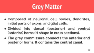
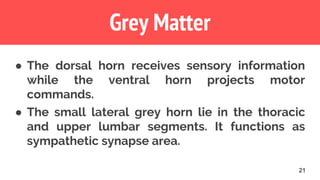
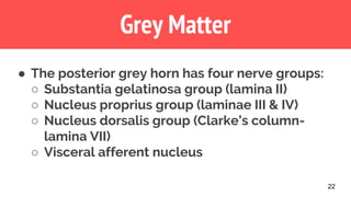
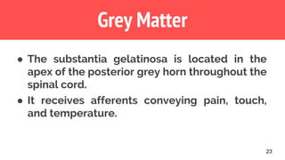

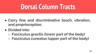
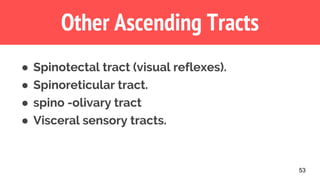


The document provides a comprehensive overview of the spinal cord, including its structure, function, and major ascending tracts. Key features discussed include the spinal cord's external anatomy, meningeal coverings, and internal organization into grey and white matter. It also highlights various ascending tracts responsible for sensory information, such as the spinothalamic and dorsal column tracts.


![PERI-PROSTHETIC FRACTURE NAIL-PLATE CONSTRUCT [NPC].pptx](https://cdn.slidesharecdn.com/ss_thumbnails/drarunkumardrmohamedashrafperiprostheticfrasturenail-plateconstructnpc-260209164459-7e9d15a1-thumbnail.jpg?width=640&height=640&fit=bounds)


