
Recommended
More Related Content
Similar to ARTERIAL SUPPLY OF HEAD AND NECK (1)-1.pptx
Similar to ARTERIAL SUPPLY OF HEAD AND NECK (1)-1.pptx (20)
Recently uploaded
Recently uploaded (20)
ARTERIAL SUPPLY OF HEAD AND NECK (1)-1.pptx
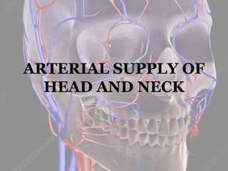
- 1. ARTERIAL SUPPLY OF HEAD AND NECK
- 2. CONTENTS ARTERIES OF HEAD AND NECK Common Carotid Artery Internal Carotid Artery External Carotid Artery Applied Anatomy
- 3. Common Carotid Artery The CCA is a large elastic artery, which provides the main blood supply to head and neck region. • There is one common carotid artery on either side of the body and these arteries differ in their origin. • The left common carotid artery arises from the aortic arch directly, whereas the right common carotid artery arises from the brachiocephalic trunk posterior to the right sterno-clavicular joint.
- 4. Course • The CCA ascends lateral to the trachea and esophagus, lies within the deep cervical fascia of the carotid sheath, along with the IJV and vagus nerve. • The left CCA is longer than the right having cervical and thoracic parts, whereas the right one has only a cervical part. • The cervical part of the common carotid artery is symmetrical on both sides.
- 5. • After separating from the aortic arch, the left CCA ascends through the superior mediastinum up to the level of the left sternoclavicular joint. • Then it ascends from behind the sternoclavicular joint up to the level of the upper border of the thyroid cartilage of the larynx, where it bifurcates into the internal and external carotid arteries. • Both the left and right CCA terminates at the level of upper border of thyroid cartilage.
- 6. Branches Internal carotid artery: It is one of the branches of CCA which is the chief artery to supply blood to the brain. External carotid artery: It is one of the terminal branches of CCA. • In general it lies anterior to ICA and is the chief artery to supply structures in front of the neck.
- 7. Lo, A., Oehley, M., Bartlett, A., Adams, D., Blyth, P. and Al‐Ali, S. (2006), ANATOMICAL VARIATIONS OF THE COMMON CAROTID ARTERY BIFURCATION. ANZ Journal of Surgery, 76: 970-972. https://doi.org/10.1111/j.1445-2197.2006.03913.x
- 8. Carotid Sheath Contents: • CCA as well as the ICA (medial) • IJV (lateral) • Vagus nerve (posterior) • Deep cervical lymph nodes
- 9. Carotid Sinus • It is also called as Carotid Bulb. • The carotid sinus is a dilation of the base of the ICA or the termination of CCA, innervated by IX cranial nerve.
- 11. Carotid body • It is also called as Carotid glomus (glomus caroticum). • Most vacularised part in body. • The carotid body is a small, flattened structure, located at the level of carotid bifurcation, typically posteromedial to the common carotid artery; measuring about 2.5 by 5mm to 4 by 7mm
- 12. • It is involved in relaying information about the arterial chemical composition to respiratory centres in the brainstem. • Also innervated by IX nerve. • The carotid body is surrounded by a fibrous capsule and consists of multiple lobules divided by septa.
- 13. • Within each lobule, there are two types of cells: glomus (type I) cells and sustenacular (type II) cells • The glomus cells are involved in storing peptides, such as neurotensin, adrenaline, noradrenaline and dopamine. • The sustentacular cells separate and support the glomus cells by forming an extensive network of fenestrated sinusoids.
- 14. • The carotid body is a chemoreceptor stimulated by hypercapnia, hypoxia and low pH. • Individuals with chronic hypoxia or who live at high altitudes may have enlarged carotid bodies
- 15. Carotid Body Tumor • Largely vascular or largely epithelial. • Referred to as glomus tumor. • A carotid body tumor that does not secrete cathecholamines is symptomless. • Only about 5% of tumors metastasize. • May embarrass the circulation. • Frequently found to surround the common or internal carotid arteries. Tumor
- 16. Applied Anatomy Carotid Pulse: is felt by palpating the CCA on either side of the neck beneath the anterior border of the sternocleidomastoid muscle by compressing the fingertips against the prominent transverse process of the sixth cervical vertebra. • During CPR this pulse is routinely checked and its absence can indicate cardiac arrest.
- 17. • The CCA is often used for measuring pulse, especially the patients in shock who lack detectable pulse in the more peripheral arteries of the body. • The carotid artery should be palpated gently because, stimulating its baroreceptors may cause severe bradycardia or even stop the heart in over sensitive persons. • A person’s both the carotid arteries should not be palpated at the same time. By doing so it may limit the blood flow to the brain and may cause fainting or cerebral ischemia.
- 18. Carotid Sinus Hypersensitivity: • Carotid sinus hypersensitivity is an increased response to carotid sinus stimulation, which can occur with advanced age, coronary artery disease or hypertension. • External pressure on the carotid sinus can cause bradycardia and hypotension, which can lead to dizziness or syncope. • Therefore, palpation of the carotid pulse is not recommended in patients with this condition.
- 19. Carotid sinus massage: • Bedside maneuver performed to terminate some supraventricular tachycardias, eg. AV reentry SVTs. • This technique is performed with the patient's neck in an extended position, the head turned away from the side being massaged. • Only one side should be massaged at a time. • Pressure is applied underneath the angle of the jaw in a gentle circular motion for about 10 seconds.
- 20. Carotid Artery Stenosis: • The common carotid artery is a common site for atherosclerosis, a degenerative arterial disease resulting in the formation of plaques. • This can lead to carotid artery stenosis, narrowing of the CCA, which increases the risk of a transient ischaemic attack or a stroke.
- 21. Treatment: Involves pharmacological management, such as aspirin or warferin therapy or surgical management- Carotid endarterectomy. • This procedure is only indicated with a stenosis greater than 50%.
- 22. Carotid Blowout Syndrome • CBS is classically defined as rupture of the extracranial carotid artery or its branches. • It is one of the most devastating complications associated with radical head and neck surgery. • Surgical management of carotid blowout is usually technically difficult for exploring and repairing the previously radiated field.
- 23. • It is an uncommon but fatal complication that occurs in patients treated for head and neck cancer(3- 4%). It is the result of necrosis of the arterial wall, which can occur following resection, after irradiation for a recurrent or primary tumor, by direct tumor invasion of the carotid artery wall or by a combination of these factors. • When associated with open surgical treatment, high mortality (40%) and neurologic morbidity (60%) have been reported.
- 24. Grading System: CBS may be categorized into four types that may involve CCA and ICA. Threatened: refers to a visibly exposed carotid artery secondary to wound breakdown that will almost inevitably rupture if it is not promptly covered with healthy tissue. Impending CBS: refers to transient carotid artery hemorrhage that resolves either spontaneously or with simple surgical packing. However, complete rupture is a certainty and may occur at any time because the carotid artery is devoid of supportive, surrounding structural elements. Acute CBS: the most emergent subtype, refers to complete rupture of the carotid artery and profuse hemorrhage that is not well controlled by surgical packing. Recurrent CBS: refers to previously treated patients with CBS who develop recurrent CBS episodes
- 25. Management Historically CBS was associated with 40-60% moratality rate but outcomes have improved with advent of various endovascular techniques. • Percutaneous Balloon Occlusion. • Embolization with coils. • Using overlapping or covered stents. • Direct carotid puncture. Benjamin Zussman, L. Fernando Gonzalez, Aaron Dumont, Stavropoula Tjoumakaris, Robert Rosenwasser, David Hasan, David Cognetti, Rita Axelrod, Pascal Jabbour;Endovascular Management of Carotid Blowout; World Neurosurgery, 2012; Volume 78, Issues 1–2,Pages 109-114
- 26. Clamping Of CCA • Clamping of CCA reduces blood flow to the brain by ICA by 50%. • Clamping of the internal or common carotid artery is followed by a high mortality rate and a high percentage of cerebral complications. • Indications: Deep neck lacerations, Carotid blowout syndrome, Carotid endarterectomy.
- 27. Internal Carotid Artery Introduction: • The Internal Carotid Artery is one of the two terminal branches of the common carotid artery. • It is a principal artery of the brain and eye. It also supplies the related bones and meninges.
- 28. Embryology • Several main and terminal branches of the ICA start developing from the 4th gestational week onwards when it bifurcates into anterior and posterior components. The anterior component will differentiate into the anterior cerebral, middle cerebral, and anterior choroidal arteries. The posterior component form the posterior cerebral and the posterior choroidal artery.
- 29. Course • The ICA arises at the level of the upper border of the thyroid cartilage opposite the disc between the third and fourth cervical vertebrae. • From its origin, the vessel passes up the neck travel through Carotid sheath in a superior direction along the neck, and enter the skull through carotid canal.
- 30. Segments & Branches. There are two main ways to categorize the segments of the internal carotid artery. The newest one (1998) which divides the artery into four parts namely: 1. Cervical part (neck) 2. Petrous part (temporal bone) 3. Cavernous part (cavernous sinus) 4. Cerebral part (after piercing the dura mater)
- 31. • According to this classification, there are seven segments: C1 – Cervical Segment C2 – Petrous Segment C3 – Lacerum Segment C4 – Cavernous Segment C5 – Clinoid Segment C6 – Ophthalmic (Supraclinoid) Segment C7 – Communicating (Terminal) Segment • An older method described in 1996, known as the Cincinnati Classification (Bouthillier et. al., 1996).
- 32. CERVICAL PART: • This part is branchless and slightly curved so that the artery can follow the movements of the neck without being stretched. • It ascends vertically in the neck from its origin to the base of the skull to reach the lower end of the carotid canal. This part is enclosed in the carotid sheath (with the internal jugular vein and the vagus). • Its initial part usually shows a dilatation, the carotid sinus which acts as a baroreceptor. • The lower part of the artery (in the carotid triangle) is comparatively superficial. • The upper part, above the posterior belly of the digastric, is deep to the parotid gland, the styloid apparatus, and many other
- 33. Relations: • Anterior or Superficial 1. In the carotid triangle: – Anterior border of sternicleidomastoid – The ECA is anteromedial to it. 2. Above the carotid triangle: – Posterior belly of digastric – Stylohyoid – Stylopharyngeus – Styloid process – Parotid gland • Posterior 1. Superior cervical ganglion 2. Carotid sheath 3. The glossopharyngeal, vagus, accessory and hypoglossal nerves at the base of the skull. • Medial 1. Pharynx 2. The ECA is anteromedial below the parotid. • Lateral 1. Internal Jugular Vein 2. Temporomandibular Joint (at the base of the skull)
- 34. PETROUS PART (C2): This segment travels superiorly before taking an anteromedial course, after which it travels along a superomedial course advancing towards foramen lacerum. Relations: • Surrounded by venousand sympathetic plexus. • Posterosuperiorly: ‒ The middle ear ‒ The cochlea • Anterolaterally ‒ The auditory tube ‒ Tensor Tympani ‒ Superiorly: Trigeminal Ganglion
- 35. BRANCHES: • Caroticotympanic artery : It originates from the petrous part (C2) and travels through the tympanic cavity via foramen within the carotid canal and anastomose with the anterior tympanic branch of the internal maxillary, and with the posterior tympanic branch of the stylomastoid artery. It supplies the tympanic cavity.
- 36. Vidian artery: • Also called as pterygoid artery. • The Vidian artery courses through the pterygoid canal together with the Vidian nerve, anastomoses with a branch of the greater palatine artery. • Supplies pharynx, pharyngo-tympanic tube. • It may not be present in some individuals, and can originate from ECA as well (branch of maxillary artery)
- 37. CAVERNOUS PART (C4): • Begins at petrolingual ligament and extends till proximal dural ring. • Within the cavernous sinus it courses superiorly along the posterior clinoid process, making its way towards the anterior clinoid process before finally emerging through the roof of the cavernous sinus.
- 38. Within the cavernous sinus, the internal carotid artery travels superomedially to CN VI and gives rise to: Cavernous branches to Trigeminal ganglion Superior and Inferior hypophyseal branches to the hypophysis cerebri
- 39. Carotid Siphon • The carotid siphon is a U or S-shaped part to the ICA that begins at the posterior bend of the cavernous part and ends at the cerebral part, at ICA bifurcation. Importance: • Several factors are related to the formation, development, and rupture of intracranial aneurysms. Hemodynamic contribution can be illustrated by the preferred location of aneurysms at arterial bifurcations and curvatures. • It is of particular importance because it is a tortuous vessel segment with sharp bends through which blood enters the anterior cerebral circulation.
- 40. CEREBRAL PART (C7): This part lies at the base of the brain after emerging from the cavernous sinus. Branches: • Ophthalmic • Posterior Communicating • Anterior Choroidal • Anterior Cerebral • Middle Cerebral
- 41. • Ophthalmic artery: passes through the optic canal, ultimately entering the orbit and supplies Frontal belly of the occipito- frontalis, Lacrimal gland, Nasalis, Procerus, Voluntary muscles of eye. • Branches: Central retinal artery Lacrimal artery Posterior ciliary arteries. • Clinical significance: Severe occlusion of the ophthalmic artery causes ocular ischemic syndrome. As with central retinal artery occlusions, ophthalmic artery occlusion can lead to rapid death of retinal cells, thereby resulting in severe loss of vision.
- 42. Circle Of Willis It is an arterial circle, situated at the base of the brain in the interpenduncular fossa. It is a part of the cerebral circulation and is formed by: • Two anterior cerebral arteries connected by anterior communicating artery; the middle and posterior cerebral arteries of the same side are united by the posterior communicating artery. It attempts to equalize the flow of blood to different parts of brain and provides a collateral circulation in the event of obstruction to one of its components.
- 43. Clinical Aspects Of ICA Basal skull fractures: • As the ICA enters the skull via the carotid canal, fractures of the base of the skull can easily tear the ICA at this particular point resulting in an Carotico-Cavernous fistula. • An abnormal communication is created between the ICA, and a vein or venous system, this being the cavernous sinus.
- 44. • An engorged cavernous sinus can put pressure on the structures passing through it, such as cranial nerves III, IV, V1, V2, and VI, ultimately producing distinct clinical signs and symptoms.
- 45. External Carotid Artery. • The external carotid artery arises from the bifurcation of the CCA when it divides into the external and internal carotid arteries. • The carotid bifurcation occurs at the level of the thyroid cartilage opposite the disc between 3rd and 4th cervical vertebrae. • It generally lies anterior to ICA and is the chief artery of supply to structures in the front of the neck and in the face.
- 46. Course • The ECA arises from CCA at the level of the superior border of the thyroid cartilage opposite the disc between the 3rd and 4th cervical vertebrae. • On each side, the artery originates in the carotid triangle, lying anteromedial to ICA. • It runs upwards and slightly backwards and laterally, and terminates behind the neck of the mandible by dividing into its terminal branches - The Maxillary and The Superficial Temporal arteries.
- 47. • In the carotid triangle, – Comparatively superficial – Lies under cover of the anterior border of the sternocleidomastoid muscle – It is crossed superficially by • The cervical branch of facial nerve • the hypoglossal nerve • the facial, lingual and superior thyroid veins. – Deep to the artery there are: • The wall of the pharynx • The superior laryngeal nerve which divides into the external and internal laryngeal nerves. • The ascending pharyngeal artery Relations: The ECA has a slightly curved course. It is anteromedial to ICA in its lower part and anterolateral to the ICA in its upper part.
- 48. • Above the carotid triangle, ECA lies deep in the substance of the parotid gland. • Within the gland, it is related superficially to the retromandibular vein and the facial nerve. • Deep to the ECA, there are: – The ICA – Styloglossus – Stylopharyngeus – Glossophayngeal nerve – Pharyngeal branch of vagus – Styloid process
- 49. Branches Anterior branches: Superior thyroid artery Lingual artery Facial artery Posterior branches: Occipital artery Posterior auricular artery Medial branch: Ascending pharyngeal artery Terminal branches: Maxillary artery Superficial temporal artery
- 50. Ligation Of External Carotid Artery There are two surgical approaches to ligate the ECA • In the carotid triangle • In the retromandibular fossa Landmarks: • Upper border of thyroid cartilage • Carotid bulb • Internal Jugular Vein • Lower border of the mandible • Anterior border of sternocleidomastoid muscle.
- 51. Ligation in the Carotid Triangle • Surgically the exposure and ligation of the external carotid artery in the carotid triangle can best be done in the following way. • The incision of the skin starts at the level of the angle of the mandible just behind the anterior border of the sternocleidomastoid muscle and is continued downward, parallel to the border of the muscle, to the level of cricoid cartilage. • After penetrating through the skin and the platysma muscle, the superficial sheath of the sternocleidomastoid muscle is incised.
- 52. • Bluntly, the anterior border of the muscle is exposed and the muscle retracted; thus the deep layer of the sterno- cleidomastoid sheath becomes visible and through it the internal jugular vein. • In front of this vein the fascia is cut to expose the arteries. • The ECA is identified by its first anterior branch, the superior thyroid artery, and then isolated and tied a few millimeters above the origin of the superior thyroid artery. • While the incision through the deep layer of the sternocleidomastoid sheath is made, care has to be taken not to injure the hypoglossal nerve. It is suggested that the incision through the fascia be started at the lowest point of the wound.
- 53. • The surgical exposure of the external carotid artery at the stylomandibular ligament is a procedure much simpler and much less dangerous than the exposure of the artery in the neck. • The skin is incised in a line which starts at the tip of the mastoid process and circles the mandibular angle, continuing forward below the mandible for about one inch. • The incision is kept at an equal distance from the posterior and inferior borders of the mandible. • After passing through the skin and some of the posterior fibers of the platysma muscle, the retromandibular vein or the external jugular vein is located, tied, and cut. • Branches of the great auricular nerve have also to be cut in order to permit the mobilization of the cervical lobe of the parotid gland. Ligation in the Retromandibular Fossa
- 54. • To this end the attachment of the parotid capsule to the anterior border of the sternocleidomastoid muscle has to be severed with the scalpel. • If this is done, the flap of soft tissues, consisting of skin and parotid gland, is retracted anteriorly and upward. • Immediately underneath the parotid gland the posterior belly of the digastric muscle becomes visible and slightly above it is the stylohyoid muscle. Above these muscles the styloid process can be palpated and also the upper border of the stylomandibular ligament, especially if at this moment the lower jaw of the patient is pulled forward. • This movement of the mandible not only widens the entrance into the retromandibular fossa, but also tenses the stylomandibular ligament. • At the stylomandibular ligament the pulse of the external carotid artery can be felt, and it is easy to isolate the artery and to tie it, even if it is accompanied by a larger vein.
- 55. Superior Thyroid Artery • The superior thyroid artery is a branch of the ECA arising from its anterior surface.
- 56. Course • The superior thyroid artery is the first branch of the ECA, arising just below the level of the greater cornua of the hyoid bone. • It runs downwards, forwards and parallel and superficial to external laryngeal nerve. • It passes deep to omohyoid, sternohyoidand sternothyroid to reach the upper pole of the lateral lobe of the thyroid gland and divides into its terminal branches.
- 57. Branches The superior thyroid artery in its course gives off the following branches: • Infrahyoid artery • Superior laryngeal artery • Sternocleidomastoid artery • Cricothyroid artery • The infrahyoid artery passes along the lower border of the hyoid bone below and anastomoses with the same artery of the contralateral side to supply the infrahyoid muscles of the neck.
- 58. • The superior laryngeal artery supplies blood to the tissues of the upper part of the larynx. It anastomoses with its fellow artery of the opposite side and with the inferior laryngeal branch of the inferior thyroid artery. • The sternocleidomastoid artery runs downward and laterally across the carotid sheath to supply the middle region of the sternocleidomastoid. • The cricothyroid artery crosses the anterior cricothyroid ligament to supply the cricothyroid muscle.
- 59. Clinical Aspects • Its relationship with external laryngeal nerve, which supplies cricothyroid muscle is very important during thyroid surgery. • The artery and nerve stays in close proximity to each other near the thyroid gland higher up. To avoid injury to the nerve the artery should be ligated as close as possible to the thyroid gland. • The thyroid has a rich blood supply; therefore, during thyroidectomy, the superior thyroid artery requires ligation.
- 60. Lingual artery. • The lingual artery is a branch of the ECA that supplies blood to the tongue and floor of the mouth.
- 61. Course • It arises medially from the ECA at the level of the greater cornua of the hyoid bone. • It may arise in common with the facial artery then becoming the linguofacial trunk. • The course is divided into three parts by hyoglossus muscle. • First part lies in carotid triangle where it forms a characteristic upward loop which is crossed by hypoglossal nerve. • Second part lies deep to hyoglossus along upper border of hyoid bone. • Third part is called deep lingual artery. It runs upwards along upper border of hyoglossus and horizontally to undersurface of tongue as fourth part. It is accompanied by lingual
- 62. Branches • On its course, the lingual artery also gives off side branches, including: • Suprahyoid branches which supplies to muscles attached to hyoid bone. • Dorsal lingual branches that supply the dorsum of the tongue till the epiglottis. • Sublingual artery supplying the sublingual gland. • The branches of the lingual artery provide blood supply to the tongue, sublingual gland, and the floor of the mouth.
- 63. LIGATION OF LINGUAL ARTERY submandibular gland palpated through skin incision circling the lower pole of the gland skin, platysma, deep fascia incised lower pole of the submandibular gland exposed gland lifted from its bed by blunt dissection flap retracted upward, tendon of the digastric exposed, mylohyoid is seen. hypoglossal nerve identified and retracted to prevent injury Digastric tendon pulled downward, Hyoglossus muscle dissected - Lingual artery is found and ligated.
- 64. • During surgical removal of tongue the first part of lingual artery is ligated before it gives off any branches to the tongue or the tonsils.
- 65. Facial Artery • The facial artery is the chief artery of the face which arises from the ECA in the carotid triangle just above the greater cornua of hyoid bone.
- 66. Course • It runs upwards in the neck as cervical part and on the face as facial part. • The course of the artery is tortuous. • This allows free movements of the pharynx during deglutition. • On the face, it allows free movements of mandible, lips and cheek during mastication and facial expressions. • The artery escapes traction and pressure during these movements.
- 67. Branches Cervical branches: • Ascending Palatine • Tonsilar • Glandular • Submental. Facial branches: • Inferior labial • Superior labial • Lateral nasal • Angular The facial artery with its branches supplies the submandibular gland, lips, chin, most of the facial muscles, tongue, pharyngotympanic tube, soft palate, and tonsils.
- 68. Variations of facial artery • The facial artery may arise with the lingual, as a liguo-facial trunk. It varies in size and supply to the face: it may end as submental artery and often extends only to the buccal angle. The deficiency is then filled by branches of neighbouring arteries • In human fetuses a common linguo- facial trunk occurred in 43%; in 42% the facial did not reach the medial orbital angle, ending as a superior (20%) or inferior (22%) labial artery (Kozielec & Jozwa 1977)
- 70. Ligation of Facial Artery • The facial artery can be easily exposed at the point where it crosses the lower border of the mandible to pass from the submandibular region into the face. • This point is situated anterior to the attachment of the masseter muscle to the mandible. • Here, the pulse of the facial artery can easily be felt, especially if the contracted masseter muscle is used as a landmark. • The artery is accompanied by the facial vein, which lies posterior to the artery. • The artery and vein are crossed superficially by the marginal mandibular branch of the facial nerve. • This rather small nerve runs approximately parallel to the lower border of the mandible, sometimes slightly above or below this border.
- 71. • The nerve and vessels are covered by the platysma muscle, the subcutaneous tissue, and the skin. • Since the mandibular branch of the facial nerve supplies the muscles of the lower lip, it is necessary to plan the operation in such a way that this nerve is not in danger of being cut. • To achieve this end, the incision is made at least one centimeter below the border of the mandible and parallel to it. • The skin, platysma muscle, and deep fascia are cut and then the soft tissues are bluntly retracted upward until the palpating finger can feel the pulse of the facial artery. • The artery then can be isolated, tied, and cut.
- 72. Occipital Artery. • The occipital artery is one of the posterior branches of the ECA that arises from the posterior aspect, opposite to the origin of Facial artery, therefore below the level of disgastric and stylohyoid.
- 73. Course
- 74. Branches • Sternocleidomastoid branches: This branch supplies SCM and hypoglossal nerve loops around this branch. • Auricular branch supplies back of concha • Mastoid branch arises along with auricular branch and supplies mastoid cells, dura by entering skull through mastoid foramen. • Meningeal branch enters skull though jugular foramen. • Occipital artery anastomoses with transverse cervical, vertebral, deep cevical artery at the base of skull which help to maintain collateral after ligation of CCA or subclavian artery. The occipital artery supplies blood to the back of the scalp, sternocleidomastoid, trapezius, deep muscles of the back, and dura mater of the posterior cranial fossa.
- 75. Posterior Auricular Artery. • The posterior auricular artery is a small branch of the ECA which arises from the posterior aspect, just above the posterior belly of digastric and stylohyoid muscle.
- 76. Course • It is largely covered by parotid gland and runs upwards and backwards deep to the gland, along the styloid process. • It crosses the base of mastoid process and ascends behind the auricle to reach auricular cartilage. • Here it divides into mainly five branches. • It may arise as a branch of occipital artery rather than an independent branch.
- 77. Branches • Stylomastoid branch • Auricular • Posterior tympanic • Parotid • Occipital branch The posterior auricular artery with its branches supplies the digastric, stylohyoid and sternocleidomastoid muscles, parotid gland, tympanic cavity, facial nerve, and mastoid antrum cells.
- 78. Ascending Pharyngeal Artery • The ascending pharyngeal artery is the smallest branch of ECA that arises from the medial side of ECA. • It arises very close to the lower end of ECA.
- 79. Course • The ascending pharyngeal artery passes vertically upwards between the lateral side of the pharynx and tonsils, middle ear and auditory tube to the base of the skull. • Further, it continues as the meningeal artery, which is the terminal branch of the ascending pharyngeal artery.
- 80. Branches • On its course, the ascending pharyngeal artery gives rise to two side branches: • Pharyngeal branches that supply the muscles and mucosa of the pharynx, as well as the pharyngotympanic tube; • Inferior tympanic branch that supplies the medial wall of the tympanic cavity. • It sends meningeal branches into the cranial cavity through foramen lacerum. • The ascending pharyngeal artery with its branches provides blood supply to the muscles and mucosa of the pharynx, pharyngotympanic tube, medial wall of the tympanic cavity, and the middle cranial fossa.
- 81. Maxillary Artery • The maxillary artery is the largest terminal branch of the ECA that arises at the back of the neck of the mandible, within the substance of parotid gland. • It supplies deep structures of the face, such as the mandible, maxilla, teeth, muscles of mastication, palate, nose, and part of the cranial dura mater.
- 82. Course. Topographically the maxillary artery can be divided into three portions: • Mandibular • Pterygoid • Pterygomandibular
- 83. • The first part runs horizontally forwards, between the neck of the mandible and sphenomandibular ligament then along the lower border of lateral pterygoid. • The second part runs upwards and forward superficial to lower head of lateral pterygoid. • The third part passes between two heads of lateral pterygoid and through pterygomaxillary fissure to enter pterygopalatine fossa.
- 84. Branches From the mandibular part, the maxillary artery gives off five branches that enter the skull to supply bones of the skull, including: I. Deep auricular artery, II. Anterior tympanic artery, III. Middle meningeal artery, IV. Accessory meningeal artery, V. Inferior alveolar artery.
- 85. Deep Auricular: • Supplies : External acoustic meatus, Tympanic membrane, TMJ Anterior tympanic: • Supplies : Middle ear, Medial surface of tympanic membrane Accessory meningeal: enters the cranial cavity through foramen ovale • Supplies: Meninges, Structures in infra temporal fossa .
- 86. Middle meningeal artery: - Course : closely related to the auriclotemporal nerve and enters the cranial cavity through the foramen spinosum running laterally over the floor of middle cranial fossa giving 2 branches. - Frontal & Parietal Supplies : Meninges, Skull bones, Middle ear, Trigeminal ganglion, Tensor tympani, Auditory tube. - Great surgical importance as it can be injured in head injuries-common source of extradural haemorrhage.
- 87. Clinical significance • An injured middle meningeal artery is the most common cause of an epidural hematoma. • A head injury can rupture the artery. Emergency treatment requires decompression of the hematoma, usually by craniotomy. • Subdural bleeding is usually venous in nature, rather than arterial. • The anterior branch of the middle meningeal artery runs beneath the Pterion. It is vulnerable to injury at this point, where the skull is thin. Rupture of the artery may give rise to an epidural hematoma.
- 88. Inferior alveolar artery: Course : runs downwards and forwards medial to the ramus to reach the mandibular foramen. Before entering the canal it gives lingual branch & mylohyoid branch. It then passes through the mandibular canal giving branches to the mandible and to the roots of each tooth attached to the bone. Then it gives mental branch that passes through the mental foramen to supply the chin.
- 89. The Pterygoid part of the maxillary artery gives off the following four branches: I. Deep temporal artery II. Pterygoid artery III. Masseteric artery IV. Buccal artery
- 90. • Deep temporal branches: supplies the temporalis muscle • Pterygoid branches: supplies the pterigoid muscles • Masseteric branches: supplies the masseter • Buccal artery: supplies the buccinator.
- 91. The Pterygopalatine part of the maxillary artery provides four branches that accompany similarly named branches of the maxillary nerve. These branches include: I. Posterior superior alveolar artery II. Infraorbital artery III. Greater palatine IV. Pharyngeal V. Artery of pterygoid canal (Vidian artery) VI. Sphenopalatine artery
- 92. Posterior superior alveolar artery: • Supplies : Maxillary Molars & Maxillary air sinus Infraorbital artery: • Supplies : Lacrimal sac, Lower eyelid, lateral part of the nose, Upper lip, Maxillary incisors, canines premolars and MB root of Maxillary 1st molar Pharyngeal: • Supplies : Nasopharynx, Auditory tube, and Sphenoidal air sinus.
- 93. Greater palatine artery : • Supplies : Palate, Gingiva • Gives off lesser palatine arteries Supplies : Soft palate, Tonsils • Can be accidently severed during minor procedures in the posterior palatine region Artery of the pterygoid canal (Vidian artery): • Supplies: Pharynx, Auditory tube, Tympanic cavity Sphenopalatine artery: o Posterolateral nasal branch • Supplies : Lateral wall of nose, Para nasal sinus o Posterior septal branch • Supplies : Nasal septum
- 94. Maxillary Artery Ligation 1) Transantral approach: • first described by Scifert in 1928, for the third division of maxillary artery. • Popularised by Chandler and Serrins in 1965 • It is currently the most widely used arterial ligation procedure for controlling posterior epistaxis. • In some cases bilateral maxillary artery ligation is required to control unilateral bleeding because of cross-anastomosis from adominant maxillary artery.
- 95. Procedure: Standard gingival incision Anterior wall of the maxillary sinus is exposed and removed with special care not to injure the infraorbital nerve. The posterior wall of the sinus is identified and a laterally based U-shaped mucosal flap elevated Positions of the posterior wall removed to gain exposure to the pterygopalatine fossa and the branches of the maxillary artery Complications – a) Hypoesthesia of the infraorbital nerve b) Persistent pain in the maxillary dentition c) Oro-antral fistula d) Damage to sphenopaltine ganglion e) Total opthalmoplegia
- 96. 2) Intraoral Approach: • This approach is described by Maceri and Makielski for the ligation of the infratemporal portion of the maxillary artery. • This approach provides access to the 1st and 2nd parts of the maxillarybartery without opening the maxillary sinus. • It doesn’t require any microscope to operate.
- 97. Procedure: • Incision at upper gingivobuccal sulcus at the level of 2nd and 3rd molar and continued inferiorly along the ramus of the mandible. • The buccal fat pad is retracted medially or removed and the attachments of the temporalis muscle to the coronoid precess of the mandible are identified. • The temporalis muscle belly may need to be split and partially dissected from the mandible to gain access to the artery. • Blunt dissection reveals the maxillary artery which is clipped or ligated.
- 98. Clinical Significance. • In 50% of all persons, maxillary artery is found lateral to lateral ptergoid muscle and remaining 50% the artery lies medial to lateral ptergoid muscle. –Lauber(US) • The maxillary artery is at risk of injury during intraoral vertical or oblique ramus osteotomy During treatment of subcondylar fracture, salivary gland tumors. Spenopalatine artery is most commonly responsible for post- operative bleeding.
- 99. Superficial Temporal Artery • The superficial temporal artery is one of the smaller terminal branches of the ECA which arises behind the neck of mandible under the cover of parotid.
- 100. Course • The superficial temporal artery separates from the ECA behind the neck of the mandible. • About 5cm above zygoma it divides into anterior and postrior roots which supply temple and scalp.
- 101. Branches • Above the zygomatic process, the superficial temporal artery divides into two branches, the frontal and parietal branches. • The frontal branch supplies the forehead area and anastomoses with the ophthalmic artery. • The parietal branch supplies the temporal and parietal regions of the scalp. It may form anastomoses with the posterior auricular artery and the occipital artery. • The superficial temporal artery also gives off the following side branches that supply the external ear, parotid gland, temporal muscle: transverse facial artery anterior auricular branches middle temporal artery
- 102. Clinical Aspects In reduction of zygomatic arch fractures : Gillie’s temporal approach the artery is often encountered. The pulsations of the artery can be felt on the zygomatic arch as it crosses the root of zygoma or pre-auricular point. The superficial temporal artery is frequently involved in a pathology called the giant-cell arteritis or temporal arteritis, which is a form of systemic inflammatory vasculitis. The temporal arteritis damages blood vessels resulting in subsequent distal ischemia. – Clinical symptoms include headache, visual disturbances, neck pain, and scalp tenderness.
Editor's Notes
- The carotid sheath is an anatomical term for the fibrous connective tissue that surrounds the main vascular components of neck. It is part of deep cervical fascia of neck. It is located on each side of the neck and deep to the SCM muscle, extending from the base of the skull to the first rib and sternum.
- The baroreceptors in carotid sinus are involved in relaying information about the arterial blood pressure by responding to change in tension of arterial wall and relaying them to the hypothalamus. It is therefore referred to as a baroreceptor and regulates arterial blood pressure. Nucleus Tractus Solitarius of medulla oblongata regulates through sympathetic and para-sympathetic system.
- Endovascular therapy with either permanent balloon occlusion or with the deployment of stent grafts has been reported as an alternative for management.
- The principles of algorithm for management of CBS patients are: (i) The initial and cardinal priorities of management are respiratory and cardiac stabilization (ie, the ABCs [airway, breathing, circulation]); (ii) diagnostic angiography can be used to identify the site and extent of the pathology; and (iii) if there is internal or common carotid system involvement and time permits, temporary balloon occlusion should be performed to determine whether or not a patient is likely to sustain a neurologic insult from carotid deconstruction. Although balloon occlusion testing ideally should be performed for all patients with CBS and common carotid or internal carotid artery involvement, insufficient time in the setting of life-threatening exsanguination presents a realistic contraindication to balloon testing. Patients who fail a balloon occlusion test may be successfully treated endovascularly with covered stents to prevent a stroke. If there is external carotid system involvement, a passed balloon occlusion test, or insufficient time to perform a balloon occlusion test, deconstructive occlusion of the carotid artery may be performed with glue embolization, coil embolization, or both. Control of the arterial hemorrhage is a fundamental aim of CBS management. Deconstructive endovascular management of CBS is preferable to traditional open surgery, which has been associated with approximately 40% intraoperative mortalit
- Ideally preceded with Balloon Occlusion Test or Angiography. Skin incision given at the level of hyoid bone. SCM retracted posteriorly, IJV avoided by opening the carotid sheath medially. Collateral circulation after ligation. Clamping of CCA reduces blood flow to the brain by ICA by 50%. Clamping of the internal or common carotid artery is followed by a high mortality rate and a high percentage of cerebral complications. Some of the patients in whom cerebral complications develop survive with residual squeal, such as varying degrees of hemiplegia, aphasia, mental changes and epilepsy. Indications: Deep neck lacerations, Carotid blowout syndrome, Carotid endarterectomy.
- During fetal development, vasculogenesis (formation of new vessels from haematoangioblastic stem cells) is responsible for the formation of two dorsal aortae, six pairs of primitive pharyngeal (branchial) arch arteries, and other vascular structures. The third pharyngeal arch artery fuses with the distal components of the dorsal aortae to give rise to the internal carotid artery. The embryological dorsal aortae (left and right) ultimately form the descending aorta. Several main and terminal branches of the internal carotid artery start developing from the 4th gestational week onwards when it bifurcates into anterior and posterior components. The anterior component will differentiate into the anterior cerebral, middle cerebral, and anterior choroidal arteries. The posterior component will form the posterior cerebral artery and the posterior choroidal artery.
- It arises in the cranial cavity by travelling via the upper part of the foramen lacerum. Before it terminates below the anterior perforated substance of the brain by separating into the anterior and middle cerebral arteries it goes inside the cavernous sinus and follows a twisted path in the cranial cavity.
- The curves increase with advancing age because of the gradual loss of elasticity of the arterial wall. In elderly persons the ICA sometimes follows a strongly tortuous course, and then its normal relation to ECA can be altered.
- Since the internal carotid artery contains arterial blood, it is under high pressure created by cardiac contractions. If an arteriovenous fistula is present, this arterial blood will flow into the low pressure system of the cavernous sinus and force blood retrogradely into its venous tributaries, which include the ophthalmic veins, which drain the orbit and its contents. If the pressure within them increases, the eyeball of the patient will protrude (pulsating exophthalmia) and the conjunctiva will be engorged with blood. An engorged cavernous sinus can put pressure on the structures passing through it, such as cranial nerves III, IV, V1, V2, and VI, ultimately producing distinct clinical signs and symptoms.
- The surgical exposure of the external carotid artery at the stylomandibular ligament is a procedure much simpler and much less dangerous than the exposure of the artery in the neck. The skin is incised in a line which starts at the tip of the mastoid process and circles the mandibular angle, continuing forward below the mandible for about one inch. The incision is kept at an equal distance from the posterior and inferior borders of the mandible. After passing through the skin and some of the posterior fibers of the platysma muscle, the retromandibular vein or the external jugular vein is located, tied, and cut. Branches of the great auricular nerve have also to be cut in order to permit the mobilization of the cervical lobe of the parotid gland. To this end the attachment of the parotid capsule to the anterior border of the sternocleidomastoid muscle has to be severed with the scalpel. If this is done, the flap of soft tissues, consisting of skin and parotid gland, is retracted anteriorly and upward. Immediately underneath the parotid gland the posterior belly of the digastric muscle becomes visible and slightly above it is the stylohyoid muscle. Above these muscles the styloid process can be palpated and also the upper border of the stylomandibular ligament, especially if at this moment the lower jaw of the patient is pulled forward. This movement of the mandible not only widens the entrance into the retromandibular fossa, but also tenses the stylomandibular ligament. At the stylomandibular ligament the pulse of the external carotid artery can be felt, and it is easy to isolate the artery and to tie it, even if it is accompanied by a larger vein.
- The cervical part runs upwards on the superior constrictor of the pharynx deep to the posterior belly of digastric. It grooves the posterior border of submandibular gland. Facial part: the vessel enters the face by winding around the base of the mandible, and by piercing the deep cervical fascia, at the anteroinferior angle of the masseter muscle. It runs upwards and forwards deep to the risorus, to a point 1,25 cm lateral to the angle of the mouth. Then it ascends by the side of the nose upto the medial angle of the eye where it terminates by anastomosing with the dorsal nasal branch of the ophthalmic artery.
- Passes posteriorly to the level of the posterior belly of the digastric muscle, crosses the carotid sheath, hypoglossal nerve, ascends between the transverse process of the atlas (C1) and the mastoid process. Then the artery crosses sternocleidomastoid, splenius capitis, longissimus capitis, and digastric, semispinalis capitis. It then goes upwards to penetrate the deep cervical fascia, ascends in the dense superficial fascia of the scalp, where it divides into many branches that supply the skin of the back of the scalp.
- Mandibular part: passes horizontally forward, between the ramus of the mandible and the sphenomandibular ligament, where it lies parallel to and a little below the auriculotemporal nerve. it crosses the inferior alveolar nerve and runsalong the lower border of the lateral pterygoid. The pterygoid part:nruns obliquely forward and upward superficial to the lower head of the lateral pterygoid. The pterygopalatine part: passes between the two heads of the lateral pterygoid and pterygomaxillary fissure, to enter into the pterygopalatine fossa where it lies in front of the sphenopalatine ganglion.
- Pterion: Frontal Temporal Parietal and Sphenoid bones join.
- Transantral approach: first described by Scifert in 1928, for the third division of maxillary artery. Popularised by Chandler and Serrins in 1965 It is currently the most widely used arterial ligation procedure for controlling posterior epistaxis. In some cases bilateral maxillary artery ligation is required to control unilateral bleeding because of cross-anastomosis from adominant maxillary artery.
- It is possible to identify the sphenopaltine artery without opening the pterygopalatine fossa, but this is technically difficult. In most cases, the pterygopalatine fossa is opened, and the branches of the maxillary artery are visualized with an operating microscope. Recurrent severe epistaxis following maxillary artery ligation occurs in 5% - 15% of patients. It is due to – a) Development of collateral circulation b) Failure to clip all of the vessels in the pterygopalatine fossa. c) Incompletely closed vascular clips. d) Bleeding from ethmoid arteries. Other factors – a) Advanced age b) Anemia c) History of hypertension The failure rates for maxillary artery ligation compare favorably with these reported for conventional packing techniques. Complications – a) Hypoesthesia of the infraorbital nerve b) Persistent pain in the maxillary dentition c) Oro-antral fistula d) Damage to sphenopaltine ganglion e) Total opthalmoplegia
- The superficial temporal artery separates from the ECA behind the neck of the mandible. It then passes upward near the auricle, over the posterior root of the zygomatic process of the temporal bone. At this point, the pulse of the superficial temporal artery is palpable. About 5cm above zygoma it divides into anterior and postrior roots which supply temple and