
Recommended
More Related Content
What's hot
What's hot (20)
Similar to cerebral aneurysm mohammad abu sad (1).pptx
Similar to cerebral aneurysm mohammad abu sad (1).pptx (20)
More from MohamadAbusaad
More from MohamadAbusaad (20)
Recently uploaded
Recently uploaded (20)
cerebral aneurysm mohammad abu sad (1).pptx
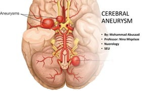
- 1. CEREBRAL ANEURYSM • By: Mohammad Abusaad • Professor: Nina Miqelaze • Nuorology • SEU
- 4. Cerebral Arteries A1-segment Anterior cerebral artery from carotid bifurcation to anterior communicating artery gives rise to the medial lenticulostriate arteries. A2-segment Part of anterior cerebral artery distal to the anterior communicating artery. P1-segment Part of the posterior cerebral artery proximal to the posterior communicating artery. The posterior communicating artery is between the carotid bifurcation and the posterior cerebral artery) P2-segment Part of the posterior cerebral artery distal to the posterior communicating artery
- 5. Middle Cerebral Artery • Horizontal M1-segment • gives rise to the lateral lenticulostriate arteries which supply part of head and body of caudate, globus pallidus, putamen and the posterior limb of the internal capsule. • Notice that the medial lenticulostriate arteries arise from the A1-segment of the anterior cerebral artery. • Sylvian M2-segment • Branches supply the temporal lobe and insular cortex (sensory language area of Wernicke), parietal lobe (sensory cortical areas) and inferolateral frontal lobe • Cortical M3-segment • Branches supply the lateral cerebral cortex
- 6. Aneurysms • They result from focal degeneration of the arterial wall. They are the most common cause of SAH. They are of 3 types : • Saccular (outpouching from arterial bifurcation) - Lacks internal elastic lamina • Fusiform (arterial dilatation) • Dissecting (pseudo-aneurysm)
- 7. SACCULAR ANEURYSM Berry like vesel outpouching arising from the bifurcation of the arteries. 80% of Intracranial aneurysms 85% - anterior circulations 15%- Posterior circulations.
- 8. FUSIFORM ANEURYSM • No definitive neck. • Long course of circumferential thickness. • Dolichoectasia . • Vertebrobasilar systemcommonly affected.
- 9. DISSECTING ANEURYSM • May be intracranial or extra-cranial. • Intracranial vertebralor posterior inferior cerebellar arteries – SAH • Extra-cranial carotid and vertebral arteries – Stroke (in young)
- 10. Presentation
- 11. INCIDENCE • General population is 0.5-5 % • Women >> Men • Increases with advancing age. • Genetic Predisposition. • Overall risk of rupture = 0.5-2 % per annum,
- 12. Risk Factor of Rupture • Daughtersac • Multi-lobulations • Irregular surface • Bottle-neck shape • Interval growth • Increase ratio of maximum • aneurysm diameter to parent vessel diameter.
- 13. Location • Most cerebral aneurysms arise from the circle of Willis and middle cerebral artery bifurcations. • Ninety percent involve the anterior circulation, and 10% the posterior circulation.
- 15. Giant Aneurysm Partially Thrombosed Giant aneurysm Leaking Giant aneurysm
- 16. Multiple aneurysm Multiple aneurysms are found in 15% to 30% of patients, Women: Men= 5:1 ratio, Most frequently involve the middle cerebral artery. Of the patients with multiple cerebral aneurysms, 75% have two, • 15% have three 10% have four or more.
- 18. • Bilateral symmetrical aneurysms are called Mirror Aneurysms, and • most often involve the ICA or the MCA bifurcations. • When one of multiple aneurysms ruptures and causes subarachnoid hemorrhage, it is most often the largest, although this is not always the case. • For instance, Nehls and colleagues have reported the propensity for anterior communicating aneurysms to rupture when several are present simultaneously.
- 19. Etiology Primary Causes Atherosclerosis • Hypertension • Smoking Abuse of cocaine, methamphetamine, ephedrine, heroin, and other drugs that produce arteritis or hypertension Vascular malformations - fibromuscular dysplasia, spontaneous cervical carotid or vertebral artery dissection, Takayasu's arteritis, neurofibromatosis I Connective tissue disorders, such as Marfan's syndrome and Ehlers- Danlos syndrome
- 20. Secondary Causes • Penetratingand nonpenetratingtrauma • Dissection (posttraumatic or otherwise) • Inflammation or mycosis due to septic • Neoplasm • infundibulumrepresents the residua of a developmental vessel that has undergone incomplete regression. • It most commonly involves the junction of the internal carotid artery and posterior communicating artery and less commonly involves the origin of the anterior choroidal artery from the internal carotidartery. • Infundibula are usually 3 mm or less in diameter.
- 21. Pathology • Pathologically, a number of patients with cerebral aneurysms demonstrate collagen type III abnormalities. • The walls of saccular cerebral aneurysms contain intima and adventitia, but the media and internal elastic membrane are thinned or absent. • Neoplasm: Pitutary adenoma – Growth hormone • Mycotic- spread of infection to vasa vasorum.
- 22. Features Need To Be Assessed • size: ideally 3 axis maximum size measurements • neck: maximal width of the neck of an aneurysm • the shape and lobulation • orientation: the direction in which the aneurysm points is often important in both endovascular and surgical planning
- 23. Cont…. • any smaller branches in the vicinity of an aneurysm • any branch taking off from the aneurysm • the presence of other aneurysms • relevant arterial variant anatomy (that may complicate or exclude endovasculartreatment
- 25. Imaging modalities • Computed tomography and CTA • MRI and MRA • DSA • Transcranial ultrasound • Earlier • Lumbar Puncture
- 26. Lumbar Puncture • Before CT, it was the only method to confirm the diagnosis of SAH but presently it is only indicated when CT is normal in patients with sudden, severe headache. • CSF is bloody not clearing with sequential tubes, Xanthrochromia is present after 1-2 days of SAH.
- 27. Computed Tomography • Acute SubarachnoidHemorrhage • The most important role for CT in the patient with a cerebral aneurysm is in the identification of ASAH, • Increased density within a cisternal space .
- 28. Cerebral Aneurysm • CT examination is secondarily important to identify cerebral aneurysms, typically 5 mm or larger in diameter. • The rate of identification of cerebral aneurysms - at least 67% for aneurysms 3 to 5 mm in diameter and to approach 100% for larger aneurysms. Giant aneurysms are most commonly identified in middle-age women; are typically located in the extradural carotid artery, the middle cerebral artery bifurcation, or the basilar summit; and usually manifest with mass effect
- 29. Cerebral Aneurysm • MRI is superior to CT for • the localization of cerebral aneurysms, • their relationship with adjacent structures, and • associated changes in neighboring brain tissue.
- 30. Management of intracranial aneurysms • The goal of preoperative management is • to stablize the patient for aneurysm obliteration and • prevent the development of systemic complications or secondary cerebral insults such as hypotension or hypoxia. • Ideally successful management of acutely ruptured aneurysm begins with adequate ventilation and oxygenation, normovolemia, hemodynamic stability, normoglycemia and ICP control.
- 31. • After initial stabilisation , a thorough radiographic evaluation is undertaken. • Surgical Methods: • Current surgical options include direct aneurysmal clippingand endovascular exclusion -- Surgical Clipping or Coil Embolization for the specific indications for treating an aneurysm surgically, endovascularly, or both).
- 32. Endovascular methods • GDC COILS - Of the various endovascular options currently available, Guglielmi detachable coils (GDCs) have had the largest influence with respect to treatment of subarachnoid hemorrhage; GDCs are first-line therapy nowdays. • These coils are soft, flexible, and can be contoured to the configuration of the aneurysm. • Sizes range from 2 to 20 mm in diameter and 2 to 30 cm in length. • Balloon embolization is efficacious in selected patients, but it has a higher incidence of complications than coil embolization.
- 33. Indications for endovascular treatment Posterior circulation aneurysms, especially basilar apex. Patients with poor clinical grade (ie, Hunt and Hess grades 4-5). Patients who are medically unstable. Symptomatic cavernous aneurysms. Small-neck aneurysms in the posterior fossa. Patients with vasospasm. Cases in which the aneurysm lacks a defined surgical neck (although these are also difficult to "coil") Patients with multiple aneurysms in different arterial territories if the surgical risk is high
- 34. Endovascular Procedure:Coiling • Step 1: Patient Preparation • Step 2: Insert the catheter
- 35. • Step 3: Locate the aneurysm
- 36. • Step 4:Insert the coil
- 37. • Stent placed in case of larger neck for the stabilization of the coil.
- 38. • Step 5: Check the coils • Step : Remove the catether
- 39. Evaluation after open /endovascular surgery MRI – MRI compatible clips Fatal cerebral hemorrhage if incompatible CTA superior to MRI DSA Catheter Angiography superior
- 40. THANK YOU