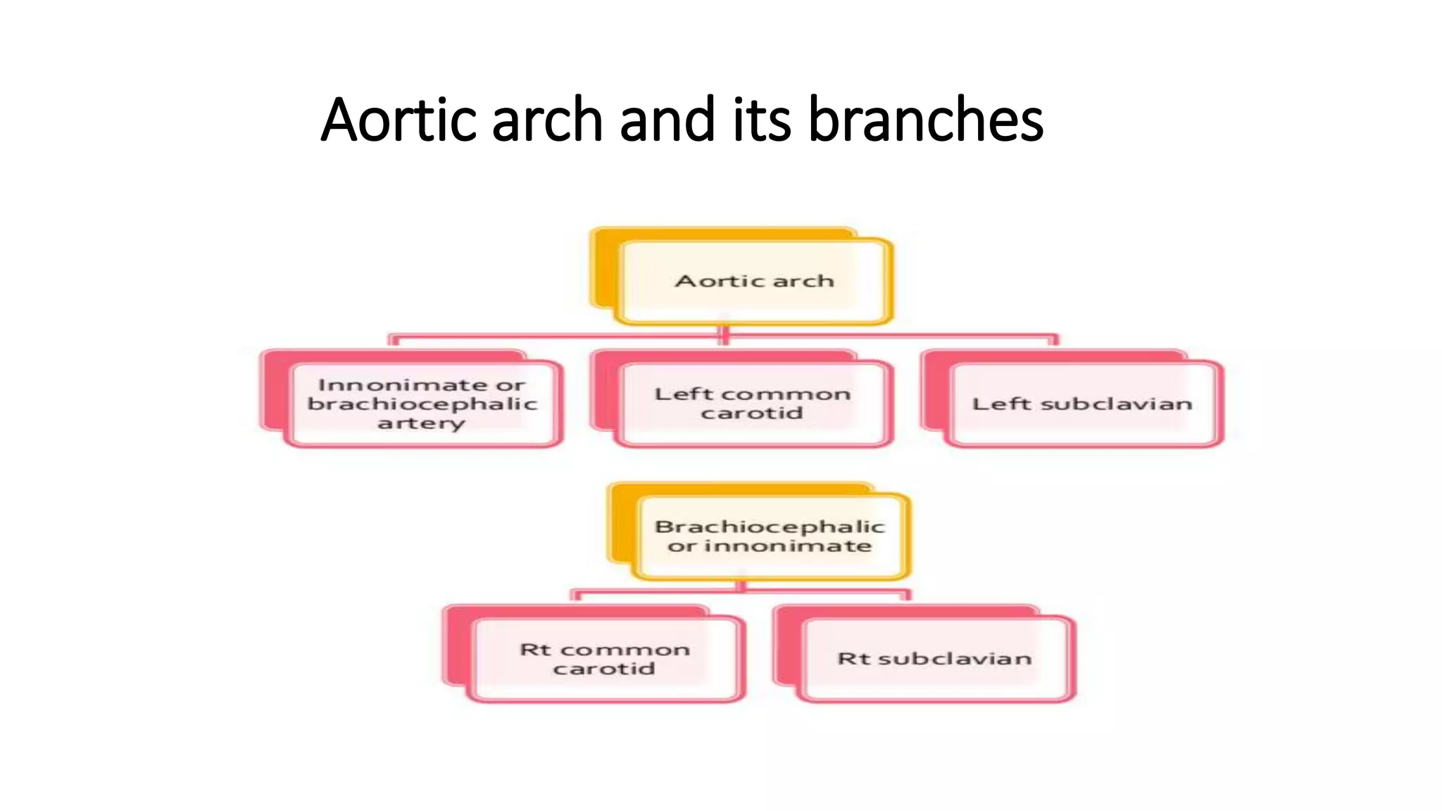
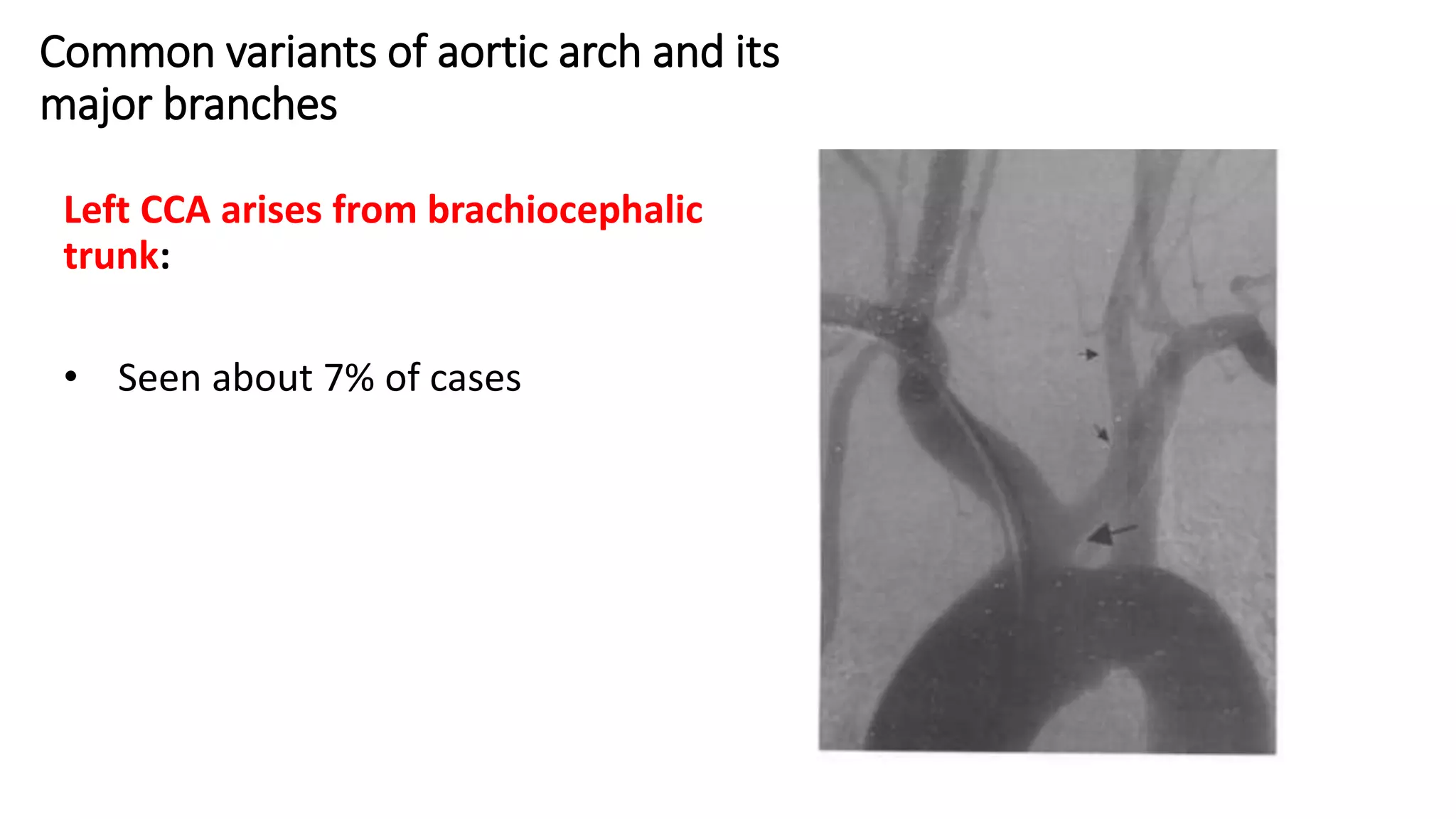
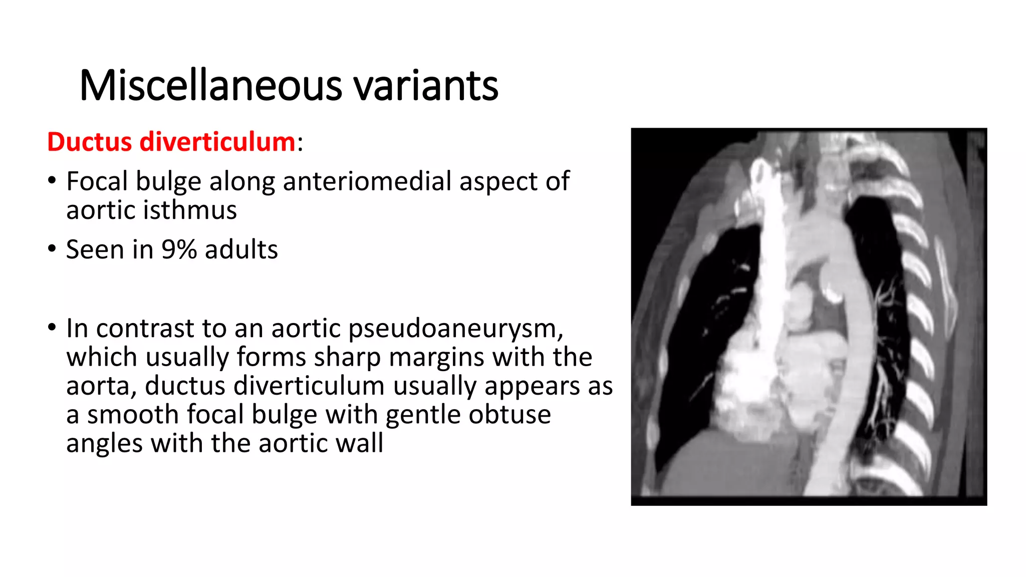
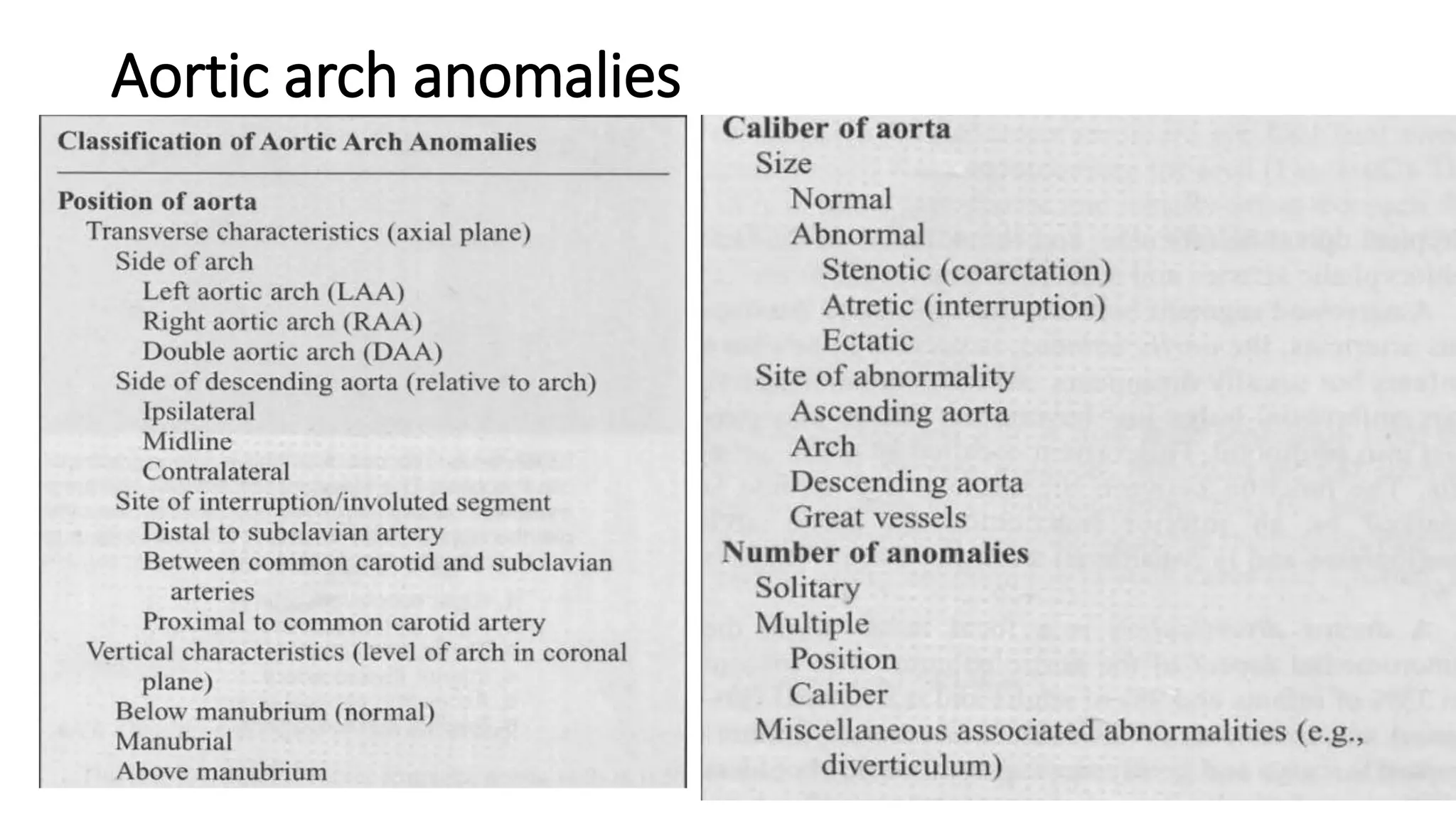
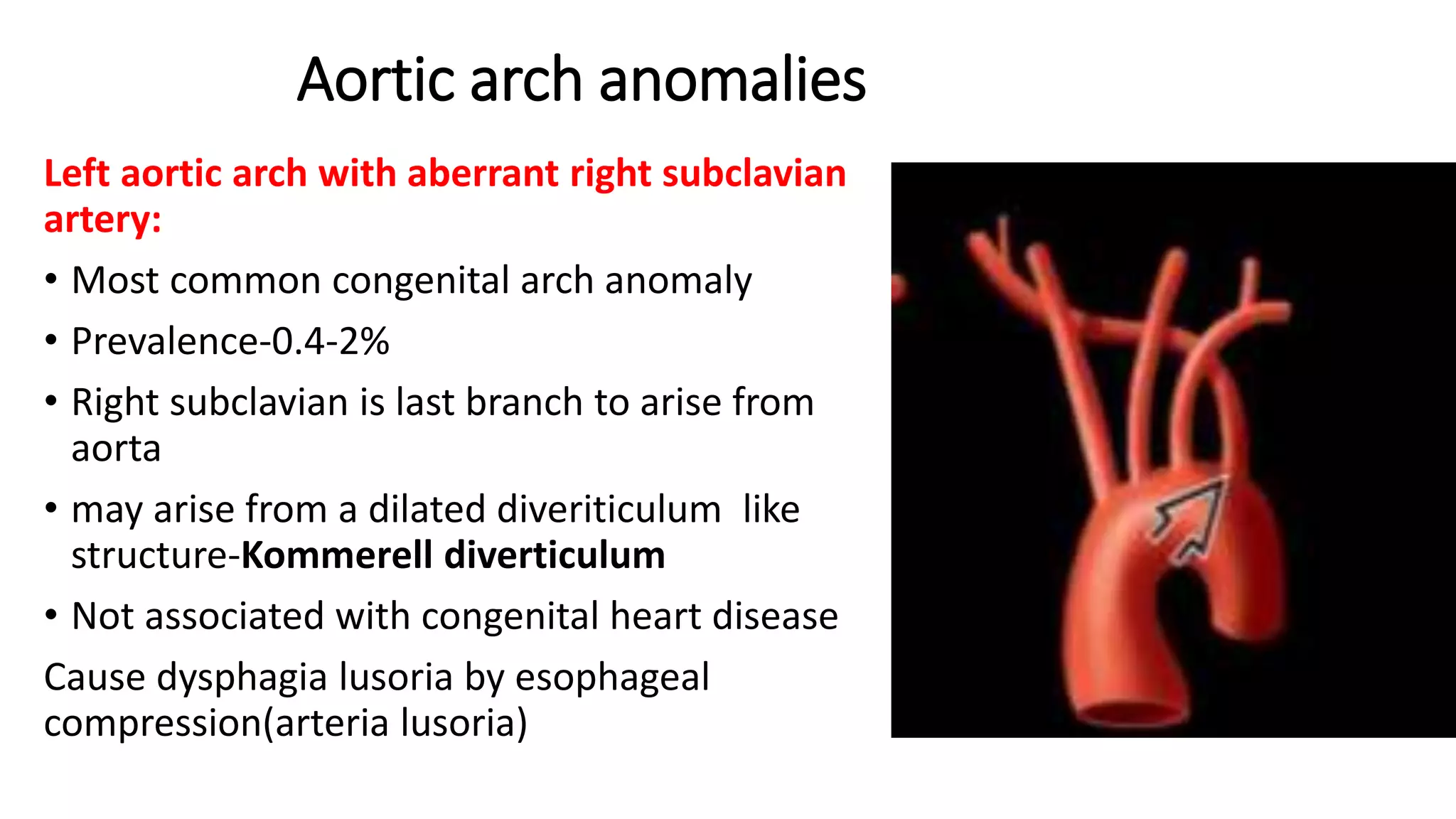
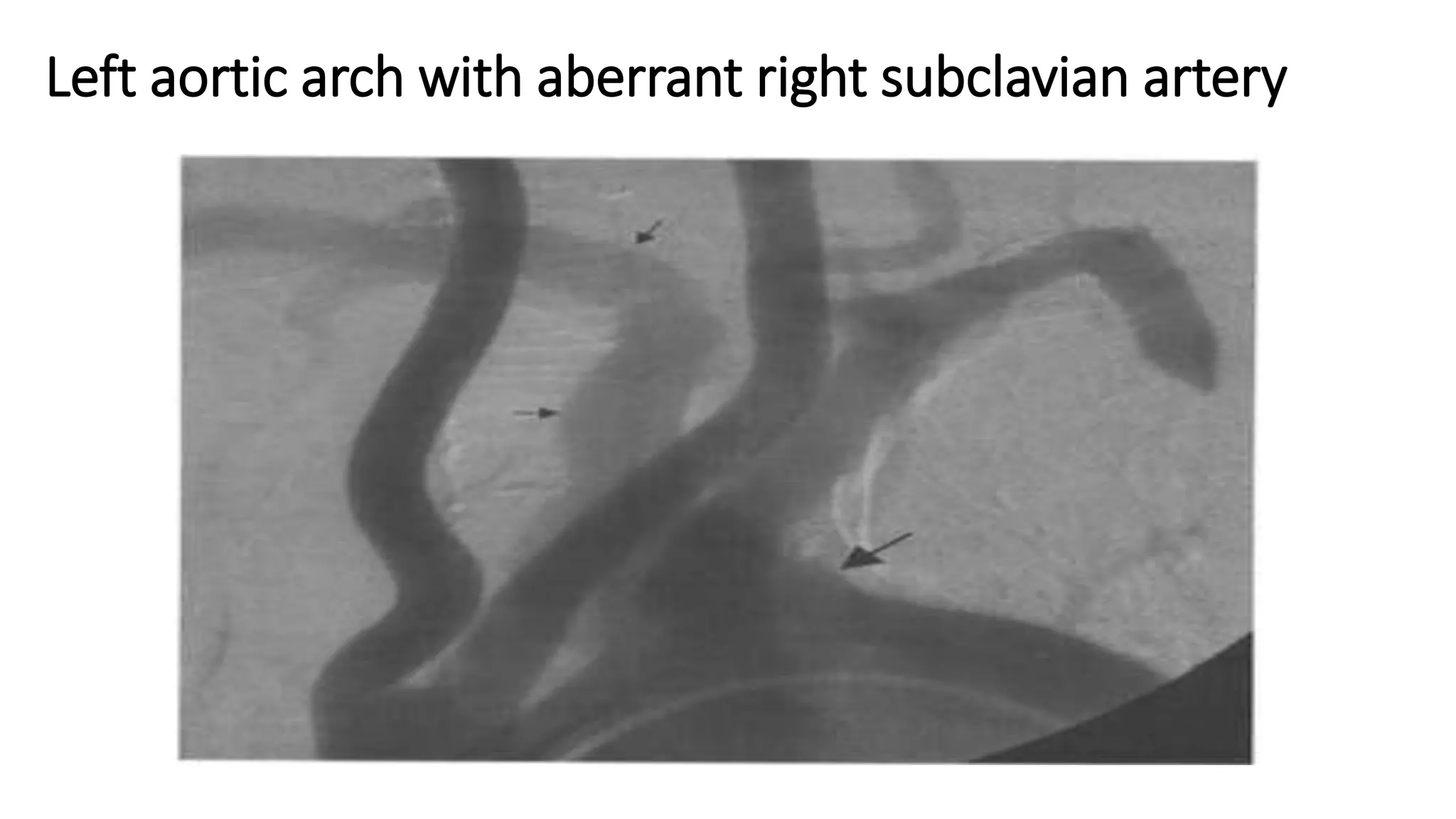
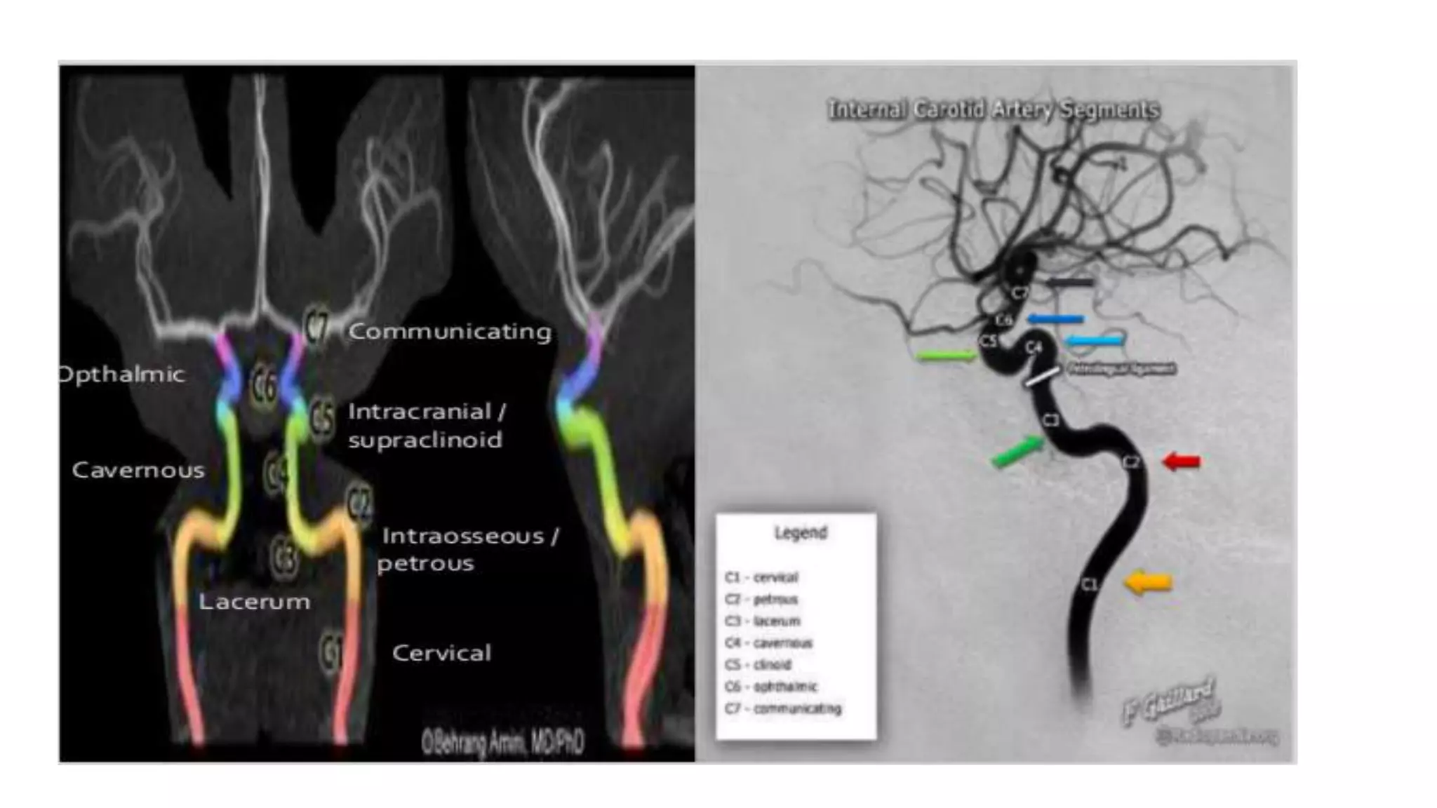
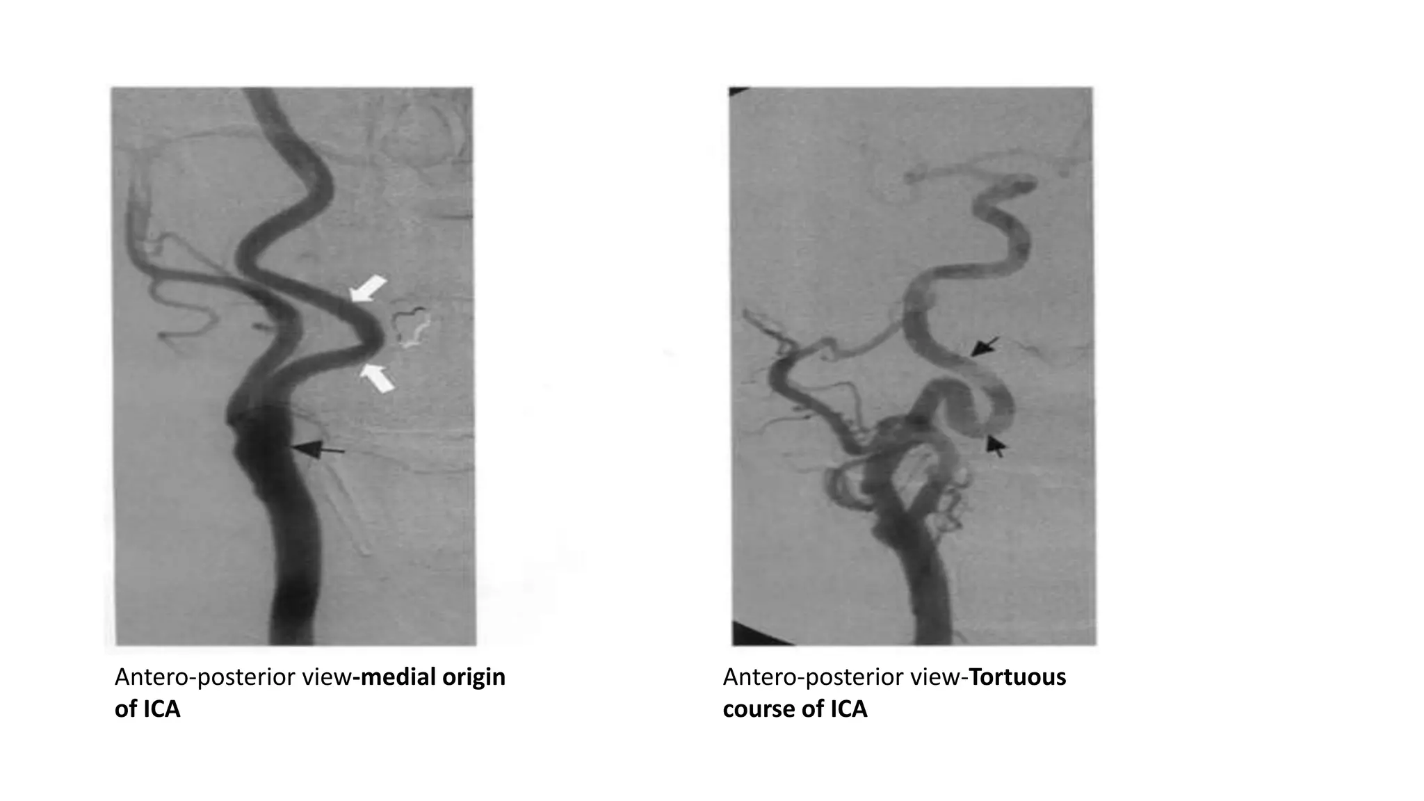
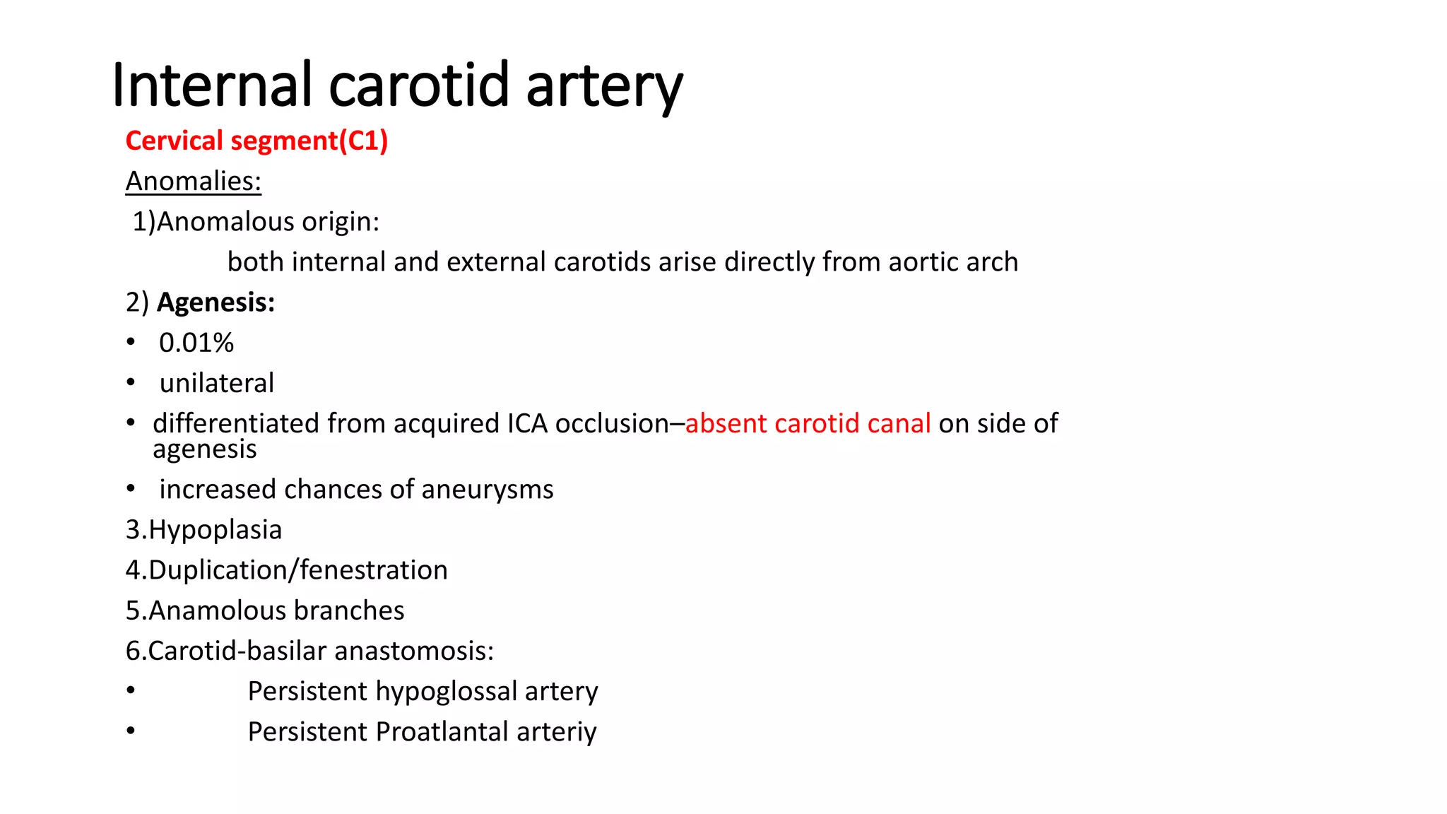
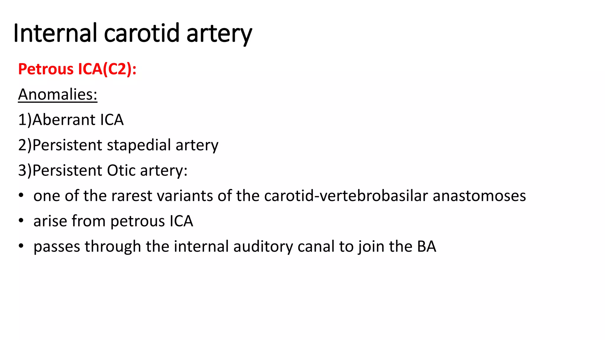
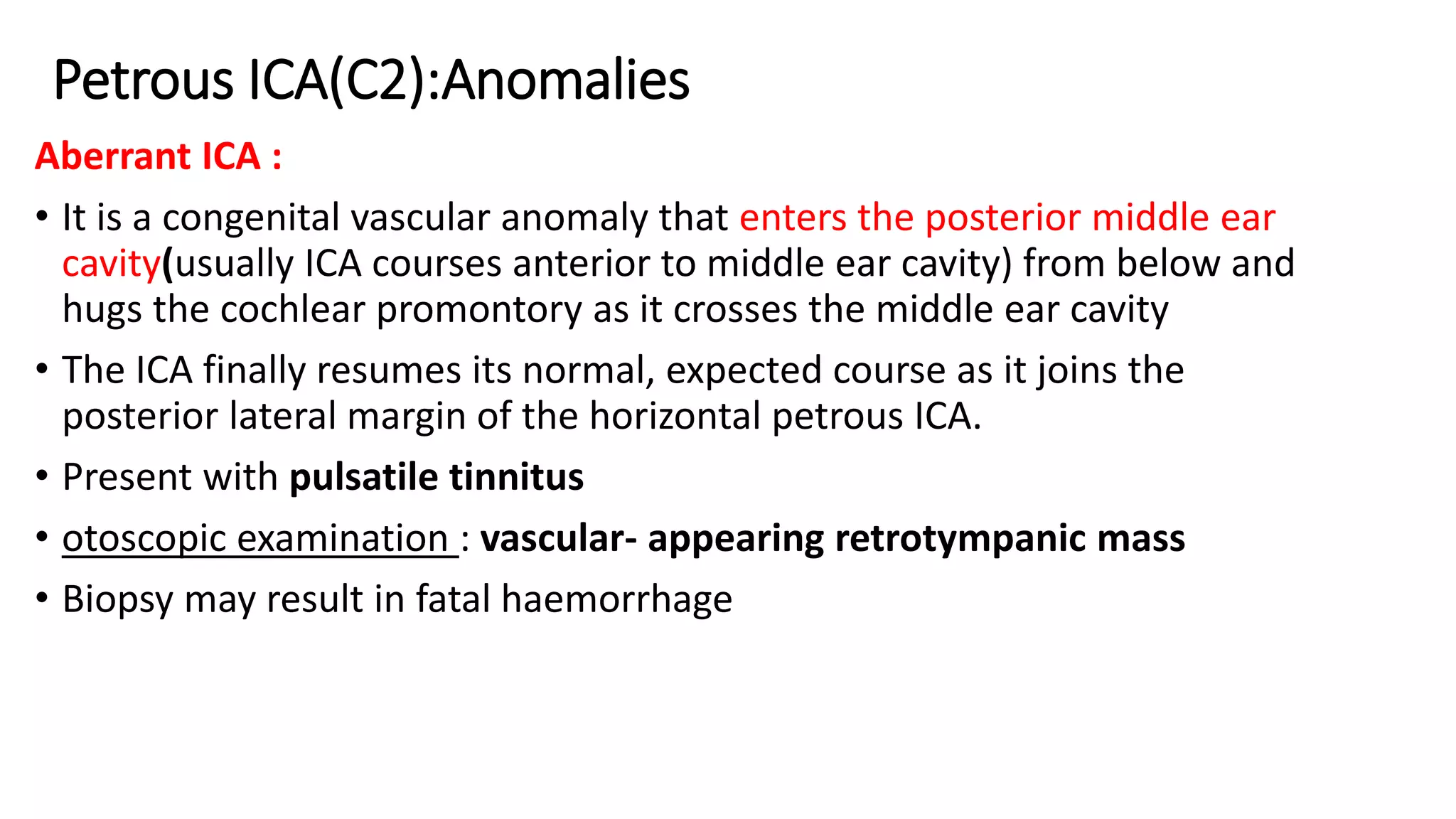
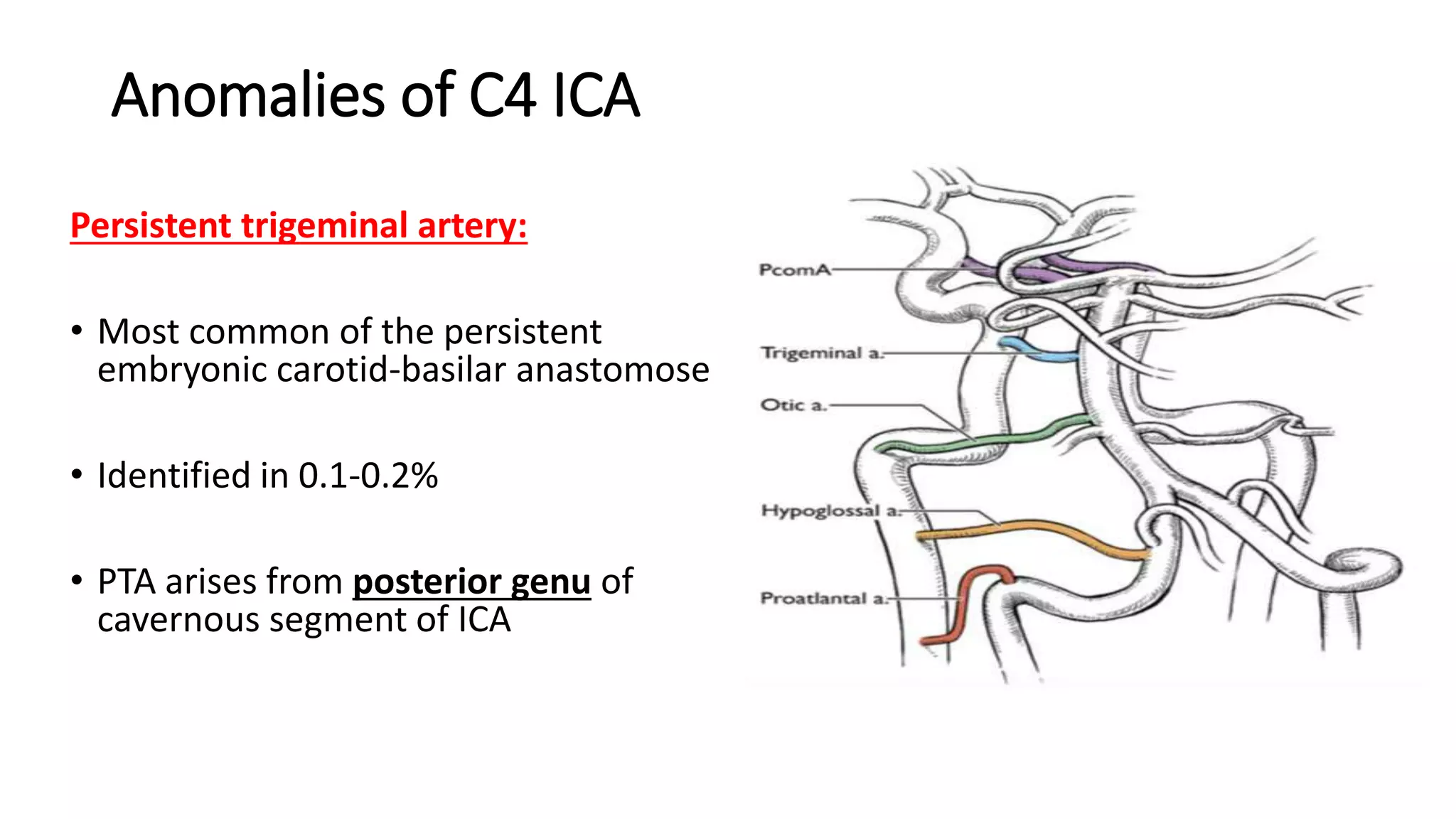
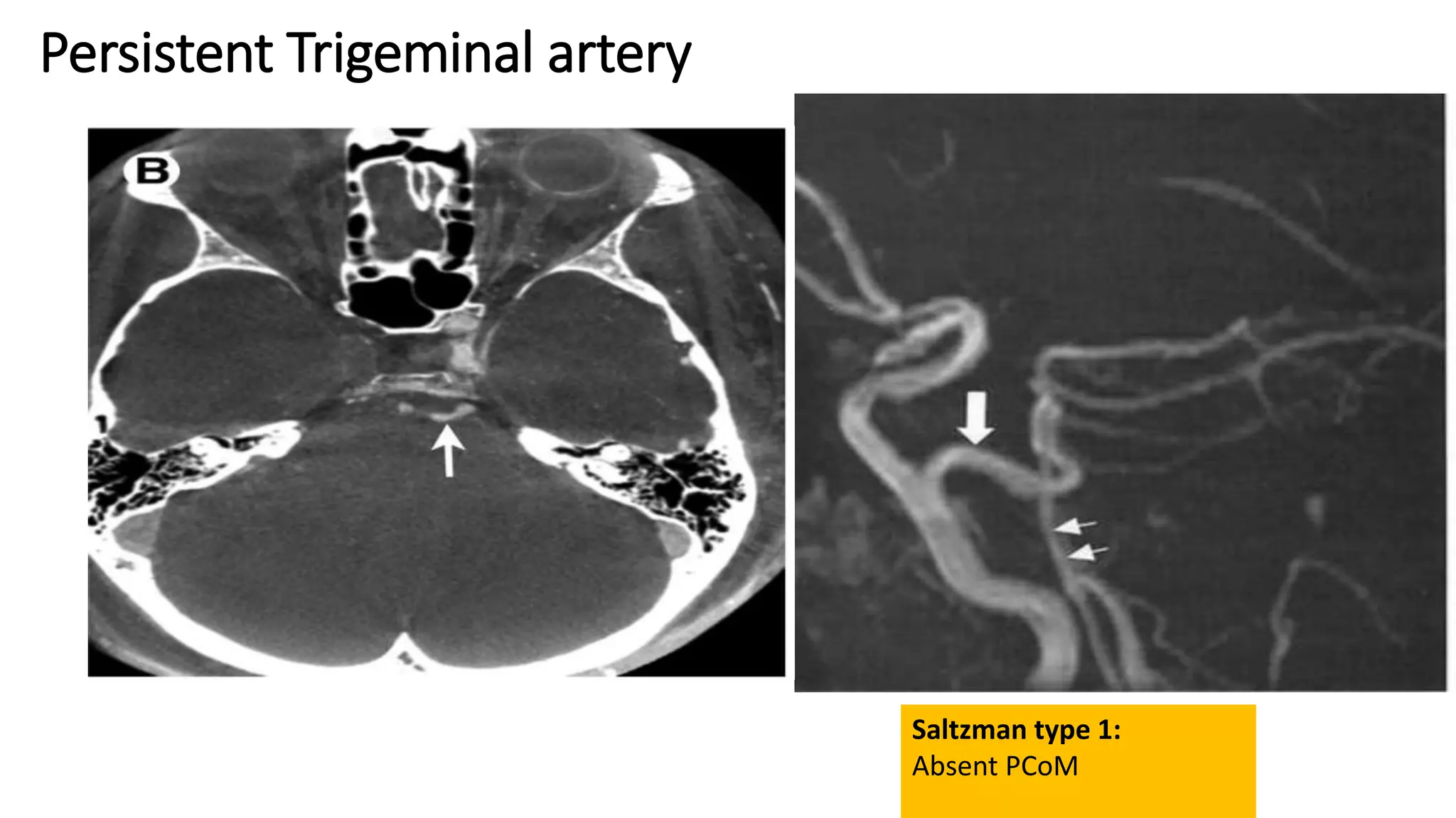
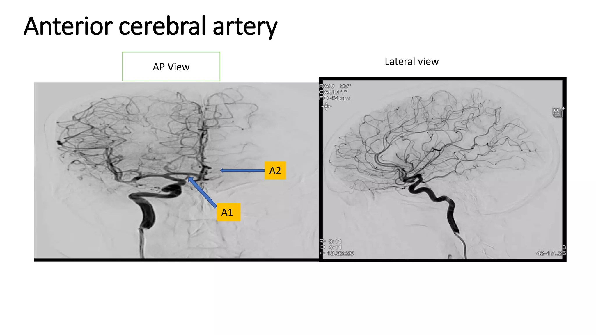
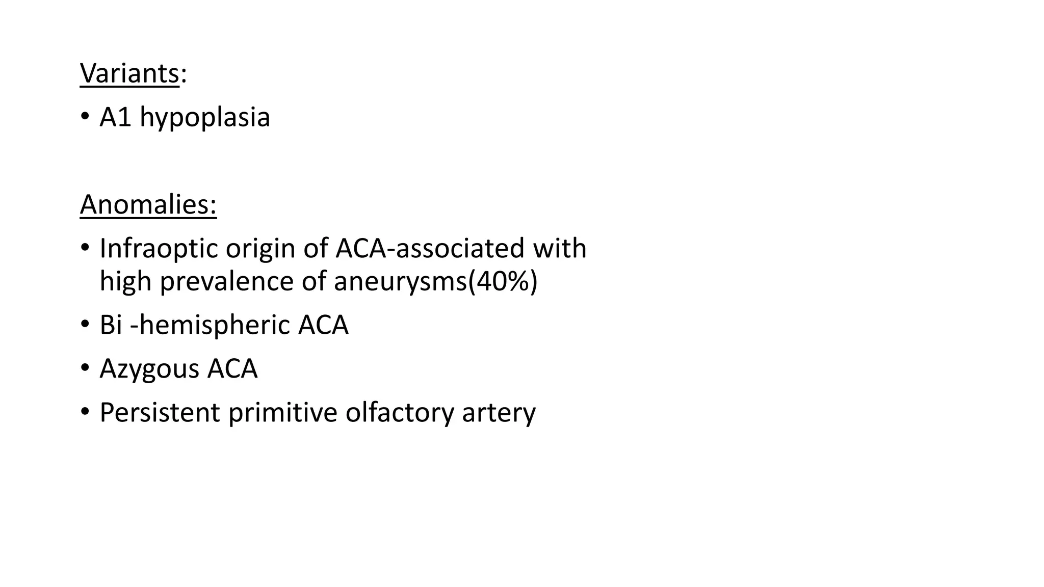
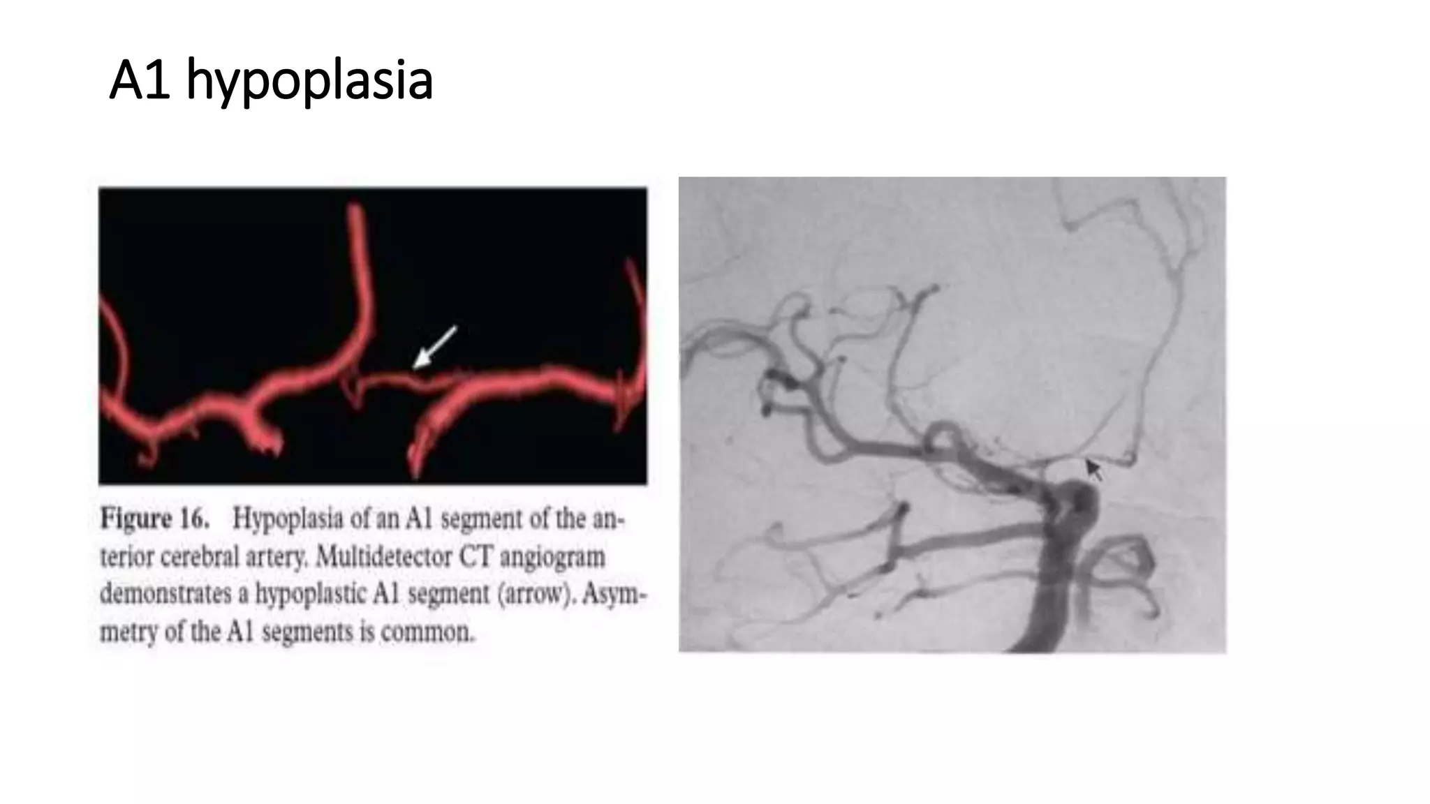
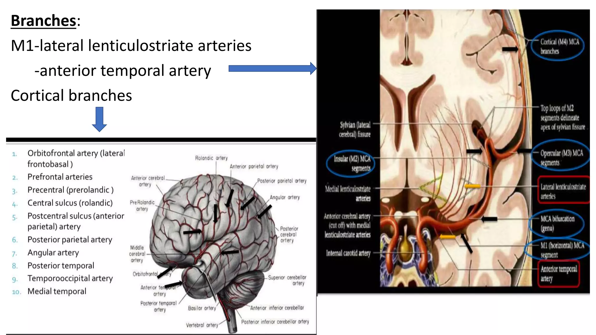
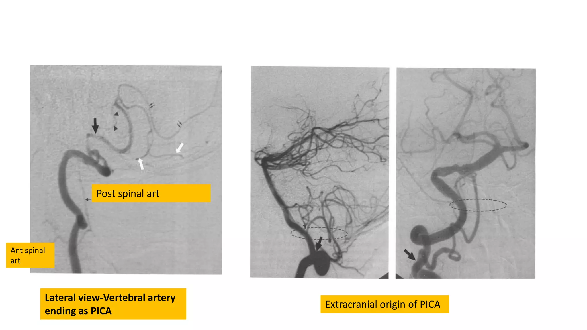
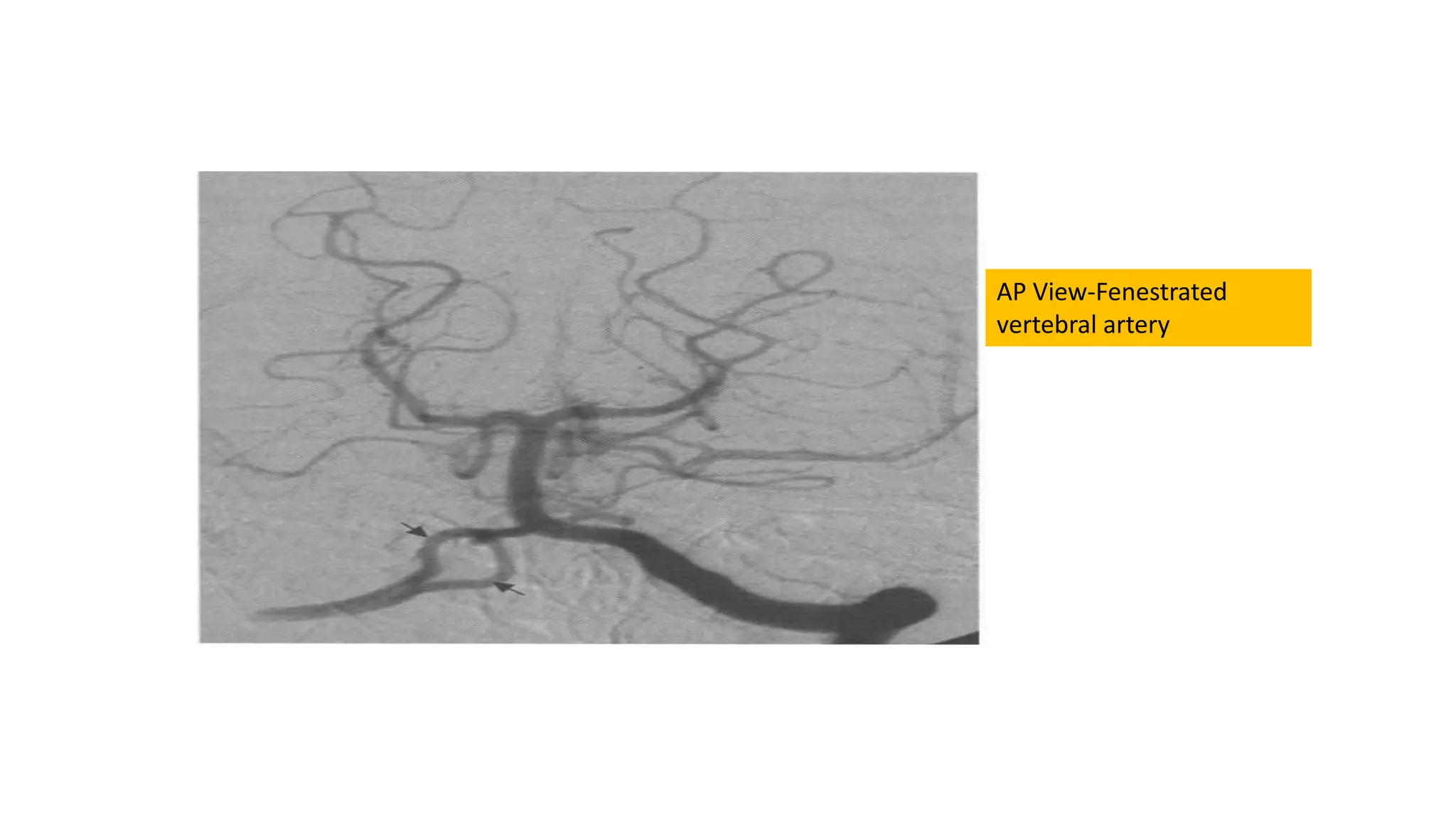
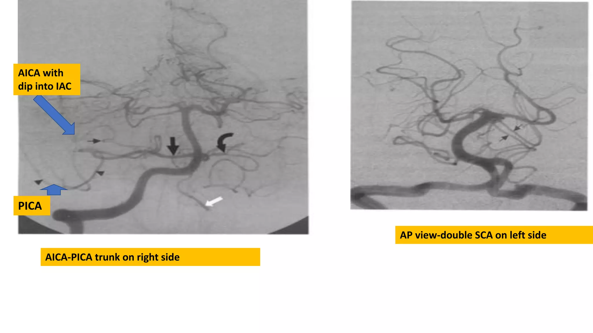
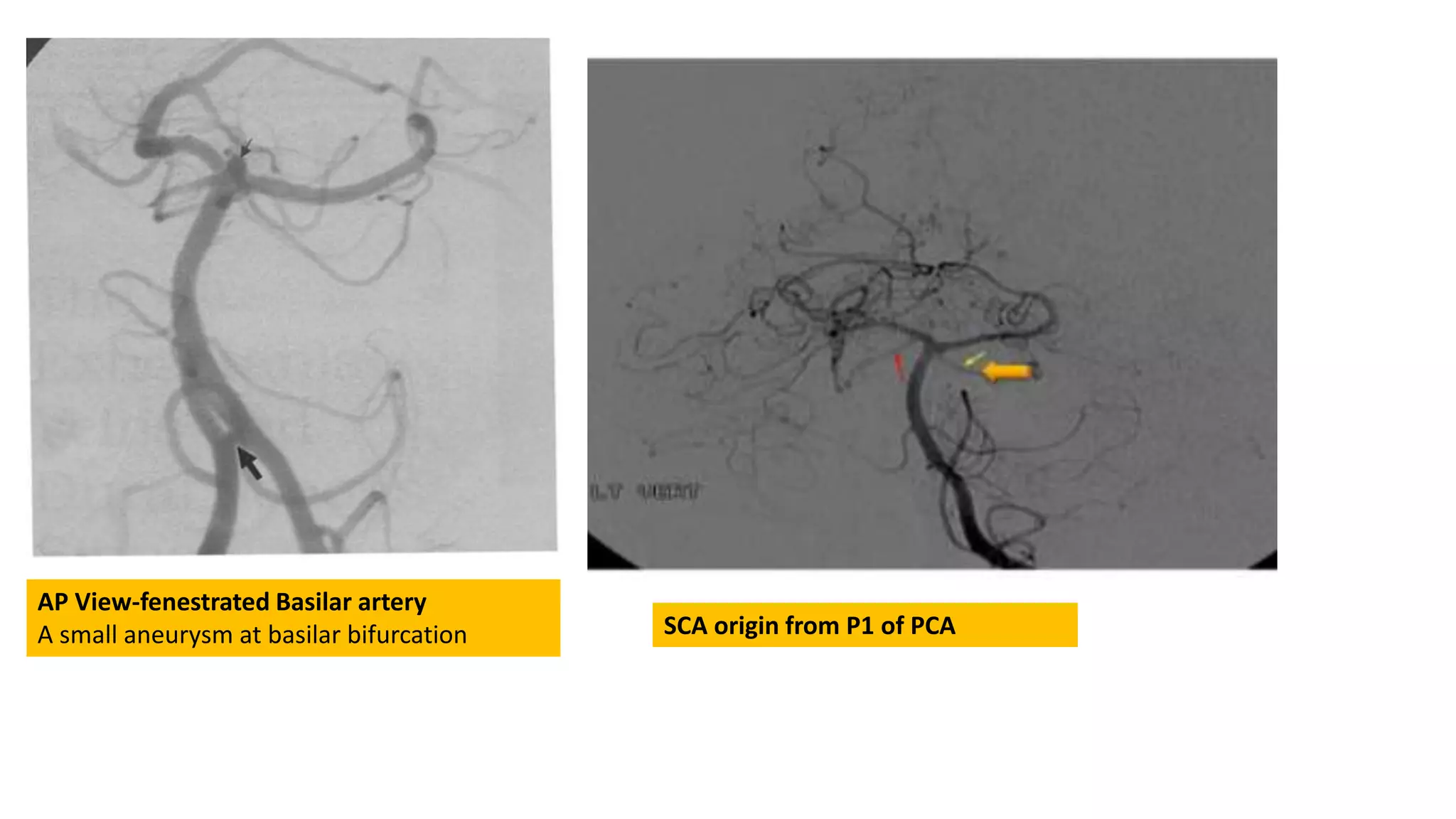
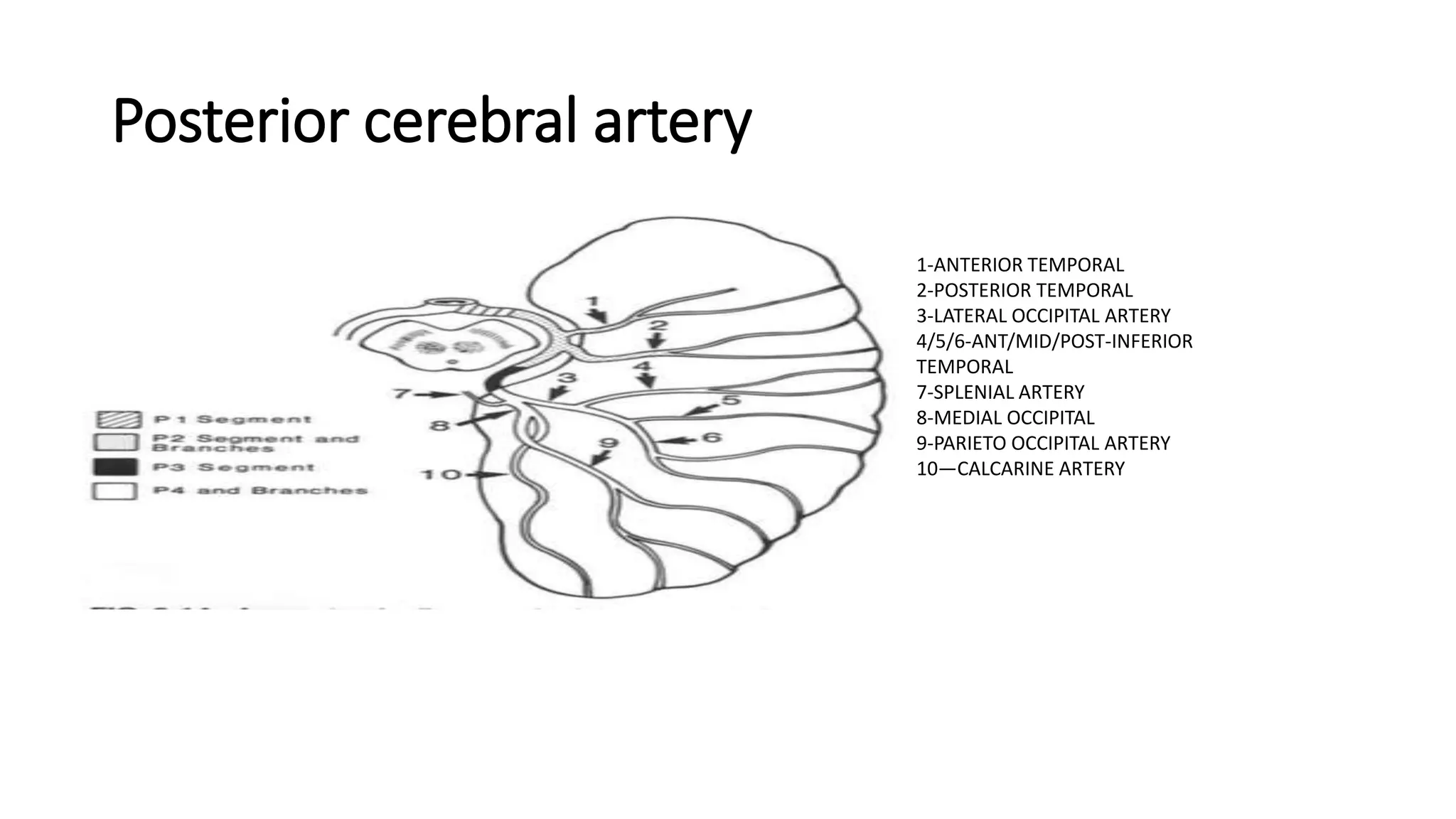
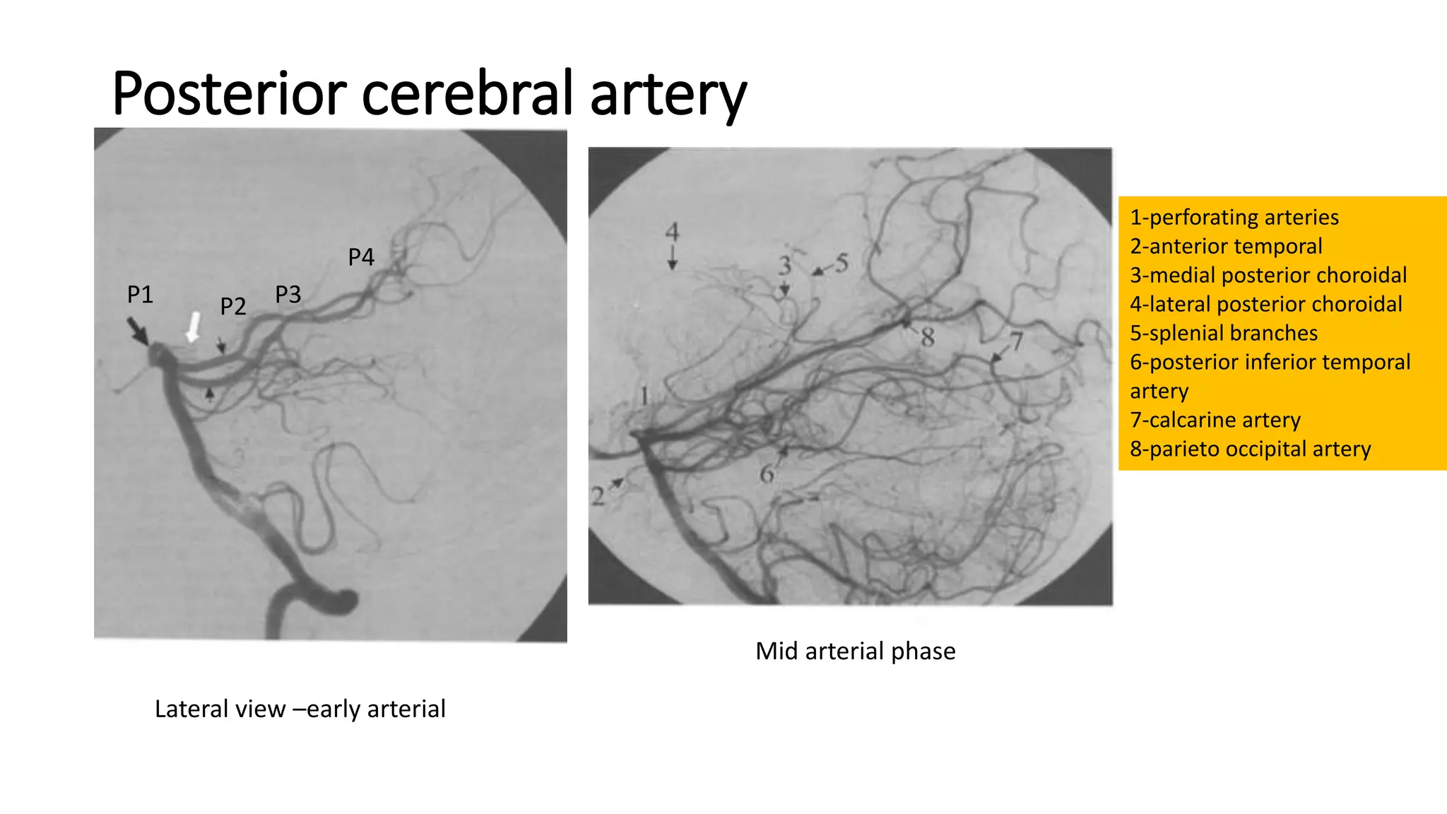
This document provides an overview of angiography and the course of blood vessels supplying the brain. It describes the aortic arch and its branches, as well as the internal carotid artery in detail. The internal carotid artery is discussed in segments from the cervical segment through the cavernous segment. Common variants and anomalies are described for various segments including the persistent trigeminal artery originating from the cavernous segment of the internal carotid artery.
imaging and anatomy of blood supply of brainSunil Kumar
The summary provides an overview of the arterial supply of the brain in 3 sentences:
The brain receives its arterial blood supply from the internal carotid and vertebral arteries. The internal carotid arteries give rise to branches that supply the anterior circulation including the anterior cerebral artery and middle cerebral artery. These arteries anastomose at the circle of Willis and give off numerous smaller branches to perfuse the brain.
This document provides an overview of the normal anatomy and variants of intracranial arteries, with a focus on the internal carotid artery. It describes the typical course and branches of the internal carotid artery and its major divisions including the anterior, middle, and posterior cerebral arteries. Various anatomical variants are also discussed, such as fenestrations, hyperplastic anterior choroidal arteries, fetal-type posterior cerebral arteries, posterior communicating artery infundibula, and persistent carotid-basilar anastomoses like the trigeminal artery. Clinical correlations including aneurysm formation and hemorrhagic risk are also mentioned.
This document provides an overview of normal variants and anatomy of the intracranial arteries, beginning with abbreviations used. It then describes the gross anatomy and specific segments of the internal carotid, basilar, vertebral, and posterior cerebral arteries. Vascular territories supplied by each artery are outlined. Finally, the document discusses normal variants and anomalies that can occur in the internal carotid artery anatomy. In particular, it notes that fenestration of the distal internal carotid artery is a rare finding associated with aneurysm formation. It also describes hyperplastic anterior choroidal arteries as a normal variant where the artery is enlarged beyond typical size.
The document describes the anatomy and course of the internal carotid artery (ICA). It discusses the anterior and posterior circulations fed by the ICA and vertebrobasilar trunk. Key points include the ICA coursing through the cavernous sinus and giving off branches like the ophthalmic artery. The posterior communicating artery connects the ICA to the vertebrobasilar system.
This document provides an overview of the vascular anatomy of the brain. It describes the arteries that make up the anterior and posterior cerebral circulations, including the internal carotid, anterior cerebral, middle cerebral, posterior cerebral and vertebrobasilar arteries. It details the branches and territories of supply of these major arteries. It also discusses variants and anomalies that can occur. Finally, it reviews the venous drainage of the brain through the dural sinuses and cerebral veins.
The document describes several types of persistent fetal carotid-vertebral anastomoses, including the persistent trigeminal artery, persistent otic artery, persistent hypoglossal artery, and persistent proatlantal artery. During typical embryonic development, these arteries regress after other anastomoses form, but in some cases they can persist into postnatal life. The persistent trigeminal artery is the most common variant and can connect the cavernous internal carotid artery to the basilar artery. The document provides detailed descriptions and images of each type of persistent fetal anastomosis.
Arterial and venous supply of brain part2 Sameeha Khan
1. The document describes the anatomy and branches of the major cerebral arteries including the anterior, middle, and posterior cerebral arteries.
2. It discusses the typical branching patterns and territories supplied by each artery and their segments.
3. Variations in arterial anatomy are also summarized such as fenestrations, duplications, hypoplasia.
This document contains an atlas of neurovascular anatomy with several figures and keys describing the major arteries of the head and neck, as well as the intracranial circulation including the anterior and posterior circulations. The figures show angiographic views of the arteries and their branches, labeled with numerical keys identifying each vessel. The document provides detailed angiographic views and labels for the major extracranial and intracranial arteries.
Vascular brain anatomy for Radiology by Dr Soumitra HalderSoumitra Halder
The document provides an overview of cerebral arterial and venous anatomy. It discusses:
1) The anterior and posterior cerebral circulations, including the internal carotid artery (ICA) and its branches that form the anterior circulation, and the vertebrobasilar system that forms the posterior circulation.
2) The branches of major arteries like the external carotid, vertebral, and basilar arteries.
3) Anatomical variations that can be seen, like hypoplastic vessels, fenestrations, and duplications.
4) Venous anatomy, including the dural venous sinuses and cerebral veins.
1. The cerebral vasculature includes the internal carotid and vertebral arteries which supply blood to the brain. Venous drainage occurs through dural sinuses and cerebral veins.
2. The Circle of Willis is a major vascular structure that connects the arteries of the brain. It includes components like the anterior and posterior cerebral arteries.
3. Imaging techniques used to evaluate the cerebral vasculature include conventional angiography, CT angiography, MR angiography, ultrasound and digital subtraction angiography. Each has advantages and disadvantages for assessing the anatomy and identifying abnormalities.
The document discusses the anterior cerebral circulation, including the internal carotid artery, anterior cerebral artery, and middle cerebral artery. It describes the typical vascular territories and clinical deficits that can result from occlusions or infarctions in different segments of these arteries. Key points include that unilateral middle cerebral artery occlusion can cause contralateral hemiplegia and homonymous hemianopia, while bilateral anterior cerebral artery occlusion can lead to paraplegia and urinary incontinence.
radiology Arterial and venous supply of brain neuroimaging part 1Sameeha Khan
The document discusses the anatomy and imaging of cerebral vasculature. It begins by covering the major vessels arising from the aortic arch, including the brachiocephalic trunk, right and left common carotid arteries, and right subclavian artery. It then details the branches and course of the external carotid artery. The remainder discusses the segments and branches of the internal carotid artery as it passes through the petrous, cavernous, and supraclinoid regions. Key branches include the ophthalmic artery and inferior hypophyseal artery. Various angiographic views and MRI/CT techniques for visualizing these vessels are also summarized.
RADIOLOGICAL ANATOMY OF ARTERIAL SUPPLY OF BRAINMohammad Naufal
1. The arterial supply of the brain comes from the internal carotid arteries and vertebral arteries, which form the circle of Willis at the base of the brain.
2. The main branches of the vertebral arteries include the posterior inferior cerebellar artery and posterior spinal arteries. The vertebral arteries join to form the basilar artery.
3. The internal carotid artery gives off branches that include the anterior cerebral artery, middle cerebral artery, and anterior choroidal artery. These arteries supply different regions of the brain.
4. The circle of Willis is formed by the anterior and posterior cerebral arteries connecting the left and right internal carotid and basilar arteries, allowing for collateral blood flow in case of arterial occlusion.
The document summarizes the arterial and venous anatomy of the brain. It describes the major arteries that supply blood to the brain, including the internal carotid, vertebral, and basilar arteries. It discusses the circle of Willis and territories supplied by the anterior, middle, and posterior cerebral arteries. It also outlines the dural venous sinuses and cerebral veins that drain blood from the brain. Key structures mentioned include the cavernous sinus and superior sagittal sinus.
The document discusses vascular anatomy of the brain. It notes that 18% of total blood volume circulates through the brain, which accounts for 2% of body weight. Loss of consciousness occurs within 15 seconds and irreversible brain damage within 5 minutes if blood flow to the brain stops. It then describes the various arteries that supply the brain, including the carotid arteries, vertebral arteries, and branches within the brain. It provides details on imaging techniques used to evaluate the vasculature such as angiography, CTA, MRA. Overall, the document provides an overview of the anatomy and imaging of brain vasculature.
The document describes the anatomy of various cerebral arteries and veins. It discusses the segments of the internal carotid artery and its branches. It also describes the arteries of the anterior, middle, and posterior cerebral circulation including their segments, branches, and territories. Additionally, it outlines the deep venous system including the internal cerebral veins, vein of Galen, straight sinus and their drainage patterns. Finally it illustrates the dural sinuses and venous connections between the brain and dura.
Brain vascular anatomy with MRA and MRI correlationArif S
This document provides an overview of the vascular anatomy of the brain. It discusses the arterial supply, venous drainage, and dural venous sinuses of the brain. For arterial supply, it describes the anterior and posterior circulations, including the internal carotid, vertebral, basilar, anterior cerebral, middle cerebral, and posterior cerebral arteries. It also discusses branches and territories of these vessels. For venous drainage, it outlines the internal cerebral veins and external cerebral veins, as well as dural venous sinuses such as the superior sagittal sinus. Watershed zones and vascular territories on cross sections are also depicted.
The common carotid artery divides into the internal and external carotid arteries. The internal carotid artery supplies structures within the cranium including the eye. The external carotid artery supplies the superficial head and neck. The ophthalmic artery, a branch of the internal carotid, gives rise to numerous branches that supply the eye and surrounding structures. These branches anastomose to form networks that provide the eye and orbit with their blood supply. Venous drainage from the eye and orbit occurs primarily through the superior and inferior ophthalmic veins which drain into the cavernous sinus.
Presentation1.pptx, radiological vascular anatomy of the head and neck.Abdellah Nazeer
This document provides an overview of the radiological vascular anatomy of the head and neck. It begins with an introduction to brain anatomy, describing its main components and vasculature. It then describes the anatomy of key blood vessels originating from the aortic arch that supply the head and neck region, including the common carotid, internal carotid, external carotid, vertebral and subclavian arteries. It details the branches and course of these vessels. It also provides an overview of cerebral and dural venous anatomy. Images are included to illustrate key anatomical structures and relationships.
This document summarizes the posterior circulation of the brain. It describes how the vertebral arteries join to form the basilar artery in the brainstem. The basilar artery then divides into the two posterior cerebral arteries. Key branches include the posterior inferior cerebellar artery and superior cerebellar artery. The posterior cerebral arteries supply blood to the occipital and temporal lobes. The vertebrobasilar system provides blood to the brainstem, cerebellum, and posterior portions of the telencephalon.
The MCA is the largest and most complex of the cerebral arteries. It arises from the internal carotid artery and has four segments - M1, M2, M3, M4. The M1 segment can be further divided into pre-bifurcation and post-bifurcation parts. Early branches and perforating arteries originate from the M1 segment. The MCA has a variable branching pattern and anomalies include duplication, accessory branches, and fenestration. Key angiographic landmarks include the Sylvian point and Sylvian triangle.
This document provides an overview of brain anatomy and vascular supply. It begins by describing the protective coverings, brain parenchyma structures, and vascular anatomy. It then discusses the arterial supply in more detail, covering the territories of the internal carotid, middle cerebral, anterior cerebral, anterior choroidal, vertebrobasilar, posterior cerebral, and circle of Willis arteries. It also reviews the vascular territories of the cerebral hemispheres, cerebellum/brainstem, and basal ganglia/internal capsule. Finally, it discusses watershed areas and their appearance on neuroimaging.
The document discusses the arterial supply of the brain. It describes the major arteries originating from the aorta including the brachiocephalic artery, common carotid arteries, vertebral arteries, and branches of these arteries. It discusses the segments and branches of the internal carotid artery as it passes through the cervical, petrous, cavernous, clinoid, and communicating segments. It also describes the circle of Willis and its variants. Key arteries discussed include the anterior, middle, and posterior cerebral arteries as well as the basilar and vertebral arteries.
The optic nerve receives its blood supply from multiple sources along its path from the eye to the brain. In the eye, it is supplied by retinal arterioles and occasionally the ciliary artery. In the prelaminar region, it receives blood from ciliary region vessels and peripapillary choroidal or short posterior ciliary vessels. Deeper regions receive supply from ciliary and retinal circulations, including recurrent pial vessels and branches of the central artery of the retina. In the orbit, it has periaxial vessels from the ophthalmic artery and axial vessels from the central retinal artery. Within the cranium, it is supplied by the pial plexus fed by branches from the ophthalmic artery.
The internal carotid artery has 7 segments from its origin at the common carotid artery bifurcation to where it enters the cranium. Each segment has unique anatomic features and branches. The segments are named cervical, petrous, lacerum, cavernous, clinoid, ophthalmic, and communicating. The petrous, cavernous, and ophthalmic segments each have important branches including the vidian artery, meningohypophyseal trunk, and ophthalmic artery respectively.
Carotid artery and ophthalmic artery2 (2)amanimohsen1
The document discusses the carotid arteries, which supply blood to the head, neck, and brain. It specifically focuses on the internal carotid artery and its branch, the ophthalmic artery (OA). The OA originates from the internal carotid artery and enters the orbit through the optic canal, supplying structures in the orbit as well as parts of the nose and face. One of its branches is the central retinal artery, which runs along the optic nerve and enters the eye to supply the inner retinal layers.
Radiologic Anatomy of the Blood Supply to the Brain.pptxWilliamsMusa1
The document summarizes the radiologic anatomy of the arterial blood supply to the brain using various imaging modalities. It describes the relevant gross anatomy of the major cerebral arteries, including branches and segments. MR angiography is discussed as the preferred noninvasive method for evaluating the cerebral vasculature. CT angiography and conventional angiography provide detailed images but are more invasive. Ultrasound can also be used to image intracranial vessels through various acoustic windows. Variations in anatomy, such as those seen in the circle of Willis, are commonly observed.
This document discusses the vascular anatomy of the head, including the common carotid artery, external carotid artery, internal carotid artery, and their branches. It provides details on the origin, course, branches, and key relationships of these arteries. The internal carotid artery is discussed in particular depth, outlining its 7 segments and typical branches within the cervical, petrous, lacerum, cavernous, clinoid, ophthalmic, and communicating segments. Key anatomy and variations are highlighted throughout.
The document summarizes the major arteries of the head and neck, including their embryological development, course, branches, and clinical significance. It describes the carotid system, internal carotid artery, and external carotid artery in detail. Key branches discussed include the superior thyroid, lingual, facial, and maxillary arteries. Variations in artery origins are also noted.
Anatomy and imaging of coronary artery disease withSarbesh Tiwari
1) Coronary CT angiography (CCTA) uses computed tomography to non-invasively image the coronary arteries. It provides high quality images of the coronary arteries and their branches.
2) CCTA is performed using either electron-beam CT or multi-detector row CT (MDCT). MDCT is now more commonly used due to its wider availability and lower cost. The latest generation 64-detector MDCT allows for very high resolution imaging.
3) CCTA requires careful preparation of the patient including medication to control heart rate and dilation of the coronary arteries. The scan itself involves ECG gating to image the heart during diastasis and injection of iodinated contrast to outline the coronary
The internal carotid artery arises from the common carotid artery and travels upward through the neck and into the skull. It has seven segments from C1-C7 as it passes through the carotid canal, cavernous sinus, and terminates by joining the circle of Willis. The internal carotid artery has few branches in the neck but gives off important branches in the cavernous sinus and within the skull such as the ophthalmic artery. Anatomical variations and injuries to the internal carotid artery during surgery can lead to serious complications if not properly managed.
1. The brain receives blood supply from the internal carotid arteries and vertebral arteries, whose branches anastomose to form the circle of Willis at the base of the brain.
2. The internal carotid artery enters the skull through the carotid canal and gives off branches including the anterior cerebral artery, middle cerebral artery, and posterior communicating artery.
3. Occlusion of the internal carotid or its major branches can cause symptoms such as hemiparesis, aphasia, or visual field defects depending on the location of occlusion.
The document discusses the vascular territories of the brain. It begins by outlining some key arteries and their branches, including the internal carotid artery, circle of Willis, middle cerebral artery, anterior cerebral artery, and persistent carotid-basilar connections. It then discusses several specific arterial territories in more detail, including the territories supplied by the posterior inferior cerebellar artery, superior cerebellar artery, branches of the vertebral and basilar arteries, and the anterior choroidal artery. The document emphasizes understanding vascular anatomy and territories to aid in diagnosing different types of strokes and infarcts.
Clinical Anatomy of the Heart, Pericardium and.pptxHafizMohd21
The document discusses the anatomy of the heart, pericardium, and coronary circulation. It describes the layers of the pericardium and clinical conditions like pericarditis and pericardial effusion. It details the structures of the heart chambers, including the atria and ventricles. It also outlines the coronary arteries and conduction system of the heart. Key points are highlighted through relevant clinical examples and sample exam questions.
Cerebral aneurysms arise from focal degeneration of arterial walls. The most common type is saccular aneurysms, which protrude from arterial bifurcations and lack an internal elastic lamina. Aneurysms can present with subarachnoid hemorrhage, cranial nerve palsy, headache or seizures. Imaging plays a key role in diagnosing aneurysms and evaluating risks. Computed tomography best identifies acute subarachnoid hemorrhage but may miss small bleeds. Catheter angiography remains the gold standard for precise aneurysm characterization to guide treatment.
Coronary angiography remains the gold standard for detecting coronary artery disease. The technique was first performed in 1958 by Dr. Mason Sones at the Cleveland Clinic. Coronary angiography allows visualization of the coronary arteries, branches, and anomalies to precisely locate lesions. It provides information needed for coronary interventions. The procedure involves accessing the femoral or radial artery and advancing a catheter into the heart to inject contrast dye and image the arteries. It can detect blockages but has limitations like vessel overlap that may obscure lesions. Complications are rare but can include artery damage, embolism, or arrhythmias.
Coronary angiography remains the gold standard for detecting coronary artery disease. The technique was first performed in 1958 by Dr. Mason Sones at the Cleveland Clinic. Coronary angiography allows visualization of the coronary arteries, branches, and anomalies to precisely locate lesions. It remains an important diagnostic tool used to evaluate patients with suspected coronary artery disease. The procedure involves accessing the femoral artery and advancing a catheter into the heart to inject contrast and obtain images of the coronary arteries under fluoroscopy. Precise technique and monitoring are required to minimize risks of potential complications.
This document discusses cerebral blood flow, its autoregulation, clinical relevance, and the role of collaterals in ischemic stroke. It begins with an overview of cerebral blood supply and drainage, then describes the autoregulation mechanism and its importance. It also discusses cerebral collaterals and their significance in acute ischemic stroke. The majority of the document provides detailed descriptions of the anatomy of cerebral arteries, veins, and sinuses. It explains factors that regulate cerebral blood flow and perfusion pressure, including metabolism, carbon dioxide, oxygen, and autoregulation.
1. Coronary artery anomalies can be classified as either normal coronary anatomy or anomalous coronary anatomy based on variations in origination, course, and termination.
2. Specific anomalous coronary artery variations include anomalous location of the coronary ostium, single coronary artery, high take-off of the coronary artery from the aorta, and origins from the opposite or improper sinus.
3. Additional intrinsic anomalies involve hypoplastic arteries, myocardial bridging, coronary artery ectasia/aneurysms, and coronary arteriovenous fistulas or other terminations such as anomalous drainage into systemic arteries.
Normal anatomy and congenital anomalies of vena cavaeGobardhan Thapa
This document discusses the normal anatomy and common congenital anomalies of the superior and inferior vena cavae. It begins with an overview of the embryological development of the vena cavae. It then describes the normal anatomy of the superior and inferior vena cavae and their major tributaries. Common congenital anomalies are then outlined, including double superior vena cavae, left-sided superior vena cava, left inferior vena cava, double inferior vena cava, azygos continuation of the inferior vena cava, and circumcaval anomalies of the left renal vein and ureter. Clinical significance is discussed for some anomalies.
The document discusses the blood supply to the inner ear (cochlea). It notes that the labyrinthine artery, a branch of the anterior inferior cerebellar artery, supplies the inner ear. This artery passes through the internal auditory meatus and divides into the anterior vestibular artery and common cochlear artery. The common cochlear artery further divides into the cochlear and vestibulocochlear arteries, which supply different parts of the cochlea. Disruptions to this blood supply can cause various complications like sensorineural hearing loss, endolymphatic hydrops, and ischemia-related hearing loss.
The document summarizes the vascular supply and drainage of the vertebrae and spinal cord. It describes the arterial supply of the vertebrae, which comes from segmental arteries, and its venous drainage through internal and external plexuses. It then discusses the arterial supply of the spinal cord from the anterior and posterior spinal arteries as well as segmental and radicular branches, and its venous drainage through anterior and posterior spinal veins. Finally, it covers ischemia of the spinal cord, including causes, presentation, management, and prognosis.
It's helpful in understanding various aspects of revascularization procedures, with good illustrations, easy to learn, no complexity, easy language, conclusion added, short descriptions
The document discusses renal vascular anatomy and its implications for surgery. It notes that the renal arteries typically enter the kidney through the hilum and branch into segmental arteries that do not anastomose. It also describes variations such as accessory renal arteries. The segmental arteries further branch within the kidney and an avascular plane exists between anterior and posterior branches. Venous drainage parallels the arterial supply. Imaging like CTA can accurately map the vasculature preoperatively to aid surgery. Understanding variations and collateral circulation is important for procedures like donor nephrectomy.
Tinnitus is the perception of sound when no external sound is present. It can be subjective, heard only by the patient, or objective, able to be heard by an examiner. Pulsatile tinnitus is a rhythmic type of tinnitus that is synchronous with the heartbeat. Imaging findings associated with pulsatile tinnitus include aberrant internal carotid arteries, persistent stapedial arteries, high or dehiscent jugular bulbs, sigmoid sinus wall anomalies, and vascular tumors or malformations. A detailed history, physical exam, audiologic evaluation, and imaging are needed to evaluate tinnitus and identify potential underlying causes.
Let's Talk About It: Breast Cancer (What is Mindset and Does it Really Matter?)bkling
Your mindset is the way you make sense of the world around you. This lens influences the way you think, the way you feel, and how you might behave in certain situations. Let's talk about mindset myths that can get us into trouble and ways to cultivate a mindset to support your cancer survivorship in authentic ways. Let’s Talk About It!
This particular slides consist of- what is Pneumothorax,what are it's causes and it's effect on body, risk factors, symptoms,complications, diagnosis and role of physiotherapy in it.
This slide is very helpful for physiotherapy students and also for other medical and healthcare students.
Here is a summary of Pneumothorax:
Pneumothorax, also known as a collapsed lung, is a condition that occurs when air leaks into the space between the lung and chest wall. This air buildup puts pressure on the lung, preventing it from expanding fully when you breathe. A pneumothorax can cause a complete or partial collapse of the lung.
DECODING THE RISKS - ALCOHOL, TOBACCO & DRUGS.pdfDr Rachana Gujar
Introduction: Substance use education is crucial due to its prevalence and societal impact.
Alcohol Use: Immediate and long-term risks include impaired judgment, health issues, and social consequences.
Tobacco Use: Immediate effects include increased heart rate, while long-term risks encompass cancer and heart disease.
Drug Use: Risks vary depending on the drug type, including health and psychological implications.
Prevention Strategies: Education, healthy coping mechanisms, community support, and policies are vital in preventing substance use.
Harm Reduction Strategies: Safe use practices, medication-assisted treatment, and naloxone availability aim to reduce harm.
Seeking Help for Addiction: Recognizing signs, available treatments, support systems, and resources are essential for recovery.
Personal Stories: Real stories of recovery emphasize hope and resilience.
Interactive Q&A: Engage the audience and encourage discussion.
Conclusion: Recap key points and emphasize the importance of awareness, prevention, and seeking help.
Resources: Provide contact information and links for further support.
The facial nerve, also known as cranial nerve VII, is one of the 12 cranial nerves originating from the brain. It's a mixed nerve, meaning it contains both sensory and motor fibres, and it plays a crucial role in controlling various facial muscles, as well as conveying sensory information from the taste buds on the anterior two-thirds of the tongue.
TEST BANK FOR Health Assessment in Nursing 7th Edition by Weber Chapters 1 - ...rightmanforbloodline
TEST BANK FOR Health Assessment in Nursing 7th Edition by Weber Chapters 1 - 34.
Exploring the Benefits of Binaural Hearing: Why Two Hearing Aids Are Better T...Ear Solutions (ESPL)
Binaural hearing using two hearing aids instead of one offers numerous advantages, including improved sound localization, enhanced sound quality, better speech understanding in noise, reduced listening effort, and greater overall satisfaction. By leveraging the brain’s natural ability to process sound from both ears, binaural hearing aids provide a more balanced, clear, and comfortable hearing experience. If you or a loved one is considering hearing aids, consult with a hearing care professional at Ear Solutions hearing aid clinic in Mumbai to explore the benefits of binaural hearing and determine the best solution for your hearing needs. Embracing binaural hearing can lead to a richer, more engaging auditory experience and significantly improve your quality of life.
The best massage spa Ajman is Chandrima Spa Ajman, which was founded in 2023 and is exclusively for men 24 hours a day. As of right now, our parent firm has been providing massage services to over 50,000+ clients in Ajman for the past 10 years. It has about 8+ branches. This demonstrates that Chandrima Spa Ajman is among the most reasonably priced spas in Ajman and the ideal place to unwind and rejuvenate. We provide a wide range of Spa massage treatments, including Indian, Pakistani, Kerala, Malayali, and body-to-body massages. Numerous massage techniques are available, including deep tissue, Swedish, Thai, Russian, and hot stone massages. Our massage therapists produce genuinely unique treatments that generate a revitalized sense of inner serenely by fusing modern techniques, the cleanest natural substances, and traditional holistic therapists.
Healthy Eating Habits:
Understanding Nutrition Labels: Teaches how to read and interpret food labels, focusing on serving sizes, calorie intake, and nutrients to limit or include.
Tips for Healthy Eating: Offers practical advice such as incorporating a variety of foods, practicing moderation, staying hydrated, and eating mindfully.
Benefits of Regular Exercise:
Physical Benefits: Discusses how exercise aids in weight management, muscle and bone health, cardiovascular health, and flexibility.
Mental Benefits: Explains the psychological advantages, including stress reduction, improved mood, and better sleep.
Tips for Staying Active:
Encourages consistency, variety in exercises, setting realistic goals, and finding enjoyable activities to maintain motivation.
Maintaining a Balanced Lifestyle:
Integrating Nutrition and Exercise: Suggests meal planning and incorporating physical activity into daily routines.
Monitoring Progress: Recommends tracking food intake and exercise, regular health check-ups, and provides tips for achieving balance, such as getting sufficient sleep, managing stress, and staying socially active.
Comprehensive Rainy Season Advisory: Safety and Preparedness Tips.pdfDr Rachana Gujar
The "Comprehensive Rainy Season Advisory: Safety and Preparedness Tips" offers essential guidance for navigating rainy weather conditions. It covers strategies for staying safe during storms, flood prevention measures, and advice on preparing for inclement weather. This advisory aims to ensure individuals are equipped with the knowledge and resources to handle the challenges of the rainy season effectively, emphasizing safety, preparedness, and resilience.
2. Objective
1) Understand course and branches of blood vessels supplying brain
2) Identifying vessels on angiograms
3) Knowing common variants of vessels
3. Overview
1)Aortic arch and its branches
2)Internal carotid
3)Anterior cerebral artery
4)Middle cerebral artery
5)Vertebral arteries
6)Basilar artery
7)Posterior cerebral arteries
8)Circle of Willis
9)External carotid artery
11. Aortic arch and its branches
1-aortic arch
2-brachiocephalic artery
3-left common carotid
4-left subclavian artery
5-right common carotid
6-right vertebral artery
7-left vertebral artery
8-left thyrocervical trunk
9-internal mammary arteries
10-right thyrocervical trunk
11-right subclavian
12-right costocervical artery
12. Common variants of aortic arch and its major branches
1)Brachiocephalic trunk and Left CCA
share common origin
2)Left CCA arises from brachiocephalic
trunk
3)Left CCA and Left Subclavian arteries
form left sided brachiocephalic trunk
Seen about 1-2% cases
4)Left vertebral artery arises directly from
arch
13. Common variants of aortic arch and its
major branches
Brachiocephalic trunk and Left CCA share
common origin
• Most frequently encountered normal
variant
• Seen in 27% cases
• Sometimes called bovine arch
14. Common variants of aortic arch and its
major branches
Left CCA arises from brachiocephalic
trunk:
• Seen about 7% of cases
15. Common variants of aortic arch and its major
branches
Left vertebral artery arises
directly from arch
• Seen in about 0.5% cases
• Results in four vessels arising
from arch
• Arises between left CCA and left
Subclavian artery
16. Miscellaneous variants
Aortic spindle:
• Circumferential bulge just beyond
ductus
• Junction between isthmus and spindle
is marked by inferior indentation
• should not be mistaken for dissection
17. Miscellaneous variants
Ductus diverticulum:
• Focal bulge along anteriomedial aspect of
aortic isthmus
• Seen in 9% adults
• In contrast to an aortic pseudoaneurysm,
which usually forms sharp margins with the
aorta, ductus diverticulum usually appears as
a smooth focal bulge with gentle obtuse
angles with the aortic wall
19. Aortic arch anomalies
Left aortic arch with aberrant right subclavian
artery:
• Most common congenital arch anomaly
• Prevalence-0.4-2%
• Right subclavian is last branch to arise from
aorta
• may arise from a dilated diveriticulum like
structure-Kommerell diverticulum
• Not associated with congenital heart disease
Cause dysphagia lusoria by esophageal
compression(arteria lusoria)
21. Internal carotid artery
Origin:
• From common carotid by
bifurcation at level of C3-C4
vertebra
• Initially lies laterally and then
courses medially crossing
external Carotid artery
28. Internal carotid artery
Cervical segment(C1)
Parts:
Carotid bulb
Ascending cervical ICA
Course:
• Common carotid bifurcates at C3-
C4
• ICA is initially posterolateral then
becomes medial to ECA
Branches: NIL
Lateral
Antero-
posterior angio
29. Internal carotid artery
Cervical segment(C1)
Normal variants:
1)High bifurcation(up to C1)
or
low bifurcation(up to T2)
2)Medial origin-
• 10% pts
• can present as retropharyngeal
pulsatile mass
3)Tortuous course
Low bifurcation at C7 level-oblique anteroposterior angio
ICA
31. Internal carotid artery
Cervical segment(C1)
Anomalies:
1)Anomalous origin:
both internal and external carotids arise directly from aortic arch
2) Agenesis:
• 0.01%
• unilateral
• differentiated from acquired ICA occlusion–absent carotid canal on side of
agenesis
• increased chances of aneurysms
3.Hypoplasia
4.Duplication/fenestration
5.Anamolous branches
6.Carotid-basilar anastomosis:
• Persistent hypoglossal artery
• Persistent Proatlantal arteriy
32. Hypoplastic ICA
Lateral view of common carotid-
hypolplastic ICA
Axial fat
saturated MR
Scan –Absent
flow void in right
ICA
CT Scan through skull Base-absent
right carotid canal
33. Cervical segment(C1)
• Anamolous branches:
Includes the vessels that normally
originate from
-ECA :ascending pharyngeal and
occipital arteries
-other segments of ICA like vidian
artery
-vertebrobasilar circulation like
cerebellar arteries
Lateral view- Occipital artery arising from ICA
38. Persistent Pro-Atlantal intersegmental arteries
Association:
one or both VA are typically hypoplastic.
Two types
Type 1:
arises from the ICA and joins the V4
segment
Type 2 :
arises from the ECA and joins the V3
segment
Both entering the skull via the foramen
magnum.
Type 1 PIA-arising from ICA
39. Internal carotid artery
Petrous ICA(C2):
Parts:
Vertical segment
Horizontal segment
Genu(knee)
Branches:
1)Caroticotympanic artery:
• supply middle ear, anastomoses with inferior
tympanic artery (branch of ascending
pharyngeal)-ECA
• Arises from genu
2)Vidian artery(artery of pterygoid canal)
arises from horizontal segment
anastomoses with ECA branches
40. Petrous ICA
Lateral view AP View
Inverted L Shape of petrous segment
White arrow-->lacerum segment
41. Petrous ICA
Lateral view- Vidian artery
Caraticotympanic
artery
Inferior
tympanic
artery
Asc
pharyngeal
art
In abberant ICA with agenesis of vertical segment
of petrous ICA Inferior tympanic artery
hypertrophies and brings blood to ICA
42. Internal carotid artery
Petrous ICA(C2):
Anomalies:
1)Aberrant ICA
2)Persistent stapedial artery
3)Persistent Otic artery:
• one of the rarest variants of the carotid-vertebrobasilar anastomoses
• arise from petrous ICA
• passes through the internal auditory canal to join the BA
43. Petrous ICA(C2):Anomalies
Aberrant ICA :
• It is a congenital vascular anomaly that enters the posterior middle ear
cavity(usually ICA courses anterior to middle ear cavity) from below and
hugs the cochlear promontory as it crosses the middle ear cavity
• The ICA finally resumes its normal, expected course as it joins the
posterior lateral margin of the horizontal petrous ICA.
• Present with pulsatile tinnitus
• otoscopic examination : vascular- appearing retrotympanic mass
• Biopsy may result in fatal haemorrhage
44. Aberrant petrous ICA
• Narrowing at entry into inferior
tympanic canaliculus
• Posterior and lateral course
45. Petrous ICA(C2):Anomalies
Persistent Stapedial Artery.
• embryonic stapedial artery persists postnatally.
• discovered incidentally at imaging or at surgery.
• arises from the C2 (petrous) ICA at the genu
• The PSA passes through the stapes footplate and doubles the size of the
anterior (tympanic) facial nerve segment
• . Intracranially, the PSA becomes the middle meningeal artery (MMA).
Pathognomonic imaging findings
• (1) the absence of the foramen spinosum (because the MMA arises from
the PSA, not the ECA)
(2) an enlarged tympanic segment of the facial nerve
• A PSA is often—but not invariably—associated with an Aberrant ICA
47. LACERUM ICA(C3)
• Small segment that extends from
petrous apex above foramen lacerum
curving upwards and then becomes the
cavernous segment
• Covered by trigeminal ganglion
• No branches
48. CAVERNOUS ICA
• Starts from petrous apex
• Terminates at its entrance into intracranial
subarachnoid space adjacent to anterior
clinoid process.
• Covered by trigeminal ganglion posteriorly.
Segments
• Ascending (posterior vertical )
• Posterior genu
• Horizontal
• Anterior genu
• Anterior vertical
49. CAVERNOUS ICA
Branches:
1)Meningohypophyseal artery:
• arises from the posterior genu
• Supplies- pituitary gland, tentorium, and clival dura
2)Inferolateral trunk:
• arises from the lateral aspect of the intracavernous ICA
• Supplies- cranial nerves and Cavernous Sinus dura.
• anastomoses freely with branches from the ECA that arise in
the pterygopalatine fossa.
• This important connection between the external and internal
carotid circulations may provide a source of collateral blood
flow in the case of ICA occlusion.
52. Anomalies of C4 ICA
Persistent trigeminal artery:
• Most common of the persistent
embryonic carotid-basilar anastomose
• Identified in 0.1-0.2%
• PTA arises from posterior genu of
cavernous segment of ICA
53. Persistent trigeminal artery
Saltzmann type I:
• PTA joins the basilar artery between the superior cerebellar arteries and
anterior inferior cerebellar arteries.
• The basilar artery proximal to the junction is usually hypoplastic
• PCoAs are absent
• PTA supplies the entire vertebrobasilar system distal to the anastomosis.
Saltzmann type II:
similar to type 1 but PCoAs are present and supply the posterior cerebral
arteries
Saltzmann type III :
which directly joins to the cerebellar artery.
54. Anomalies of C4 ICA
Persistent trigeminal artery
Importance of PTA:
• Prior to transsphenoidal surgery for pituitary
adenoma as PTA passes laterally to dorsum
sella /has intrasellar course
• Nearly one-quarter of all PTAs have associated
vascular abnormalities, such as saccular
aneurysm, moya moya, aortic coarctation, and
arterial fenestrations.
• Sagittal CTA and MRA show a "Neptune's
trident" configuration
Neptune's trident sign-
PTA with vetical and horizontal
segments of cavernous ICA
59. Superior hypophyseal artery
• Arises from posteromedial aspect of
supraclinoid ICA
Course – across the ventral surface of
optic chiasma
• Terminates- pituitary stalk and gland
• Supplies – anterior pituitary ,
Infundibulum , optic nerve and chaisma
Anastomose - with hypophyseal branch
from the contralateral ICA forms plexus
– superior hypophyseal plexus
• DSA – usually not visualized if not
enlarged
60. Communicating segment(C7)
• Begins just proximal to origin of posterior communicating artery
and ends by dividing into anterior and middle cerebral arteries
• Passes between optic nerve and oculomotor nerves
Branches:
1)Posterior communicating artery
2)Anterior choroidal artery
61. Communicating segment(C7)
Posterior communicating artery:
Origin
posterior aspect of l ICA just below anterior
choroidal artery
Course
Posterlaterally above the oculomotor nerve
to join posterior cerebral artery
Branches
Anterior thalamo-perforating arteries
Supplies -optic chiasma, pituitary stalk ,
thalamus , hypothalamus.
PCoM
Opthalmic
artery
Ant chor Art
Lateral view
62. Communicating segment(C7)
Anterior Choroidal artery:
Arises few mms above PCoA ,from postero-medial aspect of
supraclinoid ICA
Has 2 segments
1)Cisternal segment
courses posteromedially in suprasellar cistern below
optic tract and superomedial temporal lobe uncus
2)Intraventricular segment
enters temporal horn by turning sharply laterally-Plexal
point
66. Communicating segment(C7)
Variants:
PCoA:
1)Hypoplasia-seen in upto 1/3rd of
cases
2)duplication/fenestration-rare
3)Fetal origin of PCA
4)Infundibular PCoA:
• Seen in 5-15% cases
• Should be 2 mm or less
• Funnel shaped , conical
• PCoA arises from apex of
infundibulum
67. • Parts:
• A1(precommunicating segment): from its
origin to the anterior communicating artery
(AcomA)
• A2 (infracallosal segment) - runs into the
interhemispheric fissure upward in front of
the lamina terminalis to the genu of the
corpus callosum
• A3 (precallosal segment) - curves around the
genu of the corpus callosum,
• A4 (supracallosal segment) - It runs
posteriorly in the pericallosal cistern, above
the surface of the corpus callosum, toward
the splenium.
Anterior cerebral artery
76. Persistent primitive olfactory artery
• Hypoplastic proximal ACA takes
a very long anterior and
inferomedial course along the
ipsilateral olfactory tract just
above the cribriform plate. It
then makes a tight
posterosuperior "hairpin" turn
to continue as the normal distal
ACA.
• Long ACoA and absent
Recurrent art of Heubner
• associated with saccular
aneurysm, usually at the
"hairpin" turn
81. Sylvian point and sylvian triangle
-superior insular line should lie half way
between petrous ICA and inner table of
skull-displacement or deformation usually
indicates intracranial mass
89. Vertebral artery
Normal Variants :
• Variation in size:
75%cases- left vertebral dominant(large) and 25% cases right
vertebral is large i.e., dominant
• Vertebral artery terminates as PICA
Anamolies:
Direct origin from aortic arch-5% cases
PICA origin below foramen magnum
Fenestration and duplication
104. Posterior cerebral artery
Variants:
Embryonic/fetal origin of PCA:
• Usually P1 segment is larger than ipsilateral PCoA
• In 20%cases P1 segment is smaller than PCoA and blood supply to
occipital lobe is received from ICA via PCoA instead of vertebro basilar
system
• Should be distinguished from PCA occlusion which is uncommon
though.
• Injecting ipsilateral ICA confirms Fetal PCA
105.
106. Posterior cerebral artery
NORMAL PCA Partial fetal PCA
Complete Fetal
ACA
CAROTID Angio lateral
view with PCA origin
from ICA
Partial fetal PCA Complete fetal PCANORMAL PCA
107. Posterior cerebral artery
Other anomalies associated with PCA
• Persistent carotid basilar anastomosis-persistent trigeminal artery
,pro atlantal intersegmental artery
• Anamolous origin of PCA branches from ICA
108. Circle of Willis
• Anastomotic ring that surrounds the
basal brain structures and connects
the anterior and posterior
circulations with each other
• Source of potential collateral blood
flow to the occluded territory.
• COW has 10 components
Anterior Posterior
circulation
2 B/L ICAs
2ACAs
Unpaired ACoA
anteriorly
Posterior
circulation
Basilar
bifurcation
2 PCAs from Bas
B/L PCoAs
110. Circle of willis-Branches
ACAs
ACoAs
PCoA
Basilar artery
PCA’s
• Medial lenticulostriate arteries
• Recurrent artery of Heubner
• Perforating branches
• median artery of corpus callosum
• Anterior thalamo perforating
arteries
• Posterior thalamoperforating arteries
• Thalamogeniculatearteries
111. Circle of willis-variants
• Complete COW –only 20 – 25%
• Posterior circle anomalies – 50% anatomy
specimens and PCoA most commonly
effected
1) Hypoplasia of 1 or both PCoA – 34%
2) Fetal origin of PCA from ICA
• Anterior circle anomalies-
1)Hypoplasia or absent A1 ACA segment.
2)Absent , duplicate or multichannel
ACoA – 10-15%
Absent ACoA
Hypoplastic A1
112. External Carotid artery
• Smaller of the 2 carotids.
• Origin anterior and medial to
ICA.
• Supplies the extracranial
structures