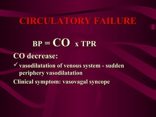
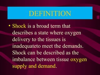
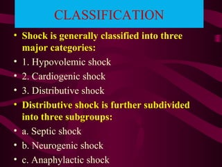
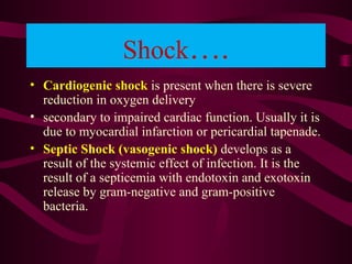
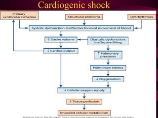
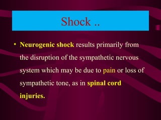
Outlines
• Circulatory failure
• Shock
• Definition
• Types
• Management
• Arrhythmias
CIRCULATORY FAILURECIRCULATORY FAILURE
• The main function of circulation is
perfusion of organs
BP = CO x TPRBP = CO x TPR
CIRCULATORY FAILURECIRCULATORY FAILURE
BPBP == COCO x TPRx TPR
CO decrease:CO decrease:
 vasodilatation of venous system - suddenvasodilatation of venous system - sudden
periphery vasodilatationperiphery vasodilatation
Clinical symptom: vasovagal syncopeClinical symptom: vasovagal syncope
introduction
• Shock is a life-threatening condition which
occurs when the circulatory system fails to
deliver oxygen and nutrients to the body
tissues and becomes unable to remove
waste products from the body. It is a
descriptive term based on the symptoms and
signs secondary to one or more of a wide
range of proble
DEFINITION
• Shock is a broad term that
describes a state where oxygen
delivery to the tissues is
inadequateto meet the demands.
Shock can be described as the
imbalance between tissue oxygen
supply and demand.
CLASSIFICATION
• Shock is generally classified into three
major categories:
• 1. Hypovolemic shock
• 2. Cardiogenic shock
• 3. Distributive shock
• Distributive shock is further subdivided
into three subgroups:
• a. Septic shock
• b. Neurogenic shock
• c. Anaphylactic shock
• Hypovolemic shock is present when marked reduction in
oxygen delivery
• results from diminished cardiac output secondary to
inadequate vascular volume.
• In general, it results from loss of fluid from circulation,
either directly or indirectly.
• A. Hemorrhage B. Loss of plasma due to burns
C. Loss of water and electrolytes in diarrhea
Shock….
• Cardiogenic shock is present when there is severe
reduction in oxygen delivery
• secondary to impaired cardiac function. Usually it is
due to myocardial infarction or pericardial tapenade.
• Septic Shock (vasogenic shock) develops as a
result of the systemic effect of infection. It is the
result of a septicemia with endotoxin and exotoxin
release by gram-negative and gram-positive
bacteria.
Septic shock
Shock ..
• Neurogenic shock results primarily from
the disruption of the sympathetic nervous
system which may be due to pain or loss of
sympathetic tone, as in spinal cord
injuries.
Neurogenic shock
• Shock stimulates a physiologic response. This
circulatory response to hypotension is to
• conserve perfusion to the vital organs (heart and
brain) at the expense of other tissues.
• Progressive vasoconstriction of skin, splanchnic
and renal vessels leads to renal cortical
• necrosis and acute renal failure. If not corrected in
time, shock leads to organ failure and
• sets up a vicious circle with hypoxia and acidosis
MANAGEMENT OF SHOCK
• General Management
Monitor the airway, breathing and circulation as first
priority
•Stop bleeding
•Fluid resuscitation, preferably crystalloids
•Head down position
•Treat the cause
•Transfusion of compatible blood if indicated
•Oxygen and other supportive measures like
inotropic agents:Dopamine
•Monitoring of resuscitation effectiveness: e.g.
determine hourly urine output,
blood pressure and pulse rate
Specific Management
 Hypovolemic Shock: The goal of treatment is to restore
vascular volume. This is effected by:
 General approach as above
 Fluid and blood replacement
 Oxygen support etc.
Specific Management
Septic Shock
• Initial management as above
• Appropriate antibiotics especially for gram-negative
microorganisms
• Inotropic support such as adrenaline and dopamine
• surgical eradication of the infection focus
• Cardiogenic shock
• Treat the causes
Neurogenic shock
• Pain relief
Treat the causes
COMPLICATIONS OF
SHOCK
• The main complications of severe shock include:
1. Shock lung (ARDS)
2. Acute renal failure
3. Gastrointestinal ulceration
4. Disseminated intravascular clotting
5. Multiorgan failure
6. Death
-therefore, a patient in shock requires immediate
emergency treatment. Early diagnosis and
immediate correction of shock prevents
permanent organ damage and death.
Arrhythmias (Cardiac)
• These are disorders of cardiac rate, rhythm
and conduction. Based on the heart rate, they
are classified into Brady arrhythmias, when
the heart rate is less than 60 per minute; and
tachyarrhythmia, when the heart rate is
greater than 100 per minute. Brady
arrhythmias include sinus bradycardia, sinus pauses
and atrioventricular blocks. The tachyarrhythmias
can further be classified into supraventricular and
ventricular arrhythmias, based on their site of origin.
Sinus bradycardia
• Regular sinus rhythm at rate below 60
beat/min.
26
Sinus tachycardia
• Regular sinus rhythm at a rate of 100 beat/min faster.
27
Sinus arrhythmia
• Irregular rhythm despite
presence of sinus process.
28
SVT
• In Supraventricular rhythm, the electrical impulse originates AT or ABOVE the AV
node.
• If QRS is narrow in all leads (nearly)
Supraventricular in origin
• Supraventricular rhythm category include:
1.Atrial flutter.
2.Atrial fibrillation.
3.paroxysmal SVT.
4.Junctional/AV nodal rythms.
29
Atrial flutter
30
Atrial flutter
• Atrial rate : 250-350 beat/min.
• Regular Atrial activity.
• Narrow QRS complex
• There is no flat base line b/w P waves.
• Typical apperance SAW TOOTH .
31
Atrial fibrillation
32
Atrial fibrillation
• Irregularly irregular rhythm.
• No P wave.
• Narrow QRS complex.
• A. Fib with :
- Rapid ventricular response= Rate:
over 120B/min.
- Controlled response= Rate: 70-
110B/min.
- Slow ventricular response=
33
Multifocal Atrial Tachycarida
34
Multifocal Atrial Tachycardia
• Irregularly irregular.
• Definite P wave.
• P wave morphology changes from beat to
beat.
• Caused by pulmonary disease or
multisystem problem .
35
Clinical features
• Palpitation, Shortness of breath,
Dizziness/syncope
• - Sensation of a pause in the heart beat
• Chest discomfort that mimics symptoms of myocardial
ischemia(angina), Heart Failure
• Rapid or slow pulse rate
• Regular pulse rate (sinus tachycardia/bradycardia
• rregularly irregular (atrial fibrillation, atrial flutter
Diagnosis of Arrhythmia
• Medical history
• Physical examination
• Laboratory test
• Anti-arrhythmia Agents
• Anti-tachycardia agents
• Anti-bradycardia agents
Anti-tachycardia agents
Modified Vaugham Williams classification
1.I class: Natrium channel blocker
lidocaine
1.II class: ß-receptor blocker
Propranolol,metoprolol
1.III class: Potassium channel blocker
dofetilide, ibutilide
1.IV class: Calcium channel blocker
verapamil
deltiazam
1.Others: Adenosine, Digital
Anti-bradycardia agents
1.ß-adrenic receptor activator
2.M-cholinergic receptor
blocker
3.Non-specific activator
Arrhythmia and shock

Arrhythmia and shock

  • 2.
    Outlines • Circulatory failure •Shock • Definition • Types • Management • Arrhythmias
  • 3.
    CIRCULATORY FAILURECIRCULATORY FAILURE •The main function of circulation is perfusion of organs BP = CO x TPRBP = CO x TPR
  • 4.
    CIRCULATORY FAILURECIRCULATORY FAILURE BPBP== COCO x TPRx TPR CO decrease:CO decrease:  vasodilatation of venous system - suddenvasodilatation of venous system - sudden periphery vasodilatationperiphery vasodilatation Clinical symptom: vasovagal syncopeClinical symptom: vasovagal syncope
  • 5.
    introduction • Shock isa life-threatening condition which occurs when the circulatory system fails to deliver oxygen and nutrients to the body tissues and becomes unable to remove waste products from the body. It is a descriptive term based on the symptoms and signs secondary to one or more of a wide range of proble
  • 6.
    DEFINITION • Shock isa broad term that describes a state where oxygen delivery to the tissues is inadequateto meet the demands. Shock can be described as the imbalance between tissue oxygen supply and demand.
  • 8.
    CLASSIFICATION • Shock isgenerally classified into three major categories: • 1. Hypovolemic shock • 2. Cardiogenic shock • 3. Distributive shock • Distributive shock is further subdivided into three subgroups: • a. Septic shock • b. Neurogenic shock • c. Anaphylactic shock
  • 9.
    • Hypovolemic shockis present when marked reduction in oxygen delivery • results from diminished cardiac output secondary to inadequate vascular volume. • In general, it results from loss of fluid from circulation, either directly or indirectly. • A. Hemorrhage B. Loss of plasma due to burns C. Loss of water and electrolytes in diarrhea
  • 11.
    Shock…. • Cardiogenic shockis present when there is severe reduction in oxygen delivery • secondary to impaired cardiac function. Usually it is due to myocardial infarction or pericardial tapenade. • Septic Shock (vasogenic shock) develops as a result of the systemic effect of infection. It is the result of a septicemia with endotoxin and exotoxin release by gram-negative and gram-positive bacteria.
  • 13.
  • 14.
    Shock .. • Neurogenicshock results primarily from the disruption of the sympathetic nervous system which may be due to pain or loss of sympathetic tone, as in spinal cord injuries.
  • 15.
  • 16.
    • Shock stimulatesa physiologic response. This circulatory response to hypotension is to • conserve perfusion to the vital organs (heart and brain) at the expense of other tissues. • Progressive vasoconstriction of skin, splanchnic and renal vessels leads to renal cortical • necrosis and acute renal failure. If not corrected in time, shock leads to organ failure and • sets up a vicious circle with hypoxia and acidosis
  • 20.
    MANAGEMENT OF SHOCK •General Management Monitor the airway, breathing and circulation as first priority •Stop bleeding •Fluid resuscitation, preferably crystalloids •Head down position •Treat the cause •Transfusion of compatible blood if indicated •Oxygen and other supportive measures like inotropic agents:Dopamine •Monitoring of resuscitation effectiveness: e.g. determine hourly urine output, blood pressure and pulse rate
  • 21.
    Specific Management  HypovolemicShock: The goal of treatment is to restore vascular volume. This is effected by:  General approach as above  Fluid and blood replacement  Oxygen support etc.
  • 22.
    Specific Management Septic Shock •Initial management as above • Appropriate antibiotics especially for gram-negative microorganisms • Inotropic support such as adrenaline and dopamine • surgical eradication of the infection focus • Cardiogenic shock • Treat the causes Neurogenic shock • Pain relief Treat the causes
  • 23.
    COMPLICATIONS OF SHOCK • Themain complications of severe shock include: 1. Shock lung (ARDS) 2. Acute renal failure 3. Gastrointestinal ulceration 4. Disseminated intravascular clotting 5. Multiorgan failure 6. Death -therefore, a patient in shock requires immediate emergency treatment. Early diagnosis and immediate correction of shock prevents permanent organ damage and death.
  • 25.
    Arrhythmias (Cardiac) • Theseare disorders of cardiac rate, rhythm and conduction. Based on the heart rate, they are classified into Brady arrhythmias, when the heart rate is less than 60 per minute; and tachyarrhythmia, when the heart rate is greater than 100 per minute. Brady arrhythmias include sinus bradycardia, sinus pauses and atrioventricular blocks. The tachyarrhythmias can further be classified into supraventricular and ventricular arrhythmias, based on their site of origin.
  • 26.
    Sinus bradycardia • Regularsinus rhythm at rate below 60 beat/min. 26
  • 27.
    Sinus tachycardia • Regularsinus rhythm at a rate of 100 beat/min faster. 27
  • 28.
    Sinus arrhythmia • Irregularrhythm despite presence of sinus process. 28
  • 29.
    SVT • In Supraventricularrhythm, the electrical impulse originates AT or ABOVE the AV node. • If QRS is narrow in all leads (nearly) Supraventricular in origin • Supraventricular rhythm category include: 1.Atrial flutter. 2.Atrial fibrillation. 3.paroxysmal SVT. 4.Junctional/AV nodal rythms. 29
  • 30.
  • 31.
    Atrial flutter • Atrialrate : 250-350 beat/min. • Regular Atrial activity. • Narrow QRS complex • There is no flat base line b/w P waves. • Typical apperance SAW TOOTH . 31
  • 32.
  • 33.
    Atrial fibrillation • Irregularlyirregular rhythm. • No P wave. • Narrow QRS complex. • A. Fib with : - Rapid ventricular response= Rate: over 120B/min. - Controlled response= Rate: 70- 110B/min. - Slow ventricular response= 33
  • 34.
  • 35.
    Multifocal Atrial Tachycardia •Irregularly irregular. • Definite P wave. • P wave morphology changes from beat to beat. • Caused by pulmonary disease or multisystem problem . 35
  • 36.
    Clinical features • Palpitation,Shortness of breath, Dizziness/syncope • - Sensation of a pause in the heart beat • Chest discomfort that mimics symptoms of myocardial ischemia(angina), Heart Failure • Rapid or slow pulse rate • Regular pulse rate (sinus tachycardia/bradycardia • rregularly irregular (atrial fibrillation, atrial flutter
  • 37.
    Diagnosis of Arrhythmia •Medical history • Physical examination • Laboratory test • Anti-arrhythmia Agents • Anti-tachycardia agents • Anti-bradycardia agents
  • 38.
    Anti-tachycardia agents Modified VaughamWilliams classification 1.I class: Natrium channel blocker lidocaine 1.II class: ß-receptor blocker Propranolol,metoprolol 1.III class: Potassium channel blocker dofetilide, ibutilide 1.IV class: Calcium channel blocker verapamil deltiazam 1.Others: Adenosine, Digital
  • 39.
    Anti-bradycardia agents 1.ß-adrenic receptoractivator 2.M-cholinergic receptor blocker 3.Non-specific activator