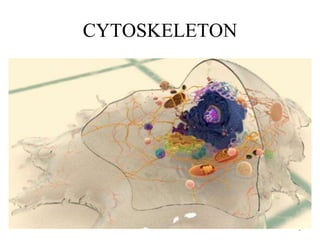
4. How and Why Cells Move
4
https://www.thoughtco.com/how-and-why-cells-move-373377
In cases involving wound injury and repair, connective tissue cells must travel to an
injury site to repair damaged tissue.
White blood cells, such as neutrophils and macrophages must quickly migrate to sites
of infection or injury to fight bacteria and other germs.
Cancer cells have the ability to metastasize or spread from one location to another
by moving through blood vessels and lymphatic vessels- Epithelial to Mesenchymal
Transition (EMT)
In the cell cycle, movement is required for the cell dividing process of cytokinesis to
occur in the formation of two daughter cells.
Movement within the cells
• Vesicle transportation
• Organelle migration
• Chromosome movement during mitosis
6. • Actin filaments determine the shape of the cell’s
surface and are necessary for whole-cell
locomotion; they also drive the pinching of one
cell into two.
• Microtubules determine the positions of
membrane-enclosed organelles, direct
intracellular transport, and form the mitotic
spindle that segregates chromosomes during cell
division.
• Intermediate filaments provide mechanical
strength.
6
8. Distribution of actin in the cell
8
https://www.mechanobio.info/cytoskeleton-dynamics/what-is-the-
cytoskeleton/what-are-actin-filaments/how-are-actin-filaments-distributed-in-cells-
and-tissues/
11. Treadmilling
Treadmilling- Simultaneous gain of monomers from
+ end and loss of monomers from – end, maintaining
the length of the actin. Rate of gain and loss of the
filament is the same. However, the monomer moves
through the filament. The monomer that is added at
the + end ultimately reaches the - end and falls off at
the – end.
11
https://www.memorangapp.com/flashcards/170868/Lecture+3%3A+Actin/
Intermediate concentrations of Actin
12. Initiation of actin filaments-Nucleation
Formins are a family of large (140-
200 kd) barbed-end tracking proteins
that both nucleate the
initial actin monomers and then
move along the growing filament,
adding new monomers to the barbed
end.
12
https://www.mechanobio.info/cytoskeleton-dynamics/what-is-the-cytoskeleton/what-are-actin-filaments/how-does-arp23-mediate-the-
nucleation-of-branched-filaments/
19. Depolymerisation
ADF/cofilin (Actin depolymerizing Factor)
These proteins bind
to actin filaments and enhance
the rate of dissociation
of actin/ADP monomers from the
pointed end.
ADF/cofilin can also sever actin filaments.
ADF/cofilin preferentially binds to ADP-
actin, so it remains bound to actin monomers
following filament disassembly and
sequesters them in the ADP-bound form,
preventing their reincorporation
into filaments.
Profilin acts by stimulating the exchange
of bound ADP for ATP, resulting in the
formation of actin/ATP monomers, which
dissociate from cofilin and are then
available for assembly into filaments. 19
23. Bundles
23
https://iopscience.iop.org/book/978-0-7503-1753-5/chapter/bk978-0-7503-1753-5ch7
Supports projections of the plasma
membrane, such as microvilli.
Fimbrin is a 68 kd protein
containing two adjacent actin-
binding domains. It binds
to actin filaments as a monomer,
holding two parallel filaments close
together. (14nm)
The second type of actin bundle is
composed of filaments that are more
widely spaced, allowing the bundle
to contract. (40nm)
Alpha-actinin binds to actin as
a dimer, each subunit of which is a
102 kd protein containing
a single actin-binding site.
55kDa globular protein
that is a component of
the crosslinking actin
filaments functional
module. Fascin
organizes F-actin into
tightly packed parallel
bundles approximately
8nm apart.
31. • Cultured fibroblasts secrete extracellular matrix proteins that stick to the
surface of the culture dish. The fibroblasts then attach to this extracellular
matrix on the culture dish via the binding of transmembrane proteins
(called integrins).
• The sites of attachment are discrete regions (called focal adhesions) that
also serve as attachment sites for large bundles of actin filaments called
stress fibers.
• Stress fibers are contractile bundles of actin filaments, cross-linked by a-
actinin and stabilized by tropomysosin, which anchor the cell and exert
tension against the substratum.
• They are attached to the plasma membrane at focal adhesions via
interactions with integrin. These complex associations are mediated by
several other proteins, including talin and vinculin.
• For example, both talin and a-actinin bind to the cytoplasmic domains of
integrins. Talin also binds to vinculin and both proteins also bind actin.
Other proteins found at focal adhesions also participate in the attachment
of actin filaments, and a combination of these interactions is responsible
for the linkage of actin filaments to the plasma membrane.
Cell-Matrix interactions
31
32. • The actin cytoskeleton is similarly anchored to regions
of cell-cell contact called adherens junctions.
• In sheets of epithelial cells, these junctions form a
continuous belt-like structure (called an adhesion belt)
around each cell in which an underlying contractile
bundle of actin filaments is linked to the plasma
membrane.
• Contact between cells at adherens junctions is mediated
by transmembrane proteins called cadherins.
• The cadherins form a complex with cytoplasmic
proteins called catenins, which associate with actin
filaments.
Cell-Cell interactions
32
35. 35
Pseudopodia are extensions of moderate
width, based on actin filaments cross-linked
into a three-dimensional network, that are
responsible for phagocytosis and for the
movement of amoebas across a surface.
Lamellipodia are broad, sheet-like
extensions at the leading edge of fibroblasts,
which similarly contain a network of actin
filaments.
Many cells also extend microspikes or
filopodia, thin projections of the plasma
membrane supported by actin bundles.
37. • Filopodia contains 15-
20 parallel filaments
tightly packed into
bundles with their
barbed membrane
facing the membrane.
37
• Lamellipodium is driven
by a growing network
of actin microfilaments
(branched actin).
• Arp2/3 and WASP
serve as nucleators.
• ADF, Profilin and
capping proteins co-
operate to help in
treadmilling.
https://www.researchgate.net/figure/Two-phases-of-the-Ecoli-movement-The-bacteria-
migrates-due-to-movement-of-the-flagella_fig4_228634410
38. 38
https://www.mechanobio.info/cytoskeleton-dynamics/what-are-invadopodia/
Invadopodia
• Invadopodia are elongated ventral
projections that extend into the underlying
ECM.
• Microtubules and intermediate filaments
have also been detected in mature
invadopodia.
• Actin filament nucleators and their
regulators such as the Arp2/3 complex, N-
WASP (neuronal Wiskott-Aldrich
syndrome protein), WASP-interacting
protein (WIP) and cortactin are also found
localized to this structure.
• Invadopodia contain a substantial number
of actin filaments arranged as parallel
bundles, akin to those observed in
filopodia.
• Cross-linking proteins such as fascin are
abundant in invadopodia
40. 40
A migrating cell needs to perform a coordinated
series of steps to move. Cdc42 regulates the
direction of migration, Rac induces membrane
protrusion at the front of the cell through stimulation
of actin polymerization and integrin adhesion
complexes, and Rho promotes actin:myosin
contraction in the cell body and at the rear.
https://www.sciencedirect.com/science/article/pii/S001216060300544X#FIG1
43. Degradation of basement membrane
Malignant cells generally secrete proteases that digest
extracellular matrix components, allowing the cancer
cells to invade adjacent normal tissues. Secretion of
collagenase, for example, appears to be an important
determinant of the ability of carcinomas to digest and
penetrate through basal laminae to invade underlying
connective tissue
43
44. https://www.omicsonline.org/articles-images/2168-9296-3-137-g002.html 44
Invadopodia in cancer migration. After tumor cells have
proceeded through EMT, invadopodia form in the cellular
cytoskeleton, giving the cells a migratory phenotype.
Degradation of basement membranes and extracellular
matrix during migration is achieved by invadopodia through
the use of metallo proteases. Cells can then migrate to the
ECM, where they form pseudopodia. Invadopodia are
involved in directional migration and chemotaxis during the
travel through the ECM. Once tumor cells reach the
endothelium, they again use invadopodia to degrade the
basement membrane and enter the blood stream.
46. 46
The core of a Z-disc consists of
actin filaments coming from
adjacent sarcomeres which are
crosslinked by α actinin
molecules. Mature Z-discs are
probably composed of hundreds
of different proteins.
48. Two additional proteins (titin and nebulin) also contribute to sarcomere
structure and stability. Titin is an extremely large protein (3000 kd), and
single titin molecules extend from the M line to the Z disc. These long
molecules of titin are thought to act like springs that keep the myosin
filaments centered in the sarcomere and maintain the resting tension that
allows a muscle to snap back if overextended.
Nebulin filaments are associated with actin and are thought to regulate the
assembly of actin filaments by acting as rulers that determine their length.
48
49. • The type of myosin present in muscle (myosin II) is a very large protein (about 500
kd) consisting of two identical heavy chains (about 200 kd each) and two pairs of light
chains (about 20 kd each).
• Each heavy chain consists of a globular head region and a long a-helical tail. The a-
helical tails of two heavy chains twist around each other in a coiled-coil structure to
form a dimer, and two light chains associate with the neck of each head region to form
the complete myosin II molecule.
• The motor activity of myosin moves its head groups along the actin filament in the
direction of the barbed end. This movement slides the actin filaments from both sides
of the sarcomere toward the M line, shortening the sarcomere and resulting in muscle
contraction.
49
51. In striated muscle each
tropomyosin molecule is
bound to troponin, which
is a complex of three
polypeptides: troponin C
(Ca2+-binding), troponin I
(inhibitory), and troponin
T (tropomyosin-binding).
51
53. Activation of myosin in smooth muscle
and non-muscle cells
• In non-muscle cells and in smooth muscle, however, contraction is
regulated primarily by phosphorylation of one of the myosin light
chains called the regulatory light chain.
• Phosphorylation of the regulatory light chain in these cells has at
least two effects: It promotes the assembly of myosin
into filaments, and it increases myosin catalytic activity enabling
contraction to proceed.
• The enzyme that catalyzes this phosphorylation, called myosin light-
chain kinase, is itself regulated by association with the Ca2+-
binding protein calmodulin.
• Increases in cytosolic Ca2+promote the binding of calmodulin to the
kinase resulting in phosphorylation of the myosin regulatory light
chain. Increase in cytosolic Ca2+ is thus responsible, albeit
indirectly, for activating myosin in smooth muscle and non muscle
cells, as well as in striated muscle.
53
57. Microtubules
• They function both to determine cell shape and in a variety of cell
movements, including some forms of cell locomotion, the intracellular
transport of organelles, and the separation of chromosomes during mitosis.
• Microtubules are composed of a single type of globular protein called tubulin.
The building blocks of microtubules are tubulin dimers consisting of two
closely related 55 kd polypeptides: α- tubulin and β-tubulin.
• A third type of tubulin (γ-tubulin) is concentrated in the centrosome where it
plays a critical role in initiating microtubule assembly.
• Tubulin dimers polymerize to form microtubules, which generally consist of
13 linear protofilaments assembled around a hollow core.
• The protofilaments, which are composed of head-to-tail arrays of tubulin
dimers, are arranged in parallel. Consequently, microtubules (like actin
filaments) are polar structures with two distinct ends: a fast-growing plus end
and a slow- growing minus end.
• Tubulin dimers can depolymerize as well as polymerize, and microtubules
can undergo rapid cycles of assembly and disassembly.
57
60. • In particular, the GTP bound to β -tubulin (though not
that bound to a-tubulin) is hydrolyzed to GDP during
or shortly after polymerization. This GTP hydrolysis
weakens the binding affinity of tubulin for adjacent
molecules, thereby favoring depolymerization and
resulting in the dynamic behavior of microtubules.
60
61. Treadmilling and the role of GTP in
microtubule polymerization
Like actin filaments, microtubules undergo treadmilling, a dynamic
behavior in which tubulin molecules bound to GDP are continually
lost from the minus end and replaced by the addition of tubulin
molecules bound to GTP to the plus end of the same microtubule.
61
62. Dynamic instability
• In microtubules, rapid GTP hydrolysis also results in the
behavior known as dynamic instability (described by Tim
Mitchison and Marc Kirschner) in which individual
microtubules alternate between cycles of growth and shrinkage.
• Whether a microtubule grows or shrinks is determined in part
by the rate of tubulin addition relative to the rate of GTP
hydrolysis.
• As long as new GTP-bound tubulin molecules are added more
rapidly than GTP is hydrolyzed, the microtubule retains a GTP
cap at its plus end and microtubule growth continues.
• However, if the rate of polymerization slows, the GTP bound to
tubulin at the plus end of the microtubule will be hydrolyzed to
GDP. If this occurs, the GDP-bound tubulin will dissociate,
resulting in rapid depolymerization and shrinkage of the
microtubule.
62
65. Assembly of Microtubules
• In animal cells, most microtubules extend outward from
the centrosome (first described by Theodor Boveri in
1888), which is located adjacent to the nucleus near the
center of interphase (non-dividing) cells.
• During mitosis, microtubules similarly extend outward
from duplicated centrosomes to form the mitotic spindle,
which is responsible for the separation and distribution of
chromosomes to daughter cells.
• Plant cells do not have centrosomes, instead the
microtubules extend from the nucleus.
65
70. Severing enzymes-Katanins
70
https://onlinelibrary.wiley.com/doi/full/10.1002/cm.21522
Control microtubules (magenta) in the absence of katanin lose dimers from the ends due to normal degradation to replenish the background concentration. This loss of
polymer is slow. (middle) In the presence of katanin (green) katanin can catalyze the loss of dimers from both the ends of control microtubules, called depolymerization,
and from the middle of control microtubules, called severing. We observe a significant amount of mobility, association, and dissociation of katanin to and from the
filaments. (right) Microtubules lacking the CTT can still bind katanin, but fewer katanins are bound and the bound katanin is less mobile. Without the CTT, katanin cannot
sever microtubules but can still catalyze the loss of dimers from the ends. CTT, C-terminal tail
71. Organization of microtubules in nerve cells
• Microtubules in axons and dendrites are organized differently and
associated with distinct MAPs.
• In axons, the microtubules are all oriented with their plus ends away
from the cell body, similar to the general orientation of microtubules in
other cell types.
• The minus ends of most of the microtubules in axons, however, are not
anchored in the centrosome; instead, both the plus and minus ends of
these microtubules terminate in the cytoplasm of the axon.
• In dendrites, the microtubules are oriented in both directions; some plus
ends point toward the cell body and some point toward the cell
periphery.
• These distinct microtubule arrangements are paralleled by differences
in MAPs: Axons contain tau proteins, but no MAP-2, whereas dendrites
contain MAP-2, but no tau proteins, and it appears that these
differences in MAP-2 and tau distribution are responsible for the
distinct organization of stable microtubules in axons and dendrites. 71
72. Axons contain tau proteins,
but no MAP-2
Dendrites contain MAP-2,
but no tau proteins
72
MAP-1, MAP-2, and tau -Neuronal cells
MAP-4 - Non-neuronal vertebrate cells
Tau- main component of the characteristic lesions found in the brains of Alzheimer's
patients.
74. Microtubule Motors and Movement
• Kinesins and the dyneins—are responsible for powering the variety of
movements in which microtubules participate.
• Kinesin and dynein move along microtubules in opposite directions—most
kinesins toward the plus end and dyneins toward the minus end.
• Kinesin I is a molecule of approximately 380 kd consisting of two heavy chains
(120 kd each) and two light chains (64 kd each).
• The heavy chains have long a-helical regions that wind around each other in a
coiled-coil structure. The amino-terminal globular head domains of the heavy
chains are the motor domains of the molecule. They bind to both microtubules
and ATP, the hydrolysis of which provides the energy required for movement.
• The tail portion of the kinesin molecule consists of the light chains in
association with the carboxy-terminal domains of the heavy chains. This portion
of kinesin is responsible for binding to other cell components (such as
membrane vesicles and organelles) that are transported along microtubules by
the action of kinesin motors.
• Cytoplasmic dynein is an extremely large molecule (up to 2000 kd), which
consists of two or three heavy chains (each about 500 kd) complexed with a
variable number of light and intermediate chains, which range from 14 to 120
kd
74
78. Cargo Transport and Intracellular
Organization
• One of the major roles of microtubules is to transport
macromolecules, membrane vesicles, and organelles
through the cytoplasm of eukaryotic cells.
• Kinesin and dynein carry their cargoes to and from the
tips of the axons, respectively.
• For example, secretory vesicles containing
neurotransmitters are carried from the Golgi apparatus to
the terminal branches of the axon by kinesin.
• In the reverse direction, cytoplasmic dynein transports
endocytic vesicles from the axon back to the cell body.
78