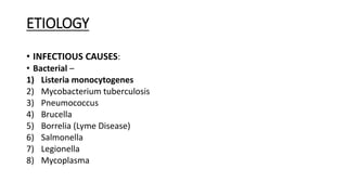
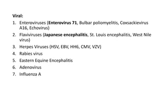
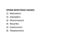
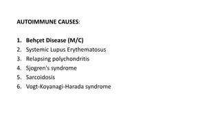
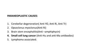
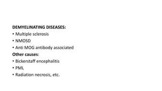
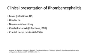
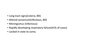
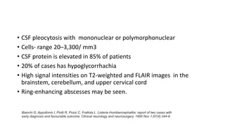
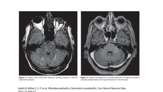
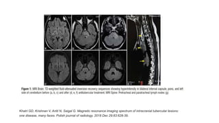
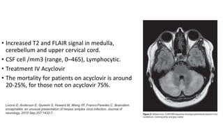
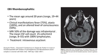
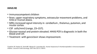
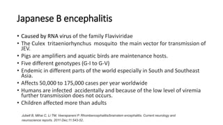
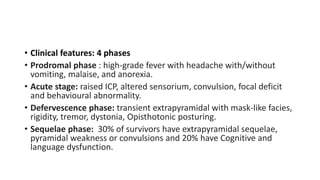
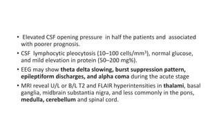
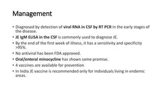
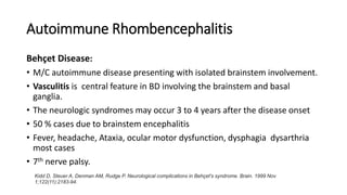
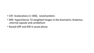
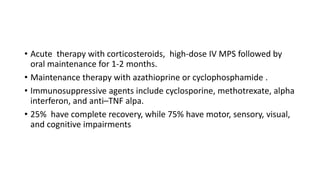
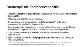
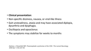
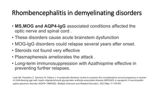
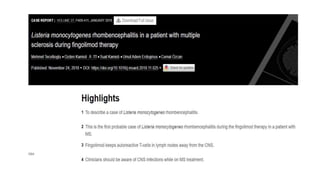
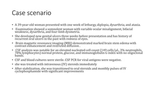
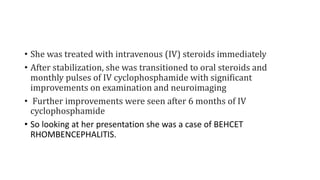
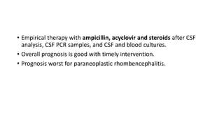
APPROACH TO
RHOMBENCHEPALITIS
DR SUMEET SINGH
SR NEUROLOGY
GMC, KOTA
INTRODUCTION
• Rhombencephalitis refers to inflammatory diseases affecting the
hindbrain (brainstem and cerebellum).
• The rhombencephalon comprises the annular protuberance, the
spinal bulb and the cerebellum.
• The term derives from the Greek, with "rhombos" meaning a
diamond shaped figure, "enkephalos", meaning the brain
• It was described for the first time by two scientists, Edwin Bickerstaff
and Philip Cloake in 1951
Jubelt B, Mihai C, Li TM, Veerapaneni P. Rhombencephalitis/brainstem encephalitis. Current neurology and
neuroscience reports. 2011 Dec;11:543-52.
THE HINDBRAIN
ETIOLOGY
• INFECTIOUS CAUSES:
• Bacterial –
1) Listeria monocytogenes
2) Mycobacterium tuberculosis
3) Pneumococcus
4) Brucella
5) Borrelia (Lyme Disease)
6) Salmonella
7) Legionella
8) Mycoplasma
Viral:
1. Enteroviruses (Enterovirus 71, Bulbar poliomyelitis, Coxsackievirus
A16, Echovirus)
2. Flaviviruses (Japanese encephalitis, St. Louis encephalitis, West Nile
virus)
3. Herpes Viruses (HSV, EBV, HH6, CMV, VZV)
4. Rabies virus
5. Eastern Equine Encephalitis
6. Adenovirus
7. Influenza A
OTHER INFECTIOUS CAUSES:
1) Melioidosis
2) Aspergillus
3) Mucormycosis
4) Nocardia
5) Cysticercosis
6) Toxoplasmosis
AUTOIMMUNE CAUSES:
1. Behçet Disease (M/C)
2. Systemic Lupus Erythematosus
3. Relapsing polychondritis
4. Sjogren's syndrome
5. Sarcoidosis
6. Vogt-Koyanagi-Harada syndrome
PARANEOPLASTIC CAUSES:
1. Cerebellar degeneration( Anti YO, Anti Ri, Anti Tr)
2. Opsoclonus myoclonus(Anti Ri)
3. Brain stem encephalitis(Anti –amphiphysin)
4. Small cell lung cancer (Anti-Hu and anti-Ma antibodies)
5. Lymphoma associated.
DEMYELINATING DISEASES:
• Multiple sclerosis
• NMOSD
• Anti MOG antibody associated
Other causes:
• Bickerstaff encephalitis
• PML
• Radiation necrosis, etc.
Clinical presentation of Rhombencephalitis
• Fever (infectious, BD)
• Headache
• Nausea and vomiting
• Cerebellar ataxia(Infectious, PNS)
• Cranial nerve palsies(65-85%)
Moragas M, Martínez-Yélamos S, Majós C, Fernández-Viladrich P, Rubio F, Arbizu T. Rhombencephalitis: a series
of 97 patients. Medicine. 2011 Jul 1;90(4):256-61.
• Long tract signs(Listeria, BD)
• Altered sensorium(infectious, BD)
• Meningismus (infectious)
• Rapidly developing respiratory failure(41% of cases)
• Locked in state to coma.
Moragas M, Martínez-Yélamos S, Majós C, Fernández-Viladrich P, Rubio F, Arbizu T. Rhombencephalitis: a series of 97
Rhombencephalitis due to Listeria
monocytogenes
• Pathogenesis:
• Gram-positive, aerobic, facultative intracellular rod.
• Associated with contaminated soft cheeses, unpasteurized milk, and
deli meats.
• Listeria is present in soil and vegetation and 1% to 5% of the general
population are asymptomatic carriers .
• Its intracellular nature shields Listeria from the immune system.
• It replicates inside the cell, and spreads from cell to cell .
Armstrong RW, Fung PC. Brainstem encephalitis (rhombencephalitis) due to Listeria monocytogenes: case report and
review. Clinical Infectious Diseases. 1993 May 1;16(5):689-702.
• Clinical presentation:
• Listerial RE is another manifestation of CNS listerosis.
• Classical CNS listerosis affects immunocompromised and extremes of ages
• Listerial RE occurs primarily in patients with intact immune systems
• Male to female ratio 1:1
• Age- 14 to 79 years
• A flu-like prodrome of headache, fever, nausea, vomiting, and malaise
lasting 1 to 15 days occurs first
• Later brainstem dysfunction.
• Two thirds of patients have dysfunction of pons and medulla
• Unilateral cranial nerve deficits(90-100%)
• Ipsilateral corticospinal tract deficits (80%)
• Cerebellar deficits (55%): hemiataxia, vertigo, cerebellar dysarthria
• Respiratory failure (40%)
• Triad of headache, fever, meningismus (45%)
Reynaud, L., Graf, M., Gentile, I., Cerini, R., Ciampi, R., Noce, S., Gentile, F., Borrelli, F., Viola, C., Briganti, F. and
Borgia, G., 2007. A rare case of brainstem encephalitis by Listeria monocytogenes with isolated mesencephalic
localization. Case report and review. Diagnostic microbiology and infectious disease, 58(1), pp.121-123.
• CSF pleocytosis with mononuclear or polymorphonuclear
• Cells- range 20–3,300/ mm3
• CSF protein is elevated in 85% of patients
• 20% of cases has hypoglycorrhachia
• High signal intensities on T2-weighted and FLAIR images in the
brainstem, cerebellum, and upper cervical cord
• Ring-enhancing abscesses may be seen.
Bianchi G, Appollonio I, Piolti R, Pozzi C, Frattola L. Listeria rhombencephalitis: report of two cases with
early diagnosis and favourable outcome. Clinical neurology and neurosurgery. 1995 Nov 1;97(4):344-8.
Jubelt B, Mihai C, Li T, et al. Rhombencephalitis / brainstem encephalitis. Curr Neurol Neurosci Rep.
Management
• CSF and blood cultures are most specific for diagnosis.
• 30-40% positive CSF cultures and 45-50% positive blood cultures.
• Empiric treatment is solely based on clinical experience.
• Ampicillin is the drug of choice, recommended dose 2 g every 4 h for
adults and 100 to 300 mg/kg per day in 4 to 6 daily doses for children for
21 days.
• Intravenous penicillin G at 24 million units per day is another alternative.
• Alternatives for penicillin allergic individuals include trimethoprim-
sulfamethoxazole, vancomycin, gentamicin and linezolid.
• The mortality rate between 20% and 30% compared to 66% in the pre-
antibiotic era
• 55% of survivors have neurologic sequelae.
Popescu GA, Saquepée M, Poisson D, Prazuck T. Treatment difficulties of a listerial rhombencephalitis in an adult patient allergic to
penicillins. Journal of clinical pathology. 2004 Jun 1;57(6):665-6.
Tubercular Rhombencephalitis
• Intracranial tuberculosis two types:
• meningeal pattern like leptomeningitis and pachymeningitis
• second pattern as parenchymal lesions like tuberculoma, cerebritis, abscess,
rhombencephalitis, and encephalopathy
• Hyperintensity of internal capsule and diffuse hyperintensity in brain
stem and cerebellum without contrast enhancement
• Managed with 12 month ATT with tapering steroids.
Duong M, Piroth L, Chavanet P, Combernoux A, Grappin M, Portier H. A case of rhombencephalitis with isolation
of cytomegalovirus and Mycobacterium avium complex in a woman with AIDS. Aids. 1994 Sep 1;8(9):1356-7.
Khatri GD, Krishnan V, Antil N, Saigal G. Magnetic resonance imaging spectrum of intracranial tubercular lesions:
one disease, many faces. Polish journal of radiology. 2018 Dec 29;83:628-39.
Enterovirus 71 Rhombencephalitis
• M/C viral cause of rhombencephalitis
• Neurologic involvement may occur in 25% of patients
• children and teenagers primarily affected.
• Aseptic meningitis, cerebellar ataxia, and various forms of
poliomyelitis, brainstem encephalitis/rhombencephalitis.
• Myoclonic jerks, tremors, ataxia, cranial nerve palsies, coma, and
respiratory failure
Hamaguchi T, Fujisawa H, Sakai K, Okino S, Kurosaki N, Nishimura Y, Shimizu H, Yamada M. Acute encephalitis caused by
intrafamilial transmission of enterovirus 71 in adult. Emerging infectious diseases. 2008 May;14(5):828.
• 70%–75% have MRI T2-weighted high-intensity lesions in the
brainstem and cerebellum.
• CSF white blood cells/mm3 (range 5–379)
• Diagnosis by virus isolation from throat, feces, or vesicles and by
antibody studies.
• Viral genomic amplification has been used.
• There is no specific therapy, there are reports of improvement with
intravenous immunoglobulin (IV Ig).
Wang Y, Zou G, Xia A, Wang X, Cai J, Gao Q, Yuan S, He G, Zhang S, Zeng M, Altmeyer R. Enterovirus 71 infection in
children with hand, foot, and mouth disease in Shanghai, China: epidemiology, clinical feature and diagnosis. Virology
journal. 2015 Dec;12:1-7.
Rhombencephalitis Caused by Herpes Viruses
• HSV Rhombencephalitis:
• Age 18–71 years.
• (80%) by HSV1 and (20%) were by HSV2.
• Encephalitis limited to the rhombencephalon in 55%.
• neuro-ophthalmology abnormalities in 80% (abnormal ocular movements, nystagmus, anisocoria,
ptosis, spasmodic movements, and oscillopsia).
• cranial nerve deficits in (70%) cases.
• Fever in (70%)
• Headache in (50%)
• Pyramidal tract findings in (45%)
• Ataxia in (40%)
• Dysphagia in (30%)
• Quadriplegia in 20%
• Increased T2 and FLAIR signal in medulla,
cerebellum and upper cervical cord.
• CSF cell /mm3 (range, 0–465), Lymphocytic.
• Treatment IV Acyclovir
• The mortality for patients on acyclovir is around
20-25%, for those not on acyclovir 75%.
Livorsi D, Anderson E, Qureshi S, Howard M, Wang YF, Franco-Paredes C. Brainstem
encephalitis: an unusual presentation of herpes simplex virus infection. Journal of
neurology. 2010 Sep;257:1432-7.
EBV Rhombencephalitis:
• The mean age around 20 years (range, 19–44
years)
• Clinical manifestations fever (75%), ataxia
(100%), and an altered level of consciousness
(50%).
• MRI 50% of the damage was infratentorial .
The mean CSF cell count 25 cells/mm3
(range, 8–55) with 100% lymphocytes.
• Treatment intravenous acyclovir.
Duong M, Piroth L, Chavanet P, Combernoux A, Grappin M, Portier H. A case of
rhombencephalitis with isolation of cytomegalovirus and Mycobacterium avium
complex in a woman with AIDS. Aids. 1994 Sep 1;8(9):1356-7.
HHV6 RE
• Immunocompetent children
• fever, upper respiratory symptoms, extraocular movement problems, and
limb or truncal ataxia.
• MRIs increased signal intensity in cerebellum , thalamus, putamen, and
insular cortex.
• CSF cells/mm3 (range, 23–157).
• Glucose normal and protein elevated. HHV6 PCR is diagnostic in both the
blood and CSF.
• Treatment with ganciclovir shows improvement.
Crawford JR, Kadom N, Santi MR, Mariani B, Lavenstein BL. Human herpesvirus 6 rhombencephalitis in immunocompetent
children. Journal of child neurology. 2007 Nov;22(11):1260-8.
Japanese B encephalitis
• Caused by RNA virus of the family Flaviviridae
• The Culex tritaeniorhynchus mosquito the main vector for transmission of
JEV.
• Pigs are amplifiers and aquatic birds are maintenance hosts.
• Five different genotypes (G-I to G-V)
• Endemic in different parts of the world especially in South and Southeast
Asia.
• Affects 50,000 to 175,000 cases per year worldwide
• Humans are infected accidentally and because of the low level of viremia
further transmission does not occurs.
• Children affected more than adults
Jubelt B, Mihai C, Li TM, Veerapaneni P. Rhombencephalitis/brainstem encephalitis. Current neurology and
neuroscience reports. 2011 Dec;11:543-52.
• Clinical features: 4 phases
• Prodromal phase : high-grade fever with headache with/without
vomiting, malaise, and anorexia.
• Acute stage: raised ICP, altered sensorium, convulsion, focal deficit
and behavioural abnormality.
• Defervescence phase: transient extrapyramidal with mask-like facies,
rigidity, tremor, dystonia, Opisthotonic posturing.
• Sequelae phase: 30% of survivors have extrapyramidal sequelae,
pyramidal weakness or convulsions and 20% have Cognitive and
language dysfunction.
• Elevated CSF opening pressure in half the patients and associated
with poorer prognosis.
• CSF lymphocytic pleocytosis (10–100 cells/mm3), normal glucose,
and mild elevation in protein (50–200 mg%).
• EEG may show theta delta slowing, burst suppression pattern,
epileptiform discharges, and alpha coma during the acute stage
• MRI reveal U/L or B/L T2 and FLAIR hyperintensities in thalami, basal
ganglia, midbrain substantia nigra, and less commonly in the pons,
medulla, cerebellum and spinal cord.
Management
• Diagnosed by detection of viral RNA in CSF by RT PCR in the early stages of
the disease.
• JE IgM ELISA in the CSF is commonly used to diagnose JE.
• By the end of the first week of illness, it has a sensitivity and specificity
>95%.
• No antiviral has been FDA approved.
• Oral/enteral minocycline has shown some promise.
• 4 vaccines are available for prevention
• In India JE vaccine is recommended only for individuals living in endemic
areas.
Autoimmune Rhombencephalitis
Behçet Disease:
• M/C autoimmune disease presenting with isolated brainstem involvement.
• Vasculitis is central feature in BD involving the brainstem and basal
ganglia.
• The neurologic syndromes may occur 3 to 4 years after the disease onset
• 50 % cases due to brainstem encephalitis
• Fever, headache, Ataxia, ocular motor dysfunction, dysphagia dysarthria
most cases
• 7th nerve palsy.
Kidd D, Steuer A, Denman AM, Rudge P. Neurological complications in Behçet's syndrome. Brain. 1999 Nov
1;122(11):2183-94.
• CSF- leukocytosis (1–360), raised protein.
• MRI hyperintense T2-weighted images in the brainstem, thalamus
,internal capsule and cerebellum
• Raised CRP and ESR in acute phase.
• Acute therapy with corticosteroids, high-dose IV MPS followed by
oral maintenance for 1-2 months.
• Maintenance therapy with azathioprine or cyclophosphamide .
• Immunosuppressive agents include cyclosporine, methotrexate, alpha
interferon, and anti–TNF alpa.
• 25% have complete recovery, while 75% have motor, sensory, visual,
and cognitive impairments
Akman-Demir G, Serdaroglu P, Tasci B, Neuro-Behçet Study Group*. Clinical patterns of neurological involvement in Behçet's
disease: evaluation of 200 patients. Brain. 1999 Nov 1;122(11):2171-82.
Paraneoplastic Rhombencephalitis
• Present as cerebellar degeneration, opsoclonus-myoclonus and brainstem
encephalitis
• PNS may antedate the onset of cancer .
• Gynaecologic and breast cancer - anti-Yo and anti-Tr antibodies
paraneoplastic cerebellar degeneration
• Breast and gynaecological cancer and small cell lung cancer - anti-Ri and
PN cerebellar degeneration and opsoclonus-myoclonus syndrome
• Lung cancer -anti-Hu and anti-Ma antibodies and in PN cerebellar
degeneration
• Small cell lung carcinoma - Amphiphysin auto-antibodies in a variety of
PNS including RE
Rojas I, Graus F, Keime-Guibert F, Rene R, Delattre JY, Ramon JM, Dalmau J, Posner JB. Long-term clinical outcome
of paraneoplastic cerebellar degeneration and anti-Yo antibodies. Neurology. 2000 Sep 12;55(5):713-5.
• Clinical presentation:
• Non-specific dizziness, nausea, or viral-like illness
• Gait unsteadiness, ataxia and may have associated diplopia,
dysarthria and dysphagia.
• Oscillopsia and opsoclonus
• The symptoms may stabilize for weeks to months
Dalmau J, Rosenfeld MR. Paraneoplastic syndromes of the CNS. The Lancet Neurology.
2008 Apr 1;7(4):327-40.
• MRI initially normal later shows cerebellar atrophy.
• CSF shows raised proteins and lymphocytic pleocytosis
• Treatment:
• Immunotherapies with steroids, IV Ig, tacrolimus, and rituximab
• Eradication of tumor.
Rhombencephalitis in demyelinating disorders
• MS,MOG and AQP4-IgG associated conditions affected the
optic nerve and spinal cord .
• These disorders cause acute brainstem dysfunction
• MOG-IgG disorders could relapse several years after onset.
• Steroids not found very effective
• Plasmapheresis ameliorates the attack .
• Long-term immunosuppression with Azathioprine effective in
preventing further relapses.
Leite MI, Panahloo Z, Harrison N, Palace J. A systematic literature review to examine the considerations around pregnancy in women
of child-bearing age with myelin oligodendrocyte glycoprotein antibody-associated disease (MOGAD) or aquaporin 4 neuromyelitis
optica spectrum disorder (AQP4+ NMOSD). Multiple Sclerosis and Related Disorders. 2023 May 11:104760.
Case scenario
• A 39-year-old woman presented with one week of lethargy, diplopia, dysarthria, and ataxia.
• Examination showed a somnolent woman with variable ocular misalignment, bifacial
weakness, dysarthria, and four-limb dysmetria.
• She developed new genital ulcers three weeks before presentation and has history of
recurrent oral ulcers in the past with redness of eyes.
• Brain magnetic resonance imaging (MRI) demonstrated marked brain stem edema with
contrast enhancement and restricted diffusion .
• CSF analysis was notable for an elevated nucleated cell count (149 cells/μL, 3% neutrophils,
78% lymphocytes) normal protein, glucose, and immunoglobulin G index with no oligoclonal
bands.
• CSF and blood cultures were sterile. CSF PCR for viral antigens were negative.
• she was treated with intravenous (IV) steroids immediately
• After stabilization, she was transitioned to oral steroids and monthly pulses of IV
cyclophosphamide with significant improvements
• She was treated with intravenous (IV) steroids immediately
• After stabilization, she was transitioned to oral steroids and
monthly pulses of IV cyclophosphamide with significant
improvements on examination and neuroimaging
• Further improvements were seen after 6 months of IV
cyclophosphamide
• So looking at her presentation she was a case of BEHCET
RHOMBENCEPHALITIS.
Conclusion
• Rhombencephalitis causes include demyelinating diseases, infections,
autoimmune, and paraneoplastic.
• Listeria and the herpes viruses are two most common infectious
causes.
• Tuberculosis though less common should be considered in Indian
population
• Behçet disease is m/c autoimmune disease causing RE with subacute
onset.
• Paraneoplastic syndromes are of slower onset with normal MRIs and
poor response to treatment.
• Empirical therapy with ampicillin, acyclovir and steroids after CSF
analysis, CSF PCR samples, and CSF and blood cultures.
• Overall prognosis is good with timely intervention.
• Prognosis worst for paraneoplastic rhombencephalitis.
REFERENCES
• Bradley 8th Edition
• Continuum journal
• De Andres C, Esquivel A, De Villoria JG, et al. Unusual magnetic resonance imaging and cerebrospinal fluid
findings in paraneoplastic cerebellar degeneration: a sequential study. JNNP. 2006;77:562–3.
• Reynauld L, Graf M, Gentile I, et al. A rare case of brainstem encephalitis by Listeria monocytogenes with
isolated mesencephalic localization. Case report and review. Diagn Microbiol Infect Dis. 2007;58:121–3.
• Duong M, Piroth L, Chavanet P, Comvernoux A, et al. A case of rhombencephalitis with isolation of
cytomegalovirus and mycobacterium avium complex in a woman with AIDS. AIDS. 1994;8:1356–7.
• Kleinschmidt-DeMasters BK, Gilden DH. The expanding spectrum of herpes virus infections of the nervous
system. Brain Pathol. 2001;11:440–51
THANK TOU

APPROACH TO RHOMBENCHEPALITIS.pptx

  • 1.
    APPROACH TO RHOMBENCHEPALITIS DR SUMEETSINGH SR NEUROLOGY GMC, KOTA
  • 2.
    INTRODUCTION • Rhombencephalitis refersto inflammatory diseases affecting the hindbrain (brainstem and cerebellum). • The rhombencephalon comprises the annular protuberance, the spinal bulb and the cerebellum. • The term derives from the Greek, with "rhombos" meaning a diamond shaped figure, "enkephalos", meaning the brain • It was described for the first time by two scientists, Edwin Bickerstaff and Philip Cloake in 1951 Jubelt B, Mihai C, Li TM, Veerapaneni P. Rhombencephalitis/brainstem encephalitis. Current neurology and neuroscience reports. 2011 Dec;11:543-52.
  • 3.
  • 4.
    ETIOLOGY • INFECTIOUS CAUSES: •Bacterial – 1) Listeria monocytogenes 2) Mycobacterium tuberculosis 3) Pneumococcus 4) Brucella 5) Borrelia (Lyme Disease) 6) Salmonella 7) Legionella 8) Mycoplasma
  • 5.
    Viral: 1. Enteroviruses (Enterovirus71, Bulbar poliomyelitis, Coxsackievirus A16, Echovirus) 2. Flaviviruses (Japanese encephalitis, St. Louis encephalitis, West Nile virus) 3. Herpes Viruses (HSV, EBV, HH6, CMV, VZV) 4. Rabies virus 5. Eastern Equine Encephalitis 6. Adenovirus 7. Influenza A
  • 6.
    OTHER INFECTIOUS CAUSES: 1)Melioidosis 2) Aspergillus 3) Mucormycosis 4) Nocardia 5) Cysticercosis 6) Toxoplasmosis
  • 7.
    AUTOIMMUNE CAUSES: 1. BehçetDisease (M/C) 2. Systemic Lupus Erythematosus 3. Relapsing polychondritis 4. Sjogren's syndrome 5. Sarcoidosis 6. Vogt-Koyanagi-Harada syndrome
  • 8.
    PARANEOPLASTIC CAUSES: 1. Cerebellardegeneration( Anti YO, Anti Ri, Anti Tr) 2. Opsoclonus myoclonus(Anti Ri) 3. Brain stem encephalitis(Anti –amphiphysin) 4. Small cell lung cancer (Anti-Hu and anti-Ma antibodies) 5. Lymphoma associated.
  • 9.
    DEMYELINATING DISEASES: • Multiplesclerosis • NMOSD • Anti MOG antibody associated Other causes: • Bickerstaff encephalitis • PML • Radiation necrosis, etc.
  • 10.
    Clinical presentation ofRhombencephalitis • Fever (infectious, BD) • Headache • Nausea and vomiting • Cerebellar ataxia(Infectious, PNS) • Cranial nerve palsies(65-85%) Moragas M, Martínez-Yélamos S, Majós C, Fernández-Viladrich P, Rubio F, Arbizu T. Rhombencephalitis: a series of 97 patients. Medicine. 2011 Jul 1;90(4):256-61.
  • 11.
    • Long tractsigns(Listeria, BD) • Altered sensorium(infectious, BD) • Meningismus (infectious) • Rapidly developing respiratory failure(41% of cases) • Locked in state to coma.
  • 12.
    Moragas M, Martínez-YélamosS, Majós C, Fernández-Viladrich P, Rubio F, Arbizu T. Rhombencephalitis: a series of 97
  • 13.
    Rhombencephalitis due toListeria monocytogenes • Pathogenesis: • Gram-positive, aerobic, facultative intracellular rod. • Associated with contaminated soft cheeses, unpasteurized milk, and deli meats. • Listeria is present in soil and vegetation and 1% to 5% of the general population are asymptomatic carriers . • Its intracellular nature shields Listeria from the immune system. • It replicates inside the cell, and spreads from cell to cell . Armstrong RW, Fung PC. Brainstem encephalitis (rhombencephalitis) due to Listeria monocytogenes: case report and review. Clinical Infectious Diseases. 1993 May 1;16(5):689-702.
  • 14.
    • Clinical presentation: •Listerial RE is another manifestation of CNS listerosis. • Classical CNS listerosis affects immunocompromised and extremes of ages • Listerial RE occurs primarily in patients with intact immune systems • Male to female ratio 1:1 • Age- 14 to 79 years • A flu-like prodrome of headache, fever, nausea, vomiting, and malaise lasting 1 to 15 days occurs first • Later brainstem dysfunction.
  • 15.
    • Two thirdsof patients have dysfunction of pons and medulla • Unilateral cranial nerve deficits(90-100%) • Ipsilateral corticospinal tract deficits (80%) • Cerebellar deficits (55%): hemiataxia, vertigo, cerebellar dysarthria • Respiratory failure (40%) • Triad of headache, fever, meningismus (45%) Reynaud, L., Graf, M., Gentile, I., Cerini, R., Ciampi, R., Noce, S., Gentile, F., Borrelli, F., Viola, C., Briganti, F. and Borgia, G., 2007. A rare case of brainstem encephalitis by Listeria monocytogenes with isolated mesencephalic localization. Case report and review. Diagnostic microbiology and infectious disease, 58(1), pp.121-123.
  • 16.
    • CSF pleocytosiswith mononuclear or polymorphonuclear • Cells- range 20–3,300/ mm3 • CSF protein is elevated in 85% of patients • 20% of cases has hypoglycorrhachia • High signal intensities on T2-weighted and FLAIR images in the brainstem, cerebellum, and upper cervical cord • Ring-enhancing abscesses may be seen. Bianchi G, Appollonio I, Piolti R, Pozzi C, Frattola L. Listeria rhombencephalitis: report of two cases with early diagnosis and favourable outcome. Clinical neurology and neurosurgery. 1995 Nov 1;97(4):344-8.
  • 17.
    Jubelt B, MihaiC, Li T, et al. Rhombencephalitis / brainstem encephalitis. Curr Neurol Neurosci Rep.
  • 18.
    Management • CSF andblood cultures are most specific for diagnosis. • 30-40% positive CSF cultures and 45-50% positive blood cultures. • Empiric treatment is solely based on clinical experience. • Ampicillin is the drug of choice, recommended dose 2 g every 4 h for adults and 100 to 300 mg/kg per day in 4 to 6 daily doses for children for 21 days. • Intravenous penicillin G at 24 million units per day is another alternative. • Alternatives for penicillin allergic individuals include trimethoprim- sulfamethoxazole, vancomycin, gentamicin and linezolid. • The mortality rate between 20% and 30% compared to 66% in the pre- antibiotic era • 55% of survivors have neurologic sequelae. Popescu GA, Saquepée M, Poisson D, Prazuck T. Treatment difficulties of a listerial rhombencephalitis in an adult patient allergic to penicillins. Journal of clinical pathology. 2004 Jun 1;57(6):665-6.
  • 19.
    Tubercular Rhombencephalitis • Intracranialtuberculosis two types: • meningeal pattern like leptomeningitis and pachymeningitis • second pattern as parenchymal lesions like tuberculoma, cerebritis, abscess, rhombencephalitis, and encephalopathy • Hyperintensity of internal capsule and diffuse hyperintensity in brain stem and cerebellum without contrast enhancement • Managed with 12 month ATT with tapering steroids. Duong M, Piroth L, Chavanet P, Combernoux A, Grappin M, Portier H. A case of rhombencephalitis with isolation of cytomegalovirus and Mycobacterium avium complex in a woman with AIDS. Aids. 1994 Sep 1;8(9):1356-7.
  • 20.
    Khatri GD, KrishnanV, Antil N, Saigal G. Magnetic resonance imaging spectrum of intracranial tubercular lesions: one disease, many faces. Polish journal of radiology. 2018 Dec 29;83:628-39.
  • 21.
    Enterovirus 71 Rhombencephalitis •M/C viral cause of rhombencephalitis • Neurologic involvement may occur in 25% of patients • children and teenagers primarily affected. • Aseptic meningitis, cerebellar ataxia, and various forms of poliomyelitis, brainstem encephalitis/rhombencephalitis. • Myoclonic jerks, tremors, ataxia, cranial nerve palsies, coma, and respiratory failure Hamaguchi T, Fujisawa H, Sakai K, Okino S, Kurosaki N, Nishimura Y, Shimizu H, Yamada M. Acute encephalitis caused by intrafamilial transmission of enterovirus 71 in adult. Emerging infectious diseases. 2008 May;14(5):828.
  • 22.
    • 70%–75% haveMRI T2-weighted high-intensity lesions in the brainstem and cerebellum. • CSF white blood cells/mm3 (range 5–379) • Diagnosis by virus isolation from throat, feces, or vesicles and by antibody studies. • Viral genomic amplification has been used. • There is no specific therapy, there are reports of improvement with intravenous immunoglobulin (IV Ig). Wang Y, Zou G, Xia A, Wang X, Cai J, Gao Q, Yuan S, He G, Zhang S, Zeng M, Altmeyer R. Enterovirus 71 infection in children with hand, foot, and mouth disease in Shanghai, China: epidemiology, clinical feature and diagnosis. Virology journal. 2015 Dec;12:1-7.
  • 23.
    Rhombencephalitis Caused byHerpes Viruses • HSV Rhombencephalitis: • Age 18–71 years. • (80%) by HSV1 and (20%) were by HSV2. • Encephalitis limited to the rhombencephalon in 55%. • neuro-ophthalmology abnormalities in 80% (abnormal ocular movements, nystagmus, anisocoria, ptosis, spasmodic movements, and oscillopsia). • cranial nerve deficits in (70%) cases. • Fever in (70%) • Headache in (50%) • Pyramidal tract findings in (45%) • Ataxia in (40%) • Dysphagia in (30%) • Quadriplegia in 20%
  • 24.
    • Increased T2and FLAIR signal in medulla, cerebellum and upper cervical cord. • CSF cell /mm3 (range, 0–465), Lymphocytic. • Treatment IV Acyclovir • The mortality for patients on acyclovir is around 20-25%, for those not on acyclovir 75%. Livorsi D, Anderson E, Qureshi S, Howard M, Wang YF, Franco-Paredes C. Brainstem encephalitis: an unusual presentation of herpes simplex virus infection. Journal of neurology. 2010 Sep;257:1432-7.
  • 25.
    EBV Rhombencephalitis: • Themean age around 20 years (range, 19–44 years) • Clinical manifestations fever (75%), ataxia (100%), and an altered level of consciousness (50%). • MRI 50% of the damage was infratentorial . The mean CSF cell count 25 cells/mm3 (range, 8–55) with 100% lymphocytes. • Treatment intravenous acyclovir. Duong M, Piroth L, Chavanet P, Combernoux A, Grappin M, Portier H. A case of rhombencephalitis with isolation of cytomegalovirus and Mycobacterium avium complex in a woman with AIDS. Aids. 1994 Sep 1;8(9):1356-7.
  • 26.
    HHV6 RE • Immunocompetentchildren • fever, upper respiratory symptoms, extraocular movement problems, and limb or truncal ataxia. • MRIs increased signal intensity in cerebellum , thalamus, putamen, and insular cortex. • CSF cells/mm3 (range, 23–157). • Glucose normal and protein elevated. HHV6 PCR is diagnostic in both the blood and CSF. • Treatment with ganciclovir shows improvement. Crawford JR, Kadom N, Santi MR, Mariani B, Lavenstein BL. Human herpesvirus 6 rhombencephalitis in immunocompetent children. Journal of child neurology. 2007 Nov;22(11):1260-8.
  • 27.
    Japanese B encephalitis •Caused by RNA virus of the family Flaviviridae • The Culex tritaeniorhynchus mosquito the main vector for transmission of JEV. • Pigs are amplifiers and aquatic birds are maintenance hosts. • Five different genotypes (G-I to G-V) • Endemic in different parts of the world especially in South and Southeast Asia. • Affects 50,000 to 175,000 cases per year worldwide • Humans are infected accidentally and because of the low level of viremia further transmission does not occurs. • Children affected more than adults Jubelt B, Mihai C, Li TM, Veerapaneni P. Rhombencephalitis/brainstem encephalitis. Current neurology and neuroscience reports. 2011 Dec;11:543-52.
  • 28.
    • Clinical features:4 phases • Prodromal phase : high-grade fever with headache with/without vomiting, malaise, and anorexia. • Acute stage: raised ICP, altered sensorium, convulsion, focal deficit and behavioural abnormality. • Defervescence phase: transient extrapyramidal with mask-like facies, rigidity, tremor, dystonia, Opisthotonic posturing. • Sequelae phase: 30% of survivors have extrapyramidal sequelae, pyramidal weakness or convulsions and 20% have Cognitive and language dysfunction.
  • 29.
    • Elevated CSFopening pressure in half the patients and associated with poorer prognosis. • CSF lymphocytic pleocytosis (10–100 cells/mm3), normal glucose, and mild elevation in protein (50–200 mg%). • EEG may show theta delta slowing, burst suppression pattern, epileptiform discharges, and alpha coma during the acute stage • MRI reveal U/L or B/L T2 and FLAIR hyperintensities in thalami, basal ganglia, midbrain substantia nigra, and less commonly in the pons, medulla, cerebellum and spinal cord.
  • 30.
    Management • Diagnosed bydetection of viral RNA in CSF by RT PCR in the early stages of the disease. • JE IgM ELISA in the CSF is commonly used to diagnose JE. • By the end of the first week of illness, it has a sensitivity and specificity >95%. • No antiviral has been FDA approved. • Oral/enteral minocycline has shown some promise. • 4 vaccines are available for prevention • In India JE vaccine is recommended only for individuals living in endemic areas.
  • 31.
    Autoimmune Rhombencephalitis Behçet Disease: •M/C autoimmune disease presenting with isolated brainstem involvement. • Vasculitis is central feature in BD involving the brainstem and basal ganglia. • The neurologic syndromes may occur 3 to 4 years after the disease onset • 50 % cases due to brainstem encephalitis • Fever, headache, Ataxia, ocular motor dysfunction, dysphagia dysarthria most cases • 7th nerve palsy. Kidd D, Steuer A, Denman AM, Rudge P. Neurological complications in Behçet's syndrome. Brain. 1999 Nov 1;122(11):2183-94.
  • 32.
    • CSF- leukocytosis(1–360), raised protein. • MRI hyperintense T2-weighted images in the brainstem, thalamus ,internal capsule and cerebellum • Raised CRP and ESR in acute phase.
  • 33.
    • Acute therapywith corticosteroids, high-dose IV MPS followed by oral maintenance for 1-2 months. • Maintenance therapy with azathioprine or cyclophosphamide . • Immunosuppressive agents include cyclosporine, methotrexate, alpha interferon, and anti–TNF alpa. • 25% have complete recovery, while 75% have motor, sensory, visual, and cognitive impairments
  • 34.
    Akman-Demir G, SerdarogluP, Tasci B, Neuro-Behçet Study Group*. Clinical patterns of neurological involvement in Behçet's disease: evaluation of 200 patients. Brain. 1999 Nov 1;122(11):2171-82.
  • 35.
    Paraneoplastic Rhombencephalitis • Presentas cerebellar degeneration, opsoclonus-myoclonus and brainstem encephalitis • PNS may antedate the onset of cancer . • Gynaecologic and breast cancer - anti-Yo and anti-Tr antibodies paraneoplastic cerebellar degeneration • Breast and gynaecological cancer and small cell lung cancer - anti-Ri and PN cerebellar degeneration and opsoclonus-myoclonus syndrome • Lung cancer -anti-Hu and anti-Ma antibodies and in PN cerebellar degeneration • Small cell lung carcinoma - Amphiphysin auto-antibodies in a variety of PNS including RE Rojas I, Graus F, Keime-Guibert F, Rene R, Delattre JY, Ramon JM, Dalmau J, Posner JB. Long-term clinical outcome of paraneoplastic cerebellar degeneration and anti-Yo antibodies. Neurology. 2000 Sep 12;55(5):713-5.
  • 36.
    • Clinical presentation: •Non-specific dizziness, nausea, or viral-like illness • Gait unsteadiness, ataxia and may have associated diplopia, dysarthria and dysphagia. • Oscillopsia and opsoclonus • The symptoms may stabilize for weeks to months Dalmau J, Rosenfeld MR. Paraneoplastic syndromes of the CNS. The Lancet Neurology. 2008 Apr 1;7(4):327-40.
  • 37.
    • MRI initiallynormal later shows cerebellar atrophy. • CSF shows raised proteins and lymphocytic pleocytosis • Treatment: • Immunotherapies with steroids, IV Ig, tacrolimus, and rituximab • Eradication of tumor.
  • 38.
    Rhombencephalitis in demyelinatingdisorders • MS,MOG and AQP4-IgG associated conditions affected the optic nerve and spinal cord . • These disorders cause acute brainstem dysfunction • MOG-IgG disorders could relapse several years after onset. • Steroids not found very effective • Plasmapheresis ameliorates the attack . • Long-term immunosuppression with Azathioprine effective in preventing further relapses. Leite MI, Panahloo Z, Harrison N, Palace J. A systematic literature review to examine the considerations around pregnancy in women of child-bearing age with myelin oligodendrocyte glycoprotein antibody-associated disease (MOGAD) or aquaporin 4 neuromyelitis optica spectrum disorder (AQP4+ NMOSD). Multiple Sclerosis and Related Disorders. 2023 May 11:104760.
  • 41.
    Case scenario • A39-year-old woman presented with one week of lethargy, diplopia, dysarthria, and ataxia. • Examination showed a somnolent woman with variable ocular misalignment, bifacial weakness, dysarthria, and four-limb dysmetria. • She developed new genital ulcers three weeks before presentation and has history of recurrent oral ulcers in the past with redness of eyes. • Brain magnetic resonance imaging (MRI) demonstrated marked brain stem edema with contrast enhancement and restricted diffusion . • CSF analysis was notable for an elevated nucleated cell count (149 cells/μL, 3% neutrophils, 78% lymphocytes) normal protein, glucose, and immunoglobulin G index with no oligoclonal bands. • CSF and blood cultures were sterile. CSF PCR for viral antigens were negative. • she was treated with intravenous (IV) steroids immediately • After stabilization, she was transitioned to oral steroids and monthly pulses of IV cyclophosphamide with significant improvements
  • 42.
    • She wastreated with intravenous (IV) steroids immediately • After stabilization, she was transitioned to oral steroids and monthly pulses of IV cyclophosphamide with significant improvements on examination and neuroimaging • Further improvements were seen after 6 months of IV cyclophosphamide • So looking at her presentation she was a case of BEHCET RHOMBENCEPHALITIS.
  • 43.
    Conclusion • Rhombencephalitis causesinclude demyelinating diseases, infections, autoimmune, and paraneoplastic. • Listeria and the herpes viruses are two most common infectious causes. • Tuberculosis though less common should be considered in Indian population • Behçet disease is m/c autoimmune disease causing RE with subacute onset. • Paraneoplastic syndromes are of slower onset with normal MRIs and poor response to treatment.
  • 44.
    • Empirical therapywith ampicillin, acyclovir and steroids after CSF analysis, CSF PCR samples, and CSF and blood cultures. • Overall prognosis is good with timely intervention. • Prognosis worst for paraneoplastic rhombencephalitis.
  • 45.
    REFERENCES • Bradley 8thEdition • Continuum journal • De Andres C, Esquivel A, De Villoria JG, et al. Unusual magnetic resonance imaging and cerebrospinal fluid findings in paraneoplastic cerebellar degeneration: a sequential study. JNNP. 2006;77:562–3. • Reynauld L, Graf M, Gentile I, et al. A rare case of brainstem encephalitis by Listeria monocytogenes with isolated mesencephalic localization. Case report and review. Diagn Microbiol Infect Dis. 2007;58:121–3. • Duong M, Piroth L, Chavanet P, Comvernoux A, et al. A case of rhombencephalitis with isolation of cytomegalovirus and mycobacterium avium complex in a woman with AIDS. AIDS. 1994;8:1356–7. • Kleinschmidt-DeMasters BK, Gilden DH. The expanding spectrum of herpes virus infections of the nervous system. Brain Pathol. 2001;11:440–51
  • 46.