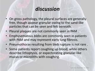
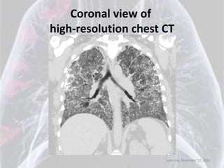
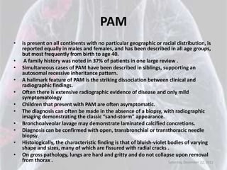
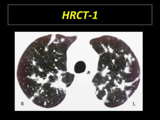
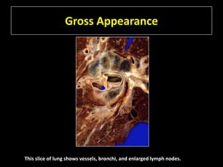
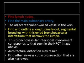
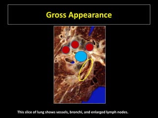
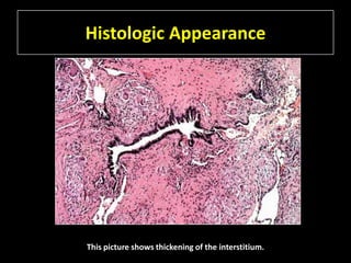
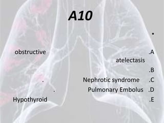
Anwser,s
    Dr :ANAS SAHLE
   1. Chest xr cases.
 2. Chest clinical case.
   3. Chest ct cases.
  4. Collicum exam.
:http://www.facebook.com/dranas224

                                     Saturday, December 22, 2012
chest xr cases
   Dr :anas sahle
 http://www.facebook.com/dranas224
CXR-26-a
CXR26-b
CXR26-c
CXR26-d
Diagnosis for CXR26 is: Ankylosing Spondylitis


                             Bamboo spine
CXR27


Diagnosis is: Aneurysm Arch of Aorta
CXR28-a
CXR28-b


Old film
CXR28:
            Aneurysm Arch of Aorta
                   Leaking Blood into Pleural Space
• Mediastinal mass
• Calcification of periphery evident along upper margin
• Loss of silhouettes of
    – aortic knob
    – left heart margin
    – left diaphragm
• Left pleural effusion
• Tracheal indentation
• Lateral view
• Middle mediastinal mass
• Calcification of periphery in lateral view
• left diaphragm not visible due to fluid in pleural space

                                                        Saturday, December 22, 2012
CXR29


Diagnosis is: Pectus Excavatum
Pectus Excavatum
• Indistinct right heart margin.
• Vague density in right lower lung field
• Lateral view
• reveals protruding sternum and flattening
  heart


                                      Saturday, December 22, 2012
CXR30-a


Diagnosis is: Pulmonary Sequestration
CXR30-b


  Aortogram
CXR30-c


Aortogram
Pulmonary Sequestration
• Anomalous Arterialization of Lung:
• Increased vascular markings at right base
• Smaller right pulmonary artery


Big anomalous artery arising from aorta supplying right lung
                                                  Saturday, December 22, 2012
Saturday, December 22, 2012
chest clinical cases
Dry Cough and Clubbing
in a 45-Year-Old Woman
Submitted by
Jamie L. Garfield, MD
Fellow
Pulmonary and Critical Care Medicine
Temple University School of Medicine
Philadelphia, Pennsylvania
Victor Kim, MD
Assistant Professor of Medicine
Temple University School of Medicine
Philadelphia, Pennsylvania
History
•   A 45-year-old white female presents to pulmonary clinic for evaluation of increasing chest
    tightness, cough and dyspnea.
•   She has a history of chronic respiratory symptoms since childhood and frequent exacerbations of
    bronchitis, but no history of respiratory failure.
•   She reports that until 1 year ago she was limited by mild pleuritic chest tightness and shortness of
    breath with heavy exertion.
•   She now reports daily symptoms of fatigue, dyspnea, and exercise intolerance with minimal
    exertion.
•   She has persistent dry cough and occasionally expectorates granular mucus.
•   She has been using 3 liters per minute of supplemental oxygen via nasal cannula for the past year.
•   Her past medical history is significant for polycythemia, gastroesophageal reflux disease,
    fibronodular breast disease, chronic tinnitus with hearing loss and genital herpes.
•   Her current medications include famotidine, diltiazem, valacyclovir, inhaled fluticasone,
    salmeterol and albuterol.
•   She has a 30 pack-year smoking history, having quit smoking 5 years ago.
•   She drinks 1-2 beers per week and she denies illicit drug use.
•   She is a homemaker and reports no occupational exposures.
•   Her parents, siblings and 2 children (ages 16 and 24) are alive and well with no respiratory disease
    or malignancy.
                                                                                Saturday, December 22, 2012
Physical Exam
• her heart rate is elevated at 112 beats per minute.
• Oxygen saturation on 2 liters of oxygen via nasal
  cannula is 89%.
• Her vital signs are otherwise normal.
• Her head and neck exam is benign.
• Her pulmonary exam is significant for diffuse
  inspiratory and expiratory dry crackles, more
  prominent at the bases bilaterally.
• Cardiac exam reveals regular rhythm with no murmurs,
  rubs or gallops.
• There is clubbing but no cyanosis or edema of her
  extremities.
                                          Saturday, December 22, 2012
Lab
• White blood cell count 12,900/mm3,
• Hemoglobin 15.5 mg/dl,
• Platelet count 473,000/mm3.
• BUN 9 mg/dl, creatinine 1.0 mg/dl,
• calcium 8.8 mg/dl and phosphate 4.9 mg/dl.
• PT, PTT, electrolytes, glucose and liver function
  tests were within normal limits.
• Arterial blood gas on 3L/min supplemental
  oxygen revealed a
    – pH of 7.39, PCO2 40 mm Hg and PaO2 62 mm Hg.
                                           Saturday, December 22, 2012
Studies
• Echocardiogram:
   –   left ventricular ejection fraction was 55-65% and
   –   grade 1 diastolic dysfunction was noted;
   –   right ventricle was mildly dilated and had mildly depressed systolic function;
   –   pulmonary artery systolic pressure was estimated to be 48-52 mm Hg; right atrium was
       mildly dilated and there was mild tricuspid regurgitation.

• Cardiac Catheterization: PAP: 30/6/13; PCWP: 2; CO: 3.38; CI: 2.16.
• Pulmonary Function Tests:
   –   FEV1/ FVC: 74%;
   –   FEV1: 0.96 L, 39%;
   –   FVC: 1.29L, 42%;
   –   TLC: 2.27L, 53%; RV: 1.14L, 79%; DLCO: 3.11L, 57%.

• 6-Minute Walk Test:
   – Resting SpO2: 86% RA; 92% on 2L/min; 8L/min was required to maintain oxygen saturation
     above 90%
   – at the end of 6 minutes; the patient was able to walk 211 meters.

                                                                          Saturday, December 22, 2012
CXR


      Saturday, December 22, 2012
Coronal view of
high-resolution chest CT


                      Saturday, December 22, 2012
Bronchoalveolar lavage


Slightly mucoid, tan fluid; cultures revealed no growth; cytology showed no evidence
of malignancy; a few white blood cells were noted, predominantly macrophages;
laminated calcified concretions and debris were also present.
Von Kossa stain positive.
                                                                 Saturday, December 22, 2012
Question 1
• What is the most likely diagnosis?
•   A. Idiopathic pulmonary fibrosis
•   B. Pulmonary alveolar proteinosis
•   C. Diffuse alveolar hemorrhage syndrome
•   D. Pulmonary alveolar microlithiasis
•   E. Broncheoloalveolar carcinomatosis


                                     Saturday, December 22, 2012
Pulmonary Alveolar Microlithiasis
                (PAM)
•   is a rare disorder of unknown etiology in which microliths occupy the alveolar space (1).
•   The mean age of diagnosis is in the fourth decade of life, although the onset of disease may
    occur in childhood (2, 3).
•   Idiopathic pulmonary fibrosis (IPF) tends to present later in life than PAM.
•   Dyspnea and hypoxemia are common to both disease entities, but the honeycombing
    classically seen on HRCT in IPF would not be expected in PAM.
•   Pulmonary alveolar proteinosis (PAS), another alveolar filling disorder, is also a rare but
    deadly entity.
•   Patients with PAS may have dry crackles and clubbing as in PAM.
•   The patchy ground glass opacities with a “crazy-paving” pattern described in PAS can
    usually be distinguished from the microcalcifications seen on HRCT in PAM. Rarely, “crazy-
    paving” can be seen in PAM (4).
•   Bronchoalveolar carcinomatosis can radiographically mimic many diseases with alveolar
    infiltrates.
•   Classically this condition is less diffuse than PAM, and as such, hypoxemia and diffusion
    impairment are less common.
•   Diffuse alveolar hemorrhage (DAH) can present with hypoxia and diffuse alveolar infiltrates
    similar to PAM.
•   The onset of DAH is usually more abrupt than PAM.
•   While frank hemoptysis can be absent in up to one third of DAH cases, hemosiderin-laden
    macrophages are commonly found in BAL.
                                                                          Saturday, December 22, 2012
Question 2
• Which of the following statements is true regarding
  pulmonary alveolar microlithiasis?
• A. Dyspnea is described in proportion to the severity of
  the radiographic abnormalities.
• B. The diagnosis can only be made by lung biopsy
  demonstrating microliths within the alveoli.
• C. Pathologically, the lungs are soft and boggy.
• D. Familial disease is inherited in an autosomal
  dominant fashion.
• E. PAM is found in all geographic regions, across all
  races, and equally between men and women.

                                             Saturday, December 22, 2012
PAM
•   is present on all continents with no particular geographic or racial distribution, is
    reported equally in males and females, and has been described in all age groups,
    but most frequently from birth to age 40.
•    A family history was noted in 37% of patients in one large review .
•   Simultaneous cases of PAM have been described in siblings, supporting an
    autosomal recessive inheritance pattern.
•   A hallmark feature of PAM is the striking dissociation between clinical and
    radiographic findings.
•   Often there is extensive radiographic evidence of disease and only mild
    symptomatology
•   Children that present with PAM are often asymptomatic.
•   The diagnosis can often be made in the absence of a biopsy, with radiographic
    imaging demonstrating the classic “sand-storm” appearance.
•   Bronchoalveolar lavage may demonstrate laminated calcified concretions.
•   Diagnosis can be confirmed with open, transbronchial or transthoracic needle
    biopsy.
•   Histologically, the characteristic finding is that of bluish-violet bodies of varying
    shape and sizes, many of which are fissured with radial cracks .
•   On gross pathology, lungs are hard and gritty and do not collapse upon removal
    from thorax .                                                       Saturday, December 22, 2012
Question 3
• PAM may be associated with all of the
  following conditions/exposures EXCEPT
  which of the following?
•   A. Milk-alkali syndrome
•   B. Calcium containing powdered tobacco
•   C. Inflammatory bowel disease
•   D. Printing ink
•   E. Renal transplant

                                     Saturday, December 22, 2012
discussion
• PAM has been described in many patients with no discernable risk
  factors for the disease.
• An increased risk of PAM has been reported in individuals with
  exposure to printing ink and among those with a history of
  smoking powdered tobacco containing calcium salts .
• Systemic diseases associated with calcium and phosphate
  deregulation, such as milk-alkali syndrome renal transplant and
  nephrolithiasis have been linked to PAM.
• It is not known if these associations are causational or
  coincidental.
• Inflammatory bowel disease, while can be associated with
  electrolyte abnormalities related to malabsorption, has not been
  described in patients with PAM.

                                                   Saturday, December 22, 2012
Question 4
• Which of the following signs/symptoms
  is not commonly associated with PAM?
•   A. Pleural plaques
•   B. Pneumothorax
•   C. Hemoptysis
•   D. Lithoptysis
•   E. Emphysema

                               Saturday, December 22, 2012
discussion
• On gross pathology, the pleural surfaces are generally
  free, though appear granular owing to the sand-like
  particles that can be seen and felt beneath.
• Pleural plaques are not commonly seen in PAM .
• Emphysematous blebs are commonly seen in patients
  with PAM and may represent early lung fibrosis.
• Pneumothorax resulting from bleb rupture is not rare .
• Some patients report coughing up blood, while others
  describe lithoptysis, or expectorating granular-like
  mucus or microliths with coughing.

                                            Saturday, December 22, 2012
Question 5
• Which of the following statements is FALSE regarding
  the underlying pulmonary process in this patient?
• A. Microliths are derived from the alveolar capillary blood
  and not a result of pathological changes in the tissue cells
  of the alveoli.
• B. Inborn errors in metabolism at the alveolar interface
  leads to local accumulation of calcium salts.
• C. Alveolar hemorrhage is commonly associated with this
  disease.
• D. PAM usually occurs in patients without evidence of
  systemic derangement in calcium metabolism.
• E. Dysfunction of SLC34A2 may reduce the clearance of
  phosphate leading to the formation of microliths.

                                                 Saturday, December 22, 2012
discussion
•   The metabolic abnormality in PAM results in precipitation of calciferous salts in an otherwise
    normal alveolus.
•   The components of the microliths are therefore thought to be derived from the alveolar capillary
    blood and not a result of pathological changes in the tissue cells of the alveoli .
•   It has been suggested that inborn errors in metabolism at the alveolar interface lead to increased
    alkalinity or mucopolysaccharide deposition, promoting the local accumulation of calcium salts.
•   Studies of calcium metabolism in affected patients have been consistently normal, suggesting it is
    unlikely that PAM is due to a systemic derangement of calcium metabolism .
•   Hemoptysis is not uncommon in PAM when inspissated microliths irritate and erode bronchial
    epithelium. Diffuse alveolar hemorrhage, however, is not known to be associated with PAM.
•   Homozygous inactivating mutations in the SLC34A2 gene, are present in patients with PAM.
•   SLC34A2 encodes a sodium phosphate transporter that is highly expressed in alveolar type II
    cells.
•   Normally, type II alveolar cells recycle and degrade surfactant and subsequently release
    phosphate into the alveolar space.
•   Dysfunction of this sodium phosphate transporter may reduce the clearance of phosphate from
    the alveolar space, thereby leading to the formation of microliths .


                                                                               Saturday, December 22, 2012
Question 6
• Treatment for PAM currently includes all
  of the following except?
•   A. Bilateral lung transplant
•   B. Gene replacement therapy
•   C. Whole lung lavage
•   D. Systemic steroids
•   E. Dibisphosphonates

                                   Saturday, December 22, 2012
discussion
• Various treatment regimens have been explored for PAM, most of which
  have not altered the natural course of the disease.
• Borrowing from the therapeutic strategy for PAS, whole lung lavage has
  been tried without success in patients with PAM.
• Systemic corticosteroids and bisphosphonates have also been used but
  fail to preserve lung function or improve symptoms.
• Bilateral lung transplant remains the only option for patients with severe
  symptoms related to PAM .
• There remains very little information on long term survival and whether
  the disease will return in the donor lungs .
• As more is learned about the SLC34A2 gene and its role in PAM, there is
  growing interest in the possibility of gene replacement therapy to treat
  PAM .
• While this therapy is not available today, early data suggest that
  targeting phosphate rather than calcium metabolism may be beneficial
  for the treatment of PAM.

                                                           Saturday, December 22, 2012
Saturday, December 22, 2012
chest ct cases-6
    Dr :anas sahle
  http://www.facebook.com/dranas224
HRCT-1
HRCT-1

• What is the distribution of the abnormal
  densities?
•   a) Bronchovascular
•   b) Interlobular septal
•   c) Pleural
•   d) Centrilobular
HRCT-1
HRCT-1

• Find an example in the right lung of combined
  bronchovascular and interlobular septal
  thickening with distortion of the architecture.
• Find an example in the left lung of thickened
  bronchovascular interstitium.
• Find a centrilobular nodule, representing
  thickening of the terminal bronchiolovascular
  interstitium, in the left lung.
HRCT-1
Gross Appearance


This slice of lung shows vessels, bronchi, and enlarged lymph nodes.
• Find lymph nodes.
• Find the main pulmonary artery.
• The adjacent thinner-walled vessel is the vein.
• Find and outline a longitudinally-cut, segmental
  bronchus with thickened bronchovascular
  interstitium that narrows the lumen.
• This bronchovascular interstitial involvement
  corresponds to that seen in the HRCT image
  above.
• Architectural distortion may result.
• Find other airways cut in cross-section that are
  also narrowed.
Gross Appearance


This slice of lung shows vessels, bronchi, and enlarged lymph nodes.
Histologic Appearance


This picture shows thickening of the interstitium.
• Find an airway and name its type.
• Find the accompanying pulmonary artery
  branch.
• Find and identify 5 rounded, interstitial,
  cellular structures.
• Find and identify the composition of the pink
  matrix.
Histologic Appearance


   Bronchiole (airway has no cartilage)


This picture shows thickening of the interstitium.
Differential diagnosis


• Differential diagnosis of nodular
  bronchovascular and interlobular septal
  thickening on HRC:............


• Histologic differential diagnosis:………
Differential diagnosis

• Differential diagnosis of nodular
  bronchovascular and interlobular septal
  thickening on HRC:
         –Sarcoidosis.
         –lymphangitic tumor.
         –Lymphoma.
         –Kaposi's sarcoma


• Histologic differential diagnosis:………
Differential diagnosis

• Differential diagnosis of nodular bronchovascular and
  interlobular septal thickening on HRC:
         – Sarcoidosis.
         – lymphangitic tumor.
         – Lymphoma.
         – Kaposi's sarcoma
• Histologic differential diagnosis:
         – Infectious granulomatous disease
           (tuberculous or fungal).
         – hypersensitivity pneumonia.
         – reaction to tumor or drug should be
           considered.
Diagnosis:


Chronic sarcoidosis
Summary of diagnostic features of
         sarcoidosis on HRCT


• Bronchovascular, interlobular septal and
  pleural thickening
• Upper lung predominance common
• Architectural distortion frequent
Saturday, December 22, 2012
Collicum EXAM
    Respiratory


                  12/22/2012
A1
.                                       •
                              IDSA
    Neutropenia
                         Cefepime .A    •
                     Ceftazidime .B     •
                        Imipenem .C     •
                     Meropenem .D       •
Tazobactam and Piperacillin sodium .E   •
A2
    . IDSA                                   •

                       :Neutropenia
                          empirical
Amoxicillin-clavulanate + Moxifloxacin .A    •
Amoxicillin-clavulanate + Ciprofloxacin .B   •
 Amoxicillin-clavulanate + Levofloxacin .C   •
Amoxicillin-clavulanate + Azithromycin .D    •
                               Cefixime .E   •
A3
       •
     .A
     .B
     .C
     .D
      .E
A4
                             •

           coccidioidal
     . Amphotericin-B .A     •
          Caspofungin .B     •
            .Fluconazol .C   •
         .Voriconazole .D    •
A5
                    •


                   .A
                   .B
                   .C
                   .D
     .Prednisone    .E
A6
                               •
                               •

exudate                        •
                               •
                          .   .A
          parapneumonic
PH                            .B
                              .C
                              .D
                              .E
A7
                                        •
                                       .A
                amebic liver abscess
Aspergillus                            .B
                                       .C
    Chylothorax                        .D
         pulmonary infarction           .E
A8
                                                 •
                                                .A
                              lupus pleuritis
 Urinothorax                    LDH             .B

                                                .C

IU/LD                              LDH          .D

                                                .E
               congestive heart failure
A9
                        •

                        •
                        •
  Cardiac tamponade .A
 .Dressler's syndrome .B
  Pulmonary Embolus .C
                      .D
                       .E
A10
                                            •

obstructive                                .A
                             atelectasis
                                      .B
         .         Nephrotic syndrome .C
              .     Pulmonary Embolus .D
Hypothyroid                            .E
Saturday, December 22, 2012

Anwser,s7

  • 1.
    Anwser,s Dr :ANAS SAHLE 1. Chest xr cases. 2. Chest clinical case. 3. Chest ct cases. 4. Collicum exam. :http://www.facebook.com/dranas224 Saturday, December 22, 2012
  • 2.
    chest xr cases Dr :anas sahle http://www.facebook.com/dranas224
  • 3.
  • 4.
  • 5.
  • 6.
    CXR26-d Diagnosis for CXR26is: Ankylosing Spondylitis Bamboo spine
  • 7.
  • 8.
  • 9.
  • 10.
    CXR28: Aneurysm Arch of Aorta Leaking Blood into Pleural Space • Mediastinal mass • Calcification of periphery evident along upper margin • Loss of silhouettes of – aortic knob – left heart margin – left diaphragm • Left pleural effusion • Tracheal indentation • Lateral view • Middle mediastinal mass • Calcification of periphery in lateral view • left diaphragm not visible due to fluid in pleural space Saturday, December 22, 2012
  • 11.
  • 12.
    Pectus Excavatum • Indistinctright heart margin. • Vague density in right lower lung field • Lateral view • reveals protruding sternum and flattening heart Saturday, December 22, 2012
  • 13.
  • 14.
  • 15.
  • 16.
    Pulmonary Sequestration • AnomalousArterialization of Lung: • Increased vascular markings at right base • Smaller right pulmonary artery Big anomalous artery arising from aorta supplying right lung Saturday, December 22, 2012
  • 17.
  • 18.
    chest clinical cases DryCough and Clubbing in a 45-Year-Old Woman Submitted by Jamie L. Garfield, MD Fellow Pulmonary and Critical Care Medicine Temple University School of Medicine Philadelphia, Pennsylvania Victor Kim, MD Assistant Professor of Medicine Temple University School of Medicine Philadelphia, Pennsylvania
  • 19.
    History • A 45-year-old white female presents to pulmonary clinic for evaluation of increasing chest tightness, cough and dyspnea. • She has a history of chronic respiratory symptoms since childhood and frequent exacerbations of bronchitis, but no history of respiratory failure. • She reports that until 1 year ago she was limited by mild pleuritic chest tightness and shortness of breath with heavy exertion. • She now reports daily symptoms of fatigue, dyspnea, and exercise intolerance with minimal exertion. • She has persistent dry cough and occasionally expectorates granular mucus. • She has been using 3 liters per minute of supplemental oxygen via nasal cannula for the past year. • Her past medical history is significant for polycythemia, gastroesophageal reflux disease, fibronodular breast disease, chronic tinnitus with hearing loss and genital herpes. • Her current medications include famotidine, diltiazem, valacyclovir, inhaled fluticasone, salmeterol and albuterol. • She has a 30 pack-year smoking history, having quit smoking 5 years ago. • She drinks 1-2 beers per week and she denies illicit drug use. • She is a homemaker and reports no occupational exposures. • Her parents, siblings and 2 children (ages 16 and 24) are alive and well with no respiratory disease or malignancy. Saturday, December 22, 2012
  • 20.
    Physical Exam • herheart rate is elevated at 112 beats per minute. • Oxygen saturation on 2 liters of oxygen via nasal cannula is 89%. • Her vital signs are otherwise normal. • Her head and neck exam is benign. • Her pulmonary exam is significant for diffuse inspiratory and expiratory dry crackles, more prominent at the bases bilaterally. • Cardiac exam reveals regular rhythm with no murmurs, rubs or gallops. • There is clubbing but no cyanosis or edema of her extremities. Saturday, December 22, 2012
  • 21.
    Lab • White bloodcell count 12,900/mm3, • Hemoglobin 15.5 mg/dl, • Platelet count 473,000/mm3. • BUN 9 mg/dl, creatinine 1.0 mg/dl, • calcium 8.8 mg/dl and phosphate 4.9 mg/dl. • PT, PTT, electrolytes, glucose and liver function tests were within normal limits. • Arterial blood gas on 3L/min supplemental oxygen revealed a – pH of 7.39, PCO2 40 mm Hg and PaO2 62 mm Hg. Saturday, December 22, 2012
  • 22.
    Studies • Echocardiogram: – left ventricular ejection fraction was 55-65% and – grade 1 diastolic dysfunction was noted; – right ventricle was mildly dilated and had mildly depressed systolic function; – pulmonary artery systolic pressure was estimated to be 48-52 mm Hg; right atrium was mildly dilated and there was mild tricuspid regurgitation. • Cardiac Catheterization: PAP: 30/6/13; PCWP: 2; CO: 3.38; CI: 2.16. • Pulmonary Function Tests: – FEV1/ FVC: 74%; – FEV1: 0.96 L, 39%; – FVC: 1.29L, 42%; – TLC: 2.27L, 53%; RV: 1.14L, 79%; DLCO: 3.11L, 57%. • 6-Minute Walk Test: – Resting SpO2: 86% RA; 92% on 2L/min; 8L/min was required to maintain oxygen saturation above 90% – at the end of 6 minutes; the patient was able to walk 211 meters. Saturday, December 22, 2012
  • 23.
    CXR Saturday, December 22, 2012
  • 24.
    Coronal view of high-resolutionchest CT Saturday, December 22, 2012
  • 25.
    Bronchoalveolar lavage Slightly mucoid,tan fluid; cultures revealed no growth; cytology showed no evidence of malignancy; a few white blood cells were noted, predominantly macrophages; laminated calcified concretions and debris were also present. Von Kossa stain positive. Saturday, December 22, 2012
  • 26.
    Question 1 • Whatis the most likely diagnosis? • A. Idiopathic pulmonary fibrosis • B. Pulmonary alveolar proteinosis • C. Diffuse alveolar hemorrhage syndrome • D. Pulmonary alveolar microlithiasis • E. Broncheoloalveolar carcinomatosis Saturday, December 22, 2012
  • 27.
    Pulmonary Alveolar Microlithiasis (PAM) • is a rare disorder of unknown etiology in which microliths occupy the alveolar space (1). • The mean age of diagnosis is in the fourth decade of life, although the onset of disease may occur in childhood (2, 3). • Idiopathic pulmonary fibrosis (IPF) tends to present later in life than PAM. • Dyspnea and hypoxemia are common to both disease entities, but the honeycombing classically seen on HRCT in IPF would not be expected in PAM. • Pulmonary alveolar proteinosis (PAS), another alveolar filling disorder, is also a rare but deadly entity. • Patients with PAS may have dry crackles and clubbing as in PAM. • The patchy ground glass opacities with a “crazy-paving” pattern described in PAS can usually be distinguished from the microcalcifications seen on HRCT in PAM. Rarely, “crazy- paving” can be seen in PAM (4). • Bronchoalveolar carcinomatosis can radiographically mimic many diseases with alveolar infiltrates. • Classically this condition is less diffuse than PAM, and as such, hypoxemia and diffusion impairment are less common. • Diffuse alveolar hemorrhage (DAH) can present with hypoxia and diffuse alveolar infiltrates similar to PAM. • The onset of DAH is usually more abrupt than PAM. • While frank hemoptysis can be absent in up to one third of DAH cases, hemosiderin-laden macrophages are commonly found in BAL. Saturday, December 22, 2012
  • 28.
    Question 2 • Whichof the following statements is true regarding pulmonary alveolar microlithiasis? • A. Dyspnea is described in proportion to the severity of the radiographic abnormalities. • B. The diagnosis can only be made by lung biopsy demonstrating microliths within the alveoli. • C. Pathologically, the lungs are soft and boggy. • D. Familial disease is inherited in an autosomal dominant fashion. • E. PAM is found in all geographic regions, across all races, and equally between men and women. Saturday, December 22, 2012
  • 29.
    PAM • is present on all continents with no particular geographic or racial distribution, is reported equally in males and females, and has been described in all age groups, but most frequently from birth to age 40. • A family history was noted in 37% of patients in one large review . • Simultaneous cases of PAM have been described in siblings, supporting an autosomal recessive inheritance pattern. • A hallmark feature of PAM is the striking dissociation between clinical and radiographic findings. • Often there is extensive radiographic evidence of disease and only mild symptomatology • Children that present with PAM are often asymptomatic. • The diagnosis can often be made in the absence of a biopsy, with radiographic imaging demonstrating the classic “sand-storm” appearance. • Bronchoalveolar lavage may demonstrate laminated calcified concretions. • Diagnosis can be confirmed with open, transbronchial or transthoracic needle biopsy. • Histologically, the characteristic finding is that of bluish-violet bodies of varying shape and sizes, many of which are fissured with radial cracks . • On gross pathology, lungs are hard and gritty and do not collapse upon removal from thorax . Saturday, December 22, 2012
  • 30.
    Question 3 • PAMmay be associated with all of the following conditions/exposures EXCEPT which of the following? • A. Milk-alkali syndrome • B. Calcium containing powdered tobacco • C. Inflammatory bowel disease • D. Printing ink • E. Renal transplant Saturday, December 22, 2012
  • 31.
    discussion • PAM hasbeen described in many patients with no discernable risk factors for the disease. • An increased risk of PAM has been reported in individuals with exposure to printing ink and among those with a history of smoking powdered tobacco containing calcium salts . • Systemic diseases associated with calcium and phosphate deregulation, such as milk-alkali syndrome renal transplant and nephrolithiasis have been linked to PAM. • It is not known if these associations are causational or coincidental. • Inflammatory bowel disease, while can be associated with electrolyte abnormalities related to malabsorption, has not been described in patients with PAM. Saturday, December 22, 2012
  • 32.
    Question 4 • Whichof the following signs/symptoms is not commonly associated with PAM? • A. Pleural plaques • B. Pneumothorax • C. Hemoptysis • D. Lithoptysis • E. Emphysema Saturday, December 22, 2012
  • 33.
    discussion • On grosspathology, the pleural surfaces are generally free, though appear granular owing to the sand-like particles that can be seen and felt beneath. • Pleural plaques are not commonly seen in PAM . • Emphysematous blebs are commonly seen in patients with PAM and may represent early lung fibrosis. • Pneumothorax resulting from bleb rupture is not rare . • Some patients report coughing up blood, while others describe lithoptysis, or expectorating granular-like mucus or microliths with coughing. Saturday, December 22, 2012
  • 34.
    Question 5 • Whichof the following statements is FALSE regarding the underlying pulmonary process in this patient? • A. Microliths are derived from the alveolar capillary blood and not a result of pathological changes in the tissue cells of the alveoli. • B. Inborn errors in metabolism at the alveolar interface leads to local accumulation of calcium salts. • C. Alveolar hemorrhage is commonly associated with this disease. • D. PAM usually occurs in patients without evidence of systemic derangement in calcium metabolism. • E. Dysfunction of SLC34A2 may reduce the clearance of phosphate leading to the formation of microliths. Saturday, December 22, 2012
  • 35.
    discussion • The metabolic abnormality in PAM results in precipitation of calciferous salts in an otherwise normal alveolus. • The components of the microliths are therefore thought to be derived from the alveolar capillary blood and not a result of pathological changes in the tissue cells of the alveoli . • It has been suggested that inborn errors in metabolism at the alveolar interface lead to increased alkalinity or mucopolysaccharide deposition, promoting the local accumulation of calcium salts. • Studies of calcium metabolism in affected patients have been consistently normal, suggesting it is unlikely that PAM is due to a systemic derangement of calcium metabolism . • Hemoptysis is not uncommon in PAM when inspissated microliths irritate and erode bronchial epithelium. Diffuse alveolar hemorrhage, however, is not known to be associated with PAM. • Homozygous inactivating mutations in the SLC34A2 gene, are present in patients with PAM. • SLC34A2 encodes a sodium phosphate transporter that is highly expressed in alveolar type II cells. • Normally, type II alveolar cells recycle and degrade surfactant and subsequently release phosphate into the alveolar space. • Dysfunction of this sodium phosphate transporter may reduce the clearance of phosphate from the alveolar space, thereby leading to the formation of microliths . Saturday, December 22, 2012
  • 36.
    Question 6 • Treatmentfor PAM currently includes all of the following except? • A. Bilateral lung transplant • B. Gene replacement therapy • C. Whole lung lavage • D. Systemic steroids • E. Dibisphosphonates Saturday, December 22, 2012
  • 37.
    discussion • Various treatmentregimens have been explored for PAM, most of which have not altered the natural course of the disease. • Borrowing from the therapeutic strategy for PAS, whole lung lavage has been tried without success in patients with PAM. • Systemic corticosteroids and bisphosphonates have also been used but fail to preserve lung function or improve symptoms. • Bilateral lung transplant remains the only option for patients with severe symptoms related to PAM . • There remains very little information on long term survival and whether the disease will return in the donor lungs . • As more is learned about the SLC34A2 gene and its role in PAM, there is growing interest in the possibility of gene replacement therapy to treat PAM . • While this therapy is not available today, early data suggest that targeting phosphate rather than calcium metabolism may be beneficial for the treatment of PAM. Saturday, December 22, 2012
  • 38.
  • 39.
    chest ct cases-6 Dr :anas sahle http://www.facebook.com/dranas224
  • 40.
  • 41.
    HRCT-1 • What isthe distribution of the abnormal densities? • a) Bronchovascular • b) Interlobular septal • c) Pleural • d) Centrilobular
  • 42.
  • 43.
    HRCT-1 • Find anexample in the right lung of combined bronchovascular and interlobular septal thickening with distortion of the architecture. • Find an example in the left lung of thickened bronchovascular interstitium. • Find a centrilobular nodule, representing thickening of the terminal bronchiolovascular interstitium, in the left lung.
  • 44.
  • 45.
    Gross Appearance This sliceof lung shows vessels, bronchi, and enlarged lymph nodes.
  • 46.
    • Find lymphnodes. • Find the main pulmonary artery. • The adjacent thinner-walled vessel is the vein. • Find and outline a longitudinally-cut, segmental bronchus with thickened bronchovascular interstitium that narrows the lumen. • This bronchovascular interstitial involvement corresponds to that seen in the HRCT image above. • Architectural distortion may result. • Find other airways cut in cross-section that are also narrowed.
  • 47.
    Gross Appearance This sliceof lung shows vessels, bronchi, and enlarged lymph nodes.
  • 48.
    Histologic Appearance This pictureshows thickening of the interstitium.
  • 49.
    • Find anairway and name its type. • Find the accompanying pulmonary artery branch. • Find and identify 5 rounded, interstitial, cellular structures. • Find and identify the composition of the pink matrix.
  • 50.
    Histologic Appearance Bronchiole (airway has no cartilage) This picture shows thickening of the interstitium.
  • 51.
    Differential diagnosis • Differentialdiagnosis of nodular bronchovascular and interlobular septal thickening on HRC:............ • Histologic differential diagnosis:………
  • 52.
    Differential diagnosis • Differentialdiagnosis of nodular bronchovascular and interlobular septal thickening on HRC: –Sarcoidosis. –lymphangitic tumor. –Lymphoma. –Kaposi's sarcoma • Histologic differential diagnosis:………
  • 53.
    Differential diagnosis • Differentialdiagnosis of nodular bronchovascular and interlobular septal thickening on HRC: – Sarcoidosis. – lymphangitic tumor. – Lymphoma. – Kaposi's sarcoma • Histologic differential diagnosis: – Infectious granulomatous disease (tuberculous or fungal). – hypersensitivity pneumonia. – reaction to tumor or drug should be considered.
  • 54.
  • 55.
    Summary of diagnosticfeatures of sarcoidosis on HRCT • Bronchovascular, interlobular septal and pleural thickening • Upper lung predominance common • Architectural distortion frequent
  • 56.
  • 57.
    Collicum EXAM Respiratory 12/22/2012
  • 58.
    A1 . • IDSA Neutropenia Cefepime .A • Ceftazidime .B • Imipenem .C • Meropenem .D • Tazobactam and Piperacillin sodium .E •
  • 59.
    A2 . IDSA • :Neutropenia empirical Amoxicillin-clavulanate + Moxifloxacin .A • Amoxicillin-clavulanate + Ciprofloxacin .B • Amoxicillin-clavulanate + Levofloxacin .C • Amoxicillin-clavulanate + Azithromycin .D • Cefixime .E •
  • 60.
    A3 • .A .B .C .D .E
  • 61.
    A4 • coccidioidal . Amphotericin-B .A • Caspofungin .B • .Fluconazol .C • .Voriconazole .D •
  • 62.
    A5 • .A .B .C .D .Prednisone .E
  • 63.
    A6 • • exudate • • . .A parapneumonic PH .B .C .D .E
  • 64.
    A7 • .A amebic liver abscess Aspergillus .B .C Chylothorax .D pulmonary infarction .E
  • 65.
    A8 • .A lupus pleuritis Urinothorax LDH .B .C IU/LD LDH .D .E congestive heart failure
  • 66.
    A9 • • • Cardiac tamponade .A .Dressler's syndrome .B Pulmonary Embolus .C .D .E
  • 67.
    A10 • obstructive .A atelectasis .B . Nephrotic syndrome .C . Pulmonary Embolus .D Hypothyroid .E
  • 68.