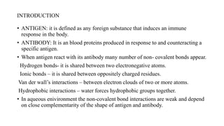
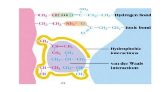
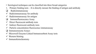
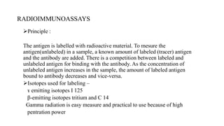
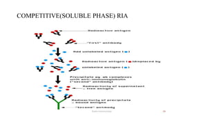
ANTIGEN ANTIBODY
BINDING TESTS
SUBMITTED BY:
MYREDDY NIKHIL
M.V.Sc 1ST YEAR
INTRODUCTION
• ANTIGEN: it is defined as any foreign substance that induces an immune
response in the body.
• ANTIBODY: It is an blood proteins produced in response to and counteracting a
specific antigen.
• When antigen react with its antibody many number of non- covalent bonds appear.
Hydrogen bonds- it is shared between two electronegative atoms.
Ionic bonds – it is shared between oppositely charged residues.
Van der wall’s interactions – between electron clouds of two or more atoms.
Hydrophobic interactions – water forces hydrophobic groups together.
• In aqueous enivironment the non-covalent bond interactions are weak and depend
on close complementarity of the shape of antigen and antibody.
• Serological techniques can be classified into three broad categories
1. Primary binding tests - It is directly mesure the binding of antigen and antibody
 Radioimmunoassay
 Radioimmunoassay for antibody
 Radioimmunoassay for antigen
 Immunofluorescence Assay
 Direct fluorescent antibody tests
 Indirect fluorescent antibody tests
 Particle concentration fluorescence immunoassay
 Immunoenzyme Assays
 Microwell Enzyme-Linked Immunosorbent Assay tests
 Western blotting
 Immunohistochemistry
 Disposable immunoassay Devices
 Immunofiltration
Immunochromatography
Antibody Labels
The flow cytometer
2. Secondary binding tests
Precipitation tests
 Immunodiffusion
Radial immunodiffusion test
Immunoelectrophoresis and related techniques
Agglutination
Antiglobulin tests
Passive agglutination tests
Viral hemagglutination an its inhibition
Complement fixation test
Antitoxin neutralization
In vivo test
 Passive cutaneous anaphylaxsis.
3. Tertiary binding test
Viral neutralization test
Animal neutralization test
RADIOIMMUNOASSAYS
Principle :
The antigen is labelled with radioactive material. To mesure the
antigen(unlabeled) in a sample, a known amount of labeled (tracer) antigen
and the antibody are added. There is a competition between labeled and
unlabeled antigen for binding with the antibody. As the concentration of
unlabeled antigen increases in the sample, the amount of labeled antigen
bound to antibody decreases and vice-versa.
Isotopes used for labeling –
ɤ emitting isotopes I 125
β-emitting isotopes tritium and C 14
Gamma radiation is easy measure and practical to use because of high
pentration power
COMPETITIVE(SOLUBLE PHASE) RIA
• By adding either 50% ammonium sulphate or 2 % PEG or
staphylococcus protein A or anti Ig the bound antigens are separated
from the unbound one.
• Conclusion:
The amount of antigen in the test sample is inversely proportion to the
amount of label measured.
Less bound radioactivity in the assay indicates more of antigen in the
test sample and vice-versa.
IMMUNOHISTOCHEMISTRY
• Enzymes conjugated to immunoglobulin ot antiglobulin can be use to
locate specific antigen in tissue section
• Horseradish peroxidase is most widely employed label
• Tissue section + enzyme labeled antibody
washing
Incubate in a solution appropriate enzyme substrate
Bound antibody is detected by developing brown stain at the site of
antibody binding.
Immunohistochemistry of intestine
NEUTRALIZATION TEST
• Its estimate the ability of antibody to neutralize the biological activity
of antigen when mixed with it in in vitro condition.
• Mice neutralization test
THANK YOU

Antigen antibody binding assay

  • 1.
    ANTIGEN ANTIBODY BINDING TESTS SUBMITTEDBY: MYREDDY NIKHIL M.V.Sc 1ST YEAR
  • 2.
    INTRODUCTION • ANTIGEN: itis defined as any foreign substance that induces an immune response in the body. • ANTIBODY: It is an blood proteins produced in response to and counteracting a specific antigen. • When antigen react with its antibody many number of non- covalent bonds appear. Hydrogen bonds- it is shared between two electronegative atoms. Ionic bonds – it is shared between oppositely charged residues. Van der wall’s interactions – between electron clouds of two or more atoms. Hydrophobic interactions – water forces hydrophobic groups together. • In aqueous enivironment the non-covalent bond interactions are weak and depend on close complementarity of the shape of antigen and antibody.
  • 4.
    • Serological techniquescan be classified into three broad categories 1. Primary binding tests - It is directly mesure the binding of antigen and antibody  Radioimmunoassay  Radioimmunoassay for antibody  Radioimmunoassay for antigen  Immunofluorescence Assay  Direct fluorescent antibody tests  Indirect fluorescent antibody tests  Particle concentration fluorescence immunoassay  Immunoenzyme Assays  Microwell Enzyme-Linked Immunosorbent Assay tests  Western blotting  Immunohistochemistry
  • 5.
     Disposable immunoassayDevices  Immunofiltration Immunochromatography Antibody Labels The flow cytometer
  • 6.
    2. Secondary bindingtests Precipitation tests  Immunodiffusion Radial immunodiffusion test Immunoelectrophoresis and related techniques Agglutination Antiglobulin tests Passive agglutination tests Viral hemagglutination an its inhibition Complement fixation test Antitoxin neutralization
  • 7.
    In vivo test Passive cutaneous anaphylaxsis. 3. Tertiary binding test Viral neutralization test Animal neutralization test
  • 8.
    RADIOIMMUNOASSAYS Principle : The antigenis labelled with radioactive material. To mesure the antigen(unlabeled) in a sample, a known amount of labeled (tracer) antigen and the antibody are added. There is a competition between labeled and unlabeled antigen for binding with the antibody. As the concentration of unlabeled antigen increases in the sample, the amount of labeled antigen bound to antibody decreases and vice-versa. Isotopes used for labeling – ɤ emitting isotopes I 125 β-emitting isotopes tritium and C 14 Gamma radiation is easy measure and practical to use because of high pentration power
  • 9.
  • 10.
    • By addingeither 50% ammonium sulphate or 2 % PEG or staphylococcus protein A or anti Ig the bound antigens are separated from the unbound one. • Conclusion: The amount of antigen in the test sample is inversely proportion to the amount of label measured. Less bound radioactivity in the assay indicates more of antigen in the test sample and vice-versa.
  • 11.
    IMMUNOHISTOCHEMISTRY • Enzymes conjugatedto immunoglobulin ot antiglobulin can be use to locate specific antigen in tissue section • Horseradish peroxidase is most widely employed label • Tissue section + enzyme labeled antibody washing Incubate in a solution appropriate enzyme substrate Bound antibody is detected by developing brown stain at the site of antibody binding.
  • 12.
  • 13.
    NEUTRALIZATION TEST • Itsestimate the ability of antibody to neutralize the biological activity of antigen when mixed with it in in vitro condition.
  • 14.
  • 15.