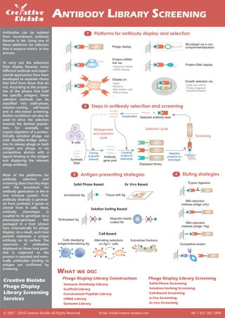
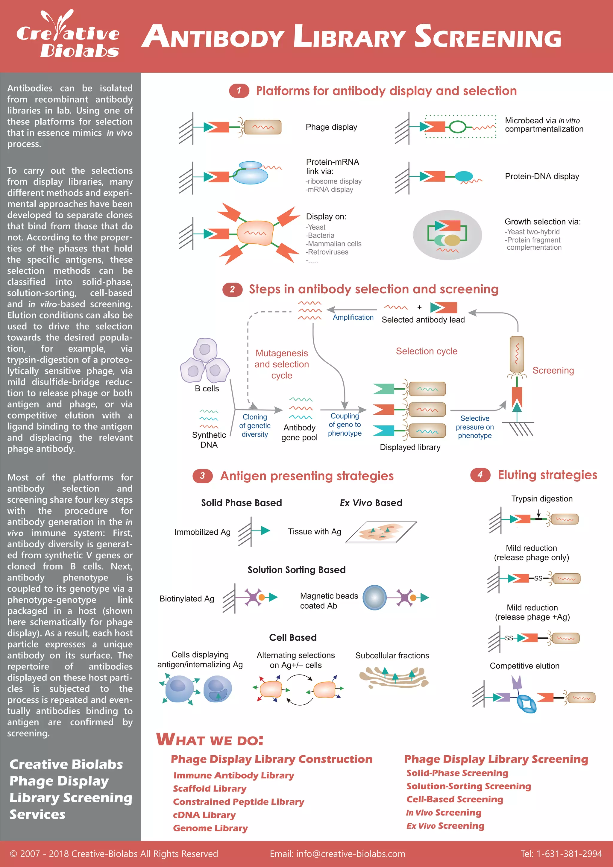
Antibodies can be isolated from recombinant antibody libraries in lab. Using one of these platforms for selection that in essence mimics in vivo process.https://www.creative-biolabs.com/phage-display-library-screening.html
Presentation of Frank Hills in 1st International Antibody Validation Forum 2014St John's Laboratory Ltd
After graduating with an honours degree in Biochemistry Dr Hills worked for several years as a Clinical Scientist at St Bartholomew's hospital in London. He was awarded his PhD in 2002 from the faculty of Medicine at Queen Mary University of London. He continued his interest in reproductive science at Imperial College London where he worked as a postdoctoral researcher before joining Middlesex in 2004 as a lecturer. Dr Hills has published many high profile original research articles on various aspects of obstetric pathology including pre-eclampsia, recurrent miscarriage, preterm labour and fetal distress as well as several articles in the area of assisted reproduction. Currently, he is research interests include investigating the role of glycosaminoglycans and proteoglycans on the development of placental pathology and breast cancer. Dr Hills teaches a range of topics in biomedical science including clinical biochemistry, cellular and developmental biology as well as statistical analysis. He is author of around 30 peer-reviewed scientific articles and has refereed manuscripts for a variety of journals in the area of reproduction and endocrinology.
For more details about 1st international antibody validation forum please check on http://www.stjohnslabs.com/ac_cms/blog
Monoclonal antibody
# Definition
# A general representation of the method used to produce monoclonal antibodies
#Applications
# Monoclonal antibodies for cancer.
#MAbs approved by the FDA for cancer include:
# Factor affecting monoclonal antibody immunogenicity
#Monoclonal Antibodies : 4 Types
# Monoclonal Antibodies : 4 Types images
Monoclonal antibodies are produced through the fusion of antibody-producing B cells with myeloma cells to form immortal hybridoma cells. This process allows for the mass production of antibodies that are identical and specifically target a single antigen or macromolecule. Monoclonal antibodies have important applications in disease diagnosis, treatment, and pregnancy testing due to their high specificity and ability to identify pathogens, tumors, and other targets.
Application of antibodies in purificationANKUR SHARMA
Immunoaffinity chromatography uses the specific binding between antigens and antibodies to purify proteins. Monoclonal antibodies are commonly used as they are highly specific and customizable. The antibodies are immobilized onto a matrix to purify target proteins from mixtures by retaining antigen-antibody complexes on the column while other molecules pass through. Various techniques are used to elute and collect the purified protein. Flow cytometry also utilizes fluorescent antibody binding to identify and separate cells based on surface markers allowing purification of cell populations.
Monoclonal antibodies (MAbs) are antibodies that are directed against a single antigen. They can be produced through hybridoma technology which involves fusing antibody-producing B cells with myeloma cells to form a hybrid cell line. This document outlines the process for producing MAbs including immunizing an animal, fusing B cells with myeloma cells, selecting antibody-producing hybridomas, cloning and mass producing the antibodies. MAbs have various diagnostic and therapeutic applications for diseases.
Antibodies, also known as immunoglobulins, are large Y-shaped glycoproteins produced by B cells that help the immune system identify and neutralize foreign objects like bacteria and viruses. There are two main types of antibodies: monoclonal antibodies, which are specifically targeted to a single antigen or epitope, and polyclonal antibodies, which recognize multiple epitopes on an antigen. Monoclonal antibodies are produced through cell fusion and cloning techniques, while polyclonal antibodies are produced by injecting an antigen into an animal and collecting the resultant serum. Both monoclonal and polyclonal antibodies have various diagnostic and therapeutic applications, such as cancer treatment and autoimmune disorder treatment.
Antibodies are proteins produced by plasma cells to target antigens like pathogens. They interact specifically with antigens through paratope regions. Antigens contain multiple epitopes that bind to different antibody clones, resulting in polyclonal antibodies targeting various epitopes. Monoclonal antibodies arise from a single clone and target the same epitope, making them highly specific. Monoclonal antibodies are produced using hybridoma technology, which fuses antibody-producing cells with myeloma cells in HAT medium for selection. They have diagnostic and therapeutic applications such as cancer detection and treatment.
Monoclonal antibodies are identical immunoglobulins generated from a single B-cell clone that recognize a unique epitope on a single antigen. They have various applications including diagnostic applications using biochemical analysis and diagnostic imaging, therapeutic applications as direct treatment agents and targeting agents, and protein purification. Monoclonal antibodies are produced by fusing B cells from an immunized animal with myeloma cells to generate hybridomas that can produce the monoclonal antibody indefinitely.
Monoclonal antibodies are identical antibodies that are produced by one type of immune cell and are directed against a specific epitope or antigen. They are typically created by fusing myeloma cells with spleen cells from a mouse that has been immunized with the desired antigen. This fusion forms a hybridoma cell line that can produce monoclonal antibodies. Monoclonal antibodies have important diagnostic uses in detecting conditions like pregnancy and infections. They are also used therapeutically for cancer treatment.
ELISPOT assays are the most sensitive technology for detecting immune cells that secrete proteins like cytokines. They directly capture the analyte secreted by each individual cell, preventing dilution or degradation, making ELISPOT more sensitive than other methods like ELISA or flow cytometry. ELISPOT assays can detect rare antigen-specific immune cells and characterize both the quantity and effector classes of the antigen-specific immune response.
Students of medical and allied subjects must be exposed to the concept of monoclonal antibodies for the efficient practice of clinical and laboratory medicine.
Monoclonal antibodies are antibodies produced by a single clone of cells that make one specific antibody. They are homogeneous, specific, and do not vary between batches as they bind to a single antigen. Monoclonal antibodies have applications in diagnostics, such as pregnancy tests, and therapeutics, such as treatments for cancer and organ transplantation. They are produced through cell fusion methods like roller bottle culture or bioreactor culture.
- Georges Köhler and Cesar Milstein developed hybridoma technology in 1975 for which they received the Nobel Prize in 1984. Hybridoma technology involves fusing myeloma cells with antibody-producing immune cells to create immortal cell lines that produce monoclonal antibodies.
- Monoclonal antibodies are identical antibodies produced by a single clone and recognize a specific antigen, whereas polyclonal antibodies are derived from different cell lines.
- Hybridoma technology involves immunizing an animal, fusing its immune cells with myeloma cells, selecting and cloning hybridoma cells that produce the desired antibody, and characterizing and storing the monoclonal antibody produced. Monoclonal antibodies have important applications in diagnostics and treatment of diseases like cancer.
Polyclonal antibodies were generated against 10 human protein targets by immunizing the same recombinant antigen in separate rabbits. Epitope mapping showed that while the antibodies often recognized similar epitopes, they did not have an identical pattern and some unique epitopes were observed for individual rabbits. Fractionation of one polyclonal antibody based on peptide affinity revealed most but not all epitopes were shared between immunizations. This suggests polyclonal antibodies from repeated immunization have related but non-identical epitope profiles.
Polyclonal antibodies are produced by injecting an antigen into an animal to elicit an immune response. They recognize and bind to multiple epitopes on an antigen. Monoclonal antibodies are produced through cell fusion and screening to identify antibodies that bind to a single epitope. Polyclonal antibodies are useful for detection applications due to binding multiple epitopes, while monoclonal antibodies are useful for research and treatment due to their high specificity for a single epitope. Both have advantages and disadvantages depending on the application.
A sandwich ELISA measures the amount of antigen between two layers of antibodies. One layer is the capture antibody, the other is the detection antibody. The antigen to be measured must contain at least two antigenic sites capable of binding to antibody, with one binding to the capture antibody and the other binding to the detection antibody.
Sandwich ELISA is very sensitive (more so than direct or indirect ELISA) and the sample does not have to be purified before analysis.
This document summarizes and compares the processes for producing monoclonal and polyclonal antibodies. Monoclonal antibodies are produced from a single clone of B cells and recognize a single epitope, while polyclonal antibodies are a heterogeneous mixture produced from different B cell clones that recognize multiple epitopes of the same antigen. The production of monoclonal antibodies involves immunizing an animal, fusing B cells with myeloma cells to create hybridomas, and screening for antibodies. Polyclonal antibodies are harvested directly from immunized animal serum. The document outlines the key similarities and differences between these two antibody types.
Monoclonal antibodies are identical antibodies produced by a single clone of B cells that recognize the same epitope on an antigen. They can be produced in a laboratory by fusing B cells that produce a desired antibody with myeloma cells to form hybridomas. Monoclonal antibodies have many diagnostic applications as they can be used to detect specific substances like proteins, pathogens, and tumor markers. They allow for rapid diagnosis of diseases like hepatitis, influenza, and cancer. Monoclonal antibodies are also used in pregnancy tests and monitoring drug levels in the body.
This document discusses monoclonal antibodies (mAbs), which are antibodies that are cloned from identical immune cells. mAbs are produced using hybridoma technology, which involves fusing antibody-producing immune cells with myeloma cells to produce immortal hybrid cell lines. These hybridomas can then be cultured to mass-produce mAbs with a single specificity. The document outlines the history, properties, production process including immunization, cell fusion and screening, and applications of mAbs in diagnostics and therapeutics. Specific examples of therapeutic mAbs are also provided.
The document describes several immunology techniques:
Flow cytometry analyzes individual cells or particles using laser light scattering and fluorescent dyes to label cellular molecules. ELISA uses antigen-coated plates and enzyme-linked antibodies to detect antibodies in serum samples. ELISPOT counts antibody-secreting cells by detecting individual antibody spots. Tissue culture methods grow cells in controlled conditions using culture medium, reagents, and equipment to maintain cells outside the body.
Monoclonal antibodies are antibodies that are directed against a specific antigen. They are produced by a single clone of cells and can be obtained from immortalized B-lymphocytes or recombinant cell lines. The production of monoclonal antibodies involves immunizing an animal, fusing its spleen cells with myeloma cells to form hybridomas, selecting hybridomas that secrete the desired antibody, and propagating these cells to produce large amounts of monoclonal antibodies. Monoclonal antibodies have various applications in cancer therapy, organ transplantation, disease diagnosis, and protein purification. Recent developments include FDA approvals of monoclonal antibodies for treating cancer and blood disorders.
Monoclonal antibodies (mAbs) are identical antibodies produced by a single clone of immune cells that are all clones of the same parent cell. mAbs can be produced against almost any substance and are important tools in biochemistry and medicine. They are produced through the fusion of antibody-producing cells with myeloma cells to form hybridomas that produce identical antibodies. mAbs have applications in research, diagnostics, and therapy due to their specificity and ability to detect or purify target substances.
Monoclonal antibodies are identical antibodies produced by identical immune cells that are clones of a single parent cell. They are produced by fusing antibody-producing cells with tumor cells to create a hybridoma cell line that continuously produces the same antibody. Monoclonal antibodies have important medical uses such as diagnosing pregnancy or HIV infection through detection of specific antigens, and treating cancer by targeting tumor-associated antigens on cancer cells. However, monoclonal antibodies produced in mice can trigger an immune response in humans, so genetically engineered antibodies are being developed to avoid this.
ELISA (enzyme-linked immunosorbent assay) is a biochemical technique used to detect the presence of antigens or antibodies in a biological sample. It uses antibodies and color changing enzymes to identify a substance in the sample. There are different types of ELISA including sandwich, indirect, and competitive ELISA. ELISA is widely used in areas like immunology, diagnostics, and food testing due to its accuracy, sensitivity, and ability to analyze multiple samples at once.
Special Investigations for Biomedical StudentsSri Lakshman
Western blot is used to detect specific proteins in tissue or cell extracts. It involves separating proteins by electrophoresis, transferring them to a membrane, and using antibodies to identify a target protein. ELISA detects specific antigens or antibodies and is used for diseases screening like HIV, syphilis, and Lyme disease. PCR amplifies specific DNA sequences and is used to detect pathogens and for disease diagnosis. Plasmid fingerprinting identifies bacteria by comparing their plasmid profiles.
Subsequent library screening will fish out the antibody mutants that have high affinity. Two library screening strategies are available. In the first "surface-panning" strategy, decreasing concentrations of antigen is surface immobilized. In the second "solution-sorting" strategy, in which a labeled antigen in solution is used, we have two approaches, selection based on the equilibrium constant (Kd) and selection based on binding kinetics. In the first approach, sub-library phage is incubated with biotinylated antigen at controlled concentrations and bound phages are captured by immobilized NeutrAvidin. Selection based on binding kinetics is also termed off-rate (Koff) selection, in which phage population is allowed to saturate the labeled antigen before a large molar excess of unlabeled antigen is added to the mix for controlled periods of time. This allows the selection of mutant antibodies that have slower off-rates. Since a reduction in Koff usually results in a higher affinity, this selection approach singles out antibody variants with improved Kd.
We use an error-prone PCR integrated DNA-shuffling approach to mutate mainly CDR regions during sub-library construction. If the potential of introducing immunogenic mutations to framework positions is not a concern, we usually use this approach to create mutations at completely random positions across the entire VH and VL fragments. In these cases, the genetic diversity of the sub-library is further increased via passage through our proprietary bacterial mutator strain, CD-affi™.
Hybridoma sequencing refers to the process of obtaining sequence information regarding the cDNA encoding the variable heavy (VH) and variable light (VL) domains of the antibody produced by your hybridoma cell line.https://www.creative-biolabs.com/antibody-sequencing-service.html
Theoretically, phage display is an exogenous gene expression method which the gene encoding the interest protein is inserted into bacteriophage coat protein gene then displaying the interest protein on the phage surfaces, resulting in a connection between genotype and phenotype.https://www.creative-biolabs.com/phage-display-service.html
At Creative Biolabs, the proprietary protocol and tailored biopanning strategy allow the increase of candidates with higher affinity and desired specificity (e.g. binders recognize conformational epitope rather than linear epitope). In general, by conducting four rounds library screening, our scientists can select scFv/Fab antibodies with the affinity of 10-7. Through constructing serial sub-libraries of the isolated scFv/Fab antibodies, we can increase the affinity from 10-8 to 10-9. Moreover, Creative Biolabs has successfully obtained a scFv antibody with an extremely high affinity of 10-12.
This document discusses various immunological techniques used to purify and detect antibodies and antigens, including affinity chromatography, immunoprecipitation, ELISA, and immunoblotting. Affinity chromatography uses immobilized antibodies or antigens to purify their binding targets from complex mixtures. Immunoprecipitation uses antibodies to precipitate and concentrate specific proteins from samples. ELISA is a common technique that uses antibody-antigen binding to detect target proteins through an enzymatic reaction, while immunoblotting detects proteins separated by gel electrophoresis using specific antibodies.
The document provides an overview of ELISA (Enzyme-Linked Immunosorbent Assay), describing it as an antibody-based method used to quantitatively or qualitatively detect specific antigens or antibodies. It discusses different types of ELISA including indirect, sandwich, and ELISPOT. The basic principles of two-step and sandwich ELISA are described, involving binding of primary and secondary antibodies and use of an enzyme to produce a color change indicating the presence of an analyte. Considerations for whether ELISA is suitable for a given experiment are also outlined.
OTECHNOLOGY IS CHALLENGING SUBJECT TO TEACH AND UNDERSTAND ALSO .....THEIR INTERESTING PART IS TO LEARN ABOUT MICROBIAL GENETICS AND THEIR METHODS OF GENE TRANSFER
This document discusses different types of DNA libraries and methods for screening libraries to identify clones containing genes of interest. It describes genomic and cDNA libraries, noting that genomic libraries contain all DNA fragments from an organism's genome while cDNA libraries contain only coding sequences. The key screening methods discussed are colony/plaque hybridization using radiolabeled probes, expression screening using antibodies, and PCR screening using gene-specific primers.
Immunoblotting techniques like ELISA, Western blotting, and Southern blotting utilize the binding specificity between antigens and antibodies. Western blotting involves separating protein mixtures by gel electrophoresis, transferring the proteins to a membrane, and detecting specific proteins using labeled antibodies. It is used to detect the presence of target proteins in complex samples. The key steps are tissue lysis and preparation, gel electrophoresis, protein transfer, membrane blocking, primary/secondary antibody probing, and colorimetric detection. This allows visualization and quantification of proteins separated by size on the membrane.
Immunoassay( theoretical basis and optimization of immunoassay)Rashmi116
An immunoassay is a biochemical test that uses antibodies and antigens to detect the presence or concentration of a molecule. It works by utilizing the binding between antibodies and antigens. There are various types including competitive and non-competitive immunoassays. Immunoassays have many applications such as disease diagnosis, drug testing, and environmental testing because they are highly sensitive, specific, and cost-effective tests.
Library Construction and Screening
Fig.1 Workflow of phage display construction
and screening (Díez et al. 2015)
Phage display is one of the most powerful and widely used laboratory technique for the study of protein-protein, protein-peptide and protein-DNA interactions. This technology is mainly based on displaying the interest protein (peptides, antibodies, scaffolds or others) on the surface of employing phage and then be used to interrogate the constructed libraries containing millions or even billions of displayed phages. Theoretically, phage display is an exogenous gene expression method which the gene encoding the interest protein is inserted into bacteriophage coat protein gene then displaying the interest protein on the phage surfaces, resulting in a connection between genotype and phenotype.
Creative Biolabs has long-term devoted to the development and application of phage display technology. With years of experience, our scientists have developed several phage display based platforms and tailored hundreds of particular libraries and thousands of specific antibody products to boost our global customers’ research and project goals. We are pleased to use our extensive experience and advanced platform to offer the best service and the most qualified products to satisfy each demand from our customers.
Production and applications of monoclonal antibodiesKaayathri Devi
production and applications of monoclonal antibodies, monoclonal antibodies ,applications of monoclonal antibodies, production of monoclonal antibodies,
protein microarray-types and approaches.pptxSachin Teotia
Protein microarrays are a high-throughput technology that allows thousands of proteins to be analyzed simultaneously. They consist of a solid support coated with thousands of different proteins in a defined array. Each protein spot represents a different protein. Protein microarrays can be used to study protein expression levels, interactions, activities, and functions on a large scale. There are different types including analytical, functional, and reverse-phase microarrays that each have their own strengths and applications.
it will help you to understand how the protein microarrays are made, what are the different types and what all purposes they are used for. its very useful ppt
ELISA is a widely used technique to detect antigens or antibodies. It works by using an enzyme-conjugated antibody that reacts with a chromogenic substrate to generate a colored product, allowing detection of antigen-antibody complexes. There are several variants of ELISA including indirect, sandwich, competitive, and ELISPOT assays that can be used qualitatively or quantitatively. ELISA has various applications such as detecting antibodies and allergens and is advantageous for being sensitive, having widely available equipment, and not using radiation.
A panel of recombinant monoclonal antibodies against zebrafishShahnaz Yusaf
This document describes the development of 10 recombinant monoclonal antibodies against neural receptors and secreted proteins in zebrafish. The antibodies were generated by expressing the extracellular domains of the target proteins in mammalian cells and using them as antigens. The antibodies were characterized, cloned into expression plasmids, and shown to specifically stain their antigens in fixed zebrafish embryo tissues. The staining patterns matched the known expression patterns of the target genes, demonstrating these antibodies will be useful tools for studying neural development in zebrafish.
The document describes the hybridoma technique for producing monoclonal antibodies. It involves immunizing an animal with an antigen, fusing the animal's antibody-producing B cells with myeloma cells to create hybridomas, and using HAT selection medium to select hybridomas that produce the desired monoclonal antibody. The hybridomas are then screened using ELISA or radioimmunoassay to identify clones that secrete antibodies targeting the specific antigen. Clones that test positive can then be cloned using limiting dilution or soft agar methods to generate stable monoclonal antibody-producing cell lines. Monoclonal antibodies have various applications in areas like diagnostics, imaging, pregnancy testing, and organ transplantation.
The document discusses Enzyme-Linked Immunosorbent Assay (ELISA), a common immunoassay technique used to detect antigens in biological samples. It describes the basic ELISA principles including immobilizing the antigen and using a detection antibody conjugated to an enzyme. The document outlines the advantages of ELISA including high sensitivity and specificity, high throughput, and ability to test various sample types. It also discusses the different types of ELISA - direct, indirect, sandwich, and competitive/inhibition - and compares their features in a table. The key information provided is an overview of the ELISA technique and a comparison of the different types of ELISA assays.
Antibody-drug conjugates employ the specific monoclonal antibodies (mAbs) to achieve targeted delivery of the conjugated cytotoxic molecules to tumor cells.
https://www.creative-biolabs.com/adc/conjugate-sites-analysis.htm
As one major component of an antibody-drug conjugate (ADC), the antibody is the key for target specificity and serves as the cargo to deliver the cytotoxic drug (payload).
https://www.creative-biolabs.com/adc/antibody-design-and-conjugation.htm
To retain antibody bioactivity, mild, near-physiological conditions are often used for conjugation reactions. Under these conditions, endogenous amino acids such as Lys and Cys are chemically reactive and can be used as conjugation sites.https://www.creative-biolabs.com/adc/antibody-design-and-conjugation.htm
As one major component of an antibody-drug conjugate (ADC), the antibody is the key for target specificity and serves as the cargo to deliver the cytotoxic drug (payload). A payload drug can be attached to different sites on an antibody using diverse conjugation chemistry. Multiple endogenous amino acids can serve as potential conjugation sites. However, to achieve more precisely controlled site-directed conjugations and subsequently a narrower distribution of drug-to-antibody ratio (DAR), special moieties with unique conjugation chemistries are engineered into antibody sequences in our antibody design services.
https://www.creative-biolabs.com/adc/antibody-design-and-conjugation.htm
CD40, also known as TNFRSF5, is a type I transmembrane protein. The molecular weight of CD40 is 48-kDa and it consists of a 193 amino acid (aa) extracellular domain, 21 aa leader sequence, 22 aa transmembrane domain, and a 62 aa intracellular domain in human (90 aa in mouse).https://www.creative-biolabs.com/adc/target-cd40-122.htm
CD30 (also known as TNFRSF8) was first identified as an antigen expressed on Hodgkin and Reed-Sternberg cells of Hodgkin's disease in 1992.https://www.creative-biolabs.com/adc/adc-development-services-targeting-cd30.htm
ADC preparation involves the chemical conjugation of the three components and depending on the conjugation strategy used, this process often yields complex and heterogeneous products.https://www.creative-biolabs.com/adc/adc-biochemical-analysis.htm
While conventional cancer therapies (surgery, chemo therapy, and radiation therapy) have shown some success in the battle again cancer, they are often accompanied by complex and sometimes, severe side-effects due to the lack of target specificity. To circumvent this flaw and improve the efficacy and safety of cancer treatment, targeted cancer therapies, especially antibody-drug conjugates (ADCs), have been actively exploited and they are gaining a significant amount of attention during the recent years.https://www.creative-biolabs.com/adc/adc-antibody-screening.htm
The elegant design of an antibody-drug conjugate is designated to achieve targeted delivery of the conjugated cytotoxic agents to tumor cells and drug release upon antigen binding and internalization, thus maximizing the antitumor effects while minimizing cytotoxicity to normal tissues. The efficacy of an ADC greatly depends on the specific antigen binding activities of the monoclonal antibody (mAb) portion of the molecule.https://www.creative-biolabs.com/adc/adc-affinity-measurement.htm
Antibody-drug conjugates (ADCs) are a unique class of novel anti-tumor agents produced by the conjugation of highly cytotoxic drug payloads with tumor specific monoclonal antibodies via elaborate chemical linkers.https://www.creative-biolabs.com/adc/adc-fc-cytotoxicity.htm
Development of 5T4-based Bispecific ADCs
A bispecific antibody can bind two different targets or two distinct epitopes on the same target. 5T4, specifically overexpressed on the cell surface of various tumors and internalized rapidly when bound to antibody, may be used as an attractive target to develop effective immunotherapy such as bispecific antibody-drug conjugate (ADC).
https://www.creative-biolabs.com/adc/development-of-5t4-based-bispecific-adcs.htm
Cancer immunotherapy is a therapy used to treat cancer patients that involves components of the immune system, commonly consisting of antibodies, vaccines, T cell infusions, and so like. https://www.creative-biolabs.com/immuno-oncology/modality.htm
✔ Registration with CTSC
✔ Preparing IND package including Cover letter, IND, 1571, 1572 form and certification form 3674.
✔ Assembling and binding volumes
✔ Submission
https://www.creative-biolabs.com/immuno-oncology/ind-publishing-and-submission.htm
For the drug development, pre-IND meeting is a critical tool to discuss the needs and challenges specific to the general product development, nonclinical testing, manufacturing information, protocol design or other regulatory questions defined in the Code of Federal Regulations (21 CFR 312.82). https://www.creative-biolabs.com/immuno-oncology/pre-ind-meeting.htm
✔ Clinical overviews (eCTD Module 2.5) including literature review and references
✔ Clinical summaries (eCTD Module 2.7) including clinical pharmacology, efficacy, and safety
✔ Clinical study report preparation and review (eCTD Module 5)
✔ Clinical justification documents for EU, US and other emerging Regulatory markets
✔ Gap analysis for dossiers in clinical module
✔ Clinical and nonclinical document support, handling queries during HA meetings and responding to them
✔ Technical review dossiers
✔ Biowaiver support and justification document services
https://www.creative-biolabs.com/immuno-oncology/medical-writing-and-translation.htm
To gain approval for clinical testing after finalizing the pre-clinical testing of innovative new therapies, it is a key milestone for pharmaceutical companies to apply for approval of Investigational New Drug (IND) with FDA or other agencies.https://www.creative-biolabs.com/immuno-oncology/regulatory-strategy-consulting.htm
Navigating the drug development process from early stage discovery to clinical stage is complex and expensive. https://www.creative-biolabs.com/immuno-oncology/ind-regulatory-services.htm
In the United States, the Current Good Manufacturing Practice (cGMP) is the Food and Drug Administration (FDA) 's formal regulations on the design, monitoring, control and maintenance of manufacturing processes and facilities.https://www.creative-biolabs.com/immuno-oncology/cgmp-manufacturing.htm
Pre-clinical toxicology is a study of the toxic effects of drugs in development based on statistical and quantitative analysis. https://www.creative-biolabs.com/immuno-oncology/antibody-and-protein-toxicology.htm
Pharmacology is a key component concerned with the study of drug action in animal models which is essential and determinant to IND approval and ultimate NDA approval for a drug candidate. https://www.creative-biolabs.com/immuno-oncology/antibody-and-protein-pharmacology.htm
- Video recording of this lecture in English language: https://youtu.be/kqbnxVAZs-0
- Video recording of this lecture in Arabic language: https://youtu.be/SINlygW1Mpc
- Link to download the book free: https://nephrotube.blogspot.com/p/nephrotube-nephrology-books.html
- Link to NephroTube website: www.NephroTube.com
- Link to NephroTube social media accounts: https://nephrotube.blogspot.com/p/join-nephrotube-on-social-media.html
Local Advanced Lung Cancer: Artificial Intelligence, Synergetics, Complex Sys...Oleg Kshivets
Overall life span (LS) was 1671.7±1721.6 days and cumulative 5YS reached 62.4%, 10 years – 50.4%, 20 years – 44.6%. 94 LCP lived more than 5 years without cancer (LS=2958.6±1723.6 days), 22 – more than 10 years (LS=5571±1841.8 days). 67 LCP died because of LC (LS=471.9±344 days). AT significantly improved 5YS (68% vs. 53.7%) (P=0.028 by log-rank test). Cox modeling displayed that 5YS of LCP significantly depended on: N0-N12, T3-4, blood cell circuit, cell ratio factors (ratio between cancer cells-CC and blood cells subpopulations), LC cell dynamics, recalcification time, heparin tolerance, prothrombin index, protein, AT, procedure type (P=0.000-0.031). Neural networks, genetic algorithm selection and bootstrap simulation revealed relationships between 5YS and N0-12 (rank=1), thrombocytes/CC (rank=2), segmented neutrophils/CC (3), eosinophils/CC (4), erythrocytes/CC (5), healthy cells/CC (6), lymphocytes/CC (7), stick neutrophils/CC (8), leucocytes/CC (9), monocytes/CC (10). Correct prediction of 5YS was 100% by neural networks computing (error=0.000; area under ROC curve=1.0).
share - Lions, tigers, AI and health misinformation, oh my!.pptxTina Purnat
• Pitfalls and pivots needed to use AI effectively in public health
• Evidence-based strategies to address health misinformation effectively
• Building trust with communities online and offline
• Equipping health professionals to address questions, concerns and health misinformation
• Assessing risk and mitigating harm from adverse health narratives in communities, health workforce and health system
Promoting Wellbeing - Applied Social Psychology - Psychology SuperNotesPsychoTech Services
A proprietary approach developed by bringing together the best of learning theories from Psychology, design principles from the world of visualization, and pedagogical methods from over a decade of training experience, that enables you to: Learn better, faster!
Osteoporosis - Definition , Evaluation and Management .pdfJim Jacob Roy
Osteoporosis is an increasing cause of morbidity among the elderly.
In this document , a brief outline of osteoporosis is given , including the risk factors of osteoporosis fractures , the indications for testing bone mineral density and the management of osteoporosis
- Video recording of this lecture in English language: https://youtu.be/Pt1nA32sdHQ
- Video recording of this lecture in Arabic language: https://youtu.be/uFdc9F0rlP0
- Link to download the book free: https://nephrotube.blogspot.com/p/nephrotube-nephrology-books.html
- Link to NephroTube website: www.NephroTube.com
- Link to NephroTube social media accounts: https://nephrotube.blogspot.com/p/join-nephrotube-on-social-media.html
Travel vaccination in Manchester offers comprehensive immunization services for individuals planning international trips. Expert healthcare providers administer vaccines tailored to your destination, ensuring you stay protected against various diseases. Conveniently located clinics and flexible appointment options make it easy to get the necessary shots before your journey. Stay healthy and travel with confidence by getting vaccinated in Manchester. Visit us: www.nxhealthcare.co.uk