
Recommended
Recommended
More Related Content
Similar to Animal Nervous System Animals To Education.pptx
Similar to Animal Nervous System Animals To Education.pptx (20)
Recently uploaded
Recently uploaded (20)
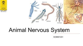
Animal Nervous System Animals To Education.pptx
- 2. Learning Objectives • able to infer the evolutionary and development of animal nervous system. • able to describe different types of nervous systems in different animal lineages. • able to identify and describe basic structures and functions of the central and peripheral nervous systems.
- 3. Functions of the Nervous System 1.Sensory input → gathering information • to monitor changes occurring inside and outside the body (stimuli). 1.Integration • to process and interpret sensory input and decide if action is needed. 1.Motor output • a response to integrated stimuli. • the response activates muscles or glands.
- 5. Diversity of Nervous Systems • All animals have a true nervous system except sea sponges. • The nervous systems of invertebrates and vertebrates is that the nerve cords of many invertebrates are located ventrally whereas the vertebrate spinal cords are located dorsally. • Compared to invertebrates, vertebrate nervous systems are more complex, centralized, and specialized. While there is great diversity among different vertebrate nervous systems, they all share a basic structure: a CNS that contains a brain and spinal cord and a PNS made up of peripheral sensory and motor nerves. • Cnidarians, such as jellyfish, lack a true brain but have a system of separate but connected neurons called a nerve net. • Echinoderms, such as sea stars, have neurons that are bundled into fibers called nerves. • Flatworms of the phylum Platyhelminthes have both a CNS made up of a small brain and two nerve cords, and PNS containing a system of nerves that extend throughout the body. • The insect nervous system is more complex but also fairly decentralized, with a brain, ventral nerve cord, and ganglia (clusters of connected neurons). These ganglia can control movements and behaviors without input from the brain. • Cephalopods, such as octopi, may have the most complicated of invertebrate nervous systems, with neurons that are organized in specialized lobes and eyes that are structurally similar to vertebrate species.
- 6. Two Alternative Hypotheses of the Origins of Nervous Systems • the evolution of animal nervous systems has focused primarily on whether they have a single origin or were independently derived in ctenophores, the comb jellies, and the common ancestor of all other animals.
- 7. The Origins of Nervous Systems: Homologous (Single Origin) If nervous systems are homologous across metazoans, and if ctenophores are the earliest- diverging animals, then nervous systems were lost in sponges and placozoans.
- 8. The Origins of Nervous Systems: Non- homologous (Independent Origins) If nervous systems are not homologous across animals then they arose more than once, a result that is not made more or less likely by any of the possible placements for ctenophores.
- 9. The Orthogon Scenario of Nervous System • defined by multiple pairs of longitudinal cords distributed along the dorsoventral axis of the animal and connected with transverse commissures. • mainly present in Platyhelminthes, but also in some representatives of other animal lineages (e.g. annelids). • the loss of the dorsal longitudinal cords of the ancestral orthogon would have originated the ventral cords found in many protostomian lineages, while the loss of the ventral cords of the orthogon would explain the dorsal location of the nerve cord in chordates.
- 10. The ‘oral nerve ring’ Scenario of Nervous System • the oral nerve ring of anthozoan cnidarians (e.g. sea anemones) directly corresponds to the longitudinal nerve cords of bilaterian animals.
- 11. The ‘nemertean’ Scenario of Nervous System • nemerteans as the starting point for the evolution of dorsal and ventral nerve cords in other bilaterian lineages. Because nemerteans have lateral nerve cords. • the movement of lateral nerve cords to the dorsal side could lead to the dorsal nerve cord of chordates, and the opposite movement would have originated the ventrally centralized longitudinal cords of Protostomia .
- 12. The ‘annelid’ Scenario of Nervous System • polychaete annelid as the closest relative of the chordates, explaining the evolution of the dorsal nerve cord of chordates by an inversion of the dorsoventral axis of the ancestral adult polychaete.
- 13. The Diversity of Neural Anatomies in Metazoa
- 14. Sponge cells hint at origins of nervous system digestive cell neuroid cell
- 15. Nerve Nets • Cnidaria and Ctenophora → the simplest types of nervous system (nerve nets) as well as relatively complex forms, i.e., radially symmetric nervous systems. • Epidermal nerve nets concentrated around the mouth and the peduncle → nerve rings • Subumbrellar → inner (large bipolar neurons), outer (small multipolar sensory neurons) (a) The nervous system of the polyp Hydra (b) radial section through the umbrella of a hydromedusa ENR=exumbrellar nerve ring, M=mesogloea, RC= ring canal, SNR=subumbrellar nerve ring, SRM=subumbrellar ring muscle, V=velum, VRM=velar ring muscle
- 16. Nerve Cords Three schemes for evolution of the chordate and hemichordate nerve cords. (a)The ancestor of chordates and ambulacrarians had a nerve net. Nerve cords in hemichordates and chordates evolved independently. (b)The ancestor of the Ambulacraria plus Chordata clade had a ventral nerve cord. A dorsal/ventral inversion occurred at the base of the ambulacraria plus Chordata. Thus, the dorsal nerve cord of hemichordates is homologous to the chordate nerve cord. (c)The ancestor of the Ambulacraria plus Chordata had a ventral nerve cord. A dorsal/ventral inversion occurred in the lineage leading to chordates. Thus, the ventral nerve cord of hemichordates is homologous to the dorsal nerve cord of chordates.
- 17. The Nervous System of Annelida • Paired “ladder-type” central nervous system → the simplest state consists of a cerebral or supraesophageal nerve ring or ganglion giving rise to paired ventral cords with a pair of ganglia per body segment connected by transverse connectives (anastomoses). • Polychaetes has developed into a three-partite brain → proto-, deuto-, and tritocerebrum • Oligochaetes → the first segments of the ventral nerve cord are often fused into a subesophageal ganglion.
- 18. The Nervous System of Nematoda • Simple nervous systems consisting of a nerve ring around the esophagus and a number of ganglia connected to this ring. • Local ganglia and nerves are found in the caudal gut and anal region. Some nerves extend from the esophageal nerve ring to the sense organs in the “head” region such as sensory papillae and bristles. Other sense organs are chemoreceptive organs called “amphidia”.
- 19. The Nervous System of Arthropods • Ventral, regularly segmented, paired “ladder-type” nerve cord. • The first ganglia have fused into a complex brain. In mandibulates there are three major brain divisions, i.e., a proto-, deuto-, and tritocerebrum. The protocerebrum is associated with the paired optic lobes, the deutocerebrum with the first and the tritocerebrum with the second pair of antennae. • Mandibulates display a subesophageal ganglion having formed by fusion of the three first ventral ganglia. They supply the mouth region and mandibles, in crustaceans the first and second maxillae, in insects the maxillae, mandibles and labium. • Caudal ganglia of the ventral cords exhibit a strong tendency to fuse and to form specialized abdominal structures. (a) Lateral view and (b) ventral view of the brain and nerves in the scorpion fly Panorpa (c) Schematic of the brain of a honey bee
- 20. Structural Classification of the Nervous System 1. Central nervous system (CNS) → Brain & Spinal cord 2. Peripheral nervous system (PNS) → Cranial nerves & Spinal nerves
- 21. Peripheral Nervous System (PNS) 1. Sensory (afferent): • Nerve fibers that carry information to the central nervous system from: ❏ sensory receptors in the skin, skeletal muscles and joints (somatic sensory fibers). ❏ Sensory receptors in the visceral organs (visceral sensory 1. Motor (efferent) division: • Nerve fibers that carry impulses away from the central nervous system (to Muscles & glands). ❏ Somatic nervous system = voluntary , it controls skeletal muscles → skeletal muscle reflexes are involuntary ❏ Autonomic nervous system = involuntary , it controls smooth & cardiac muscles & glands → Sympathetic & parasympathetic
- 22. Organization of the Nervous System
- 23. Histology of Nervous Tissue • Neurons → excitable nerve cells that transmit electrical signals. • Neuroglia ( glial) cells Astrocytes, Microglia, Ependymal cells, Oligodendrocytes ) are the supporting cells.
- 24. Neuron (Nerve cell) • Cells specialized to transmit messages: 1. A Cell body with nucleus and the usual organelles, except centrioles. 2. Extensions processes outside the cell body → Dendrites conduct impulses toward the cell body Axons conduct impulses away from the cell body.
- 25. Multipolar neurons many extensions from the cell body; three or more processes.
- 26. Bipolar neurons one axon and one dendrite; two processes (axon and dendrite).
- 27. Unipolar neurons have a short single process leaving the cell body.
- 28. Structural Variation of Neurons
- 29. Neuroglia in CNS: Astrocytes • Abundant, star shaped cells • Brace neurons • Form barrier between capillaries and neurons • Control the chemical environment of the brain (CNS)
- 30. Neuroglia in CNS: Microglia & Ependymal cells • Microglia → Spider like phagocytes, dispose of debris • Ependymal cells → Line cavities of the brain and spinal cord, circulate cerebrospinal fluid Microglia
- 31. Neuroglia in CNS: Oligodendrocytes • Produce myelin sheath around nerve fibers in the central nervous system.
- 32. Neuroglia in PNS: Satellite & Schwann cells • Satellite cells → surround neuron cell bodies in the periphery, protect neuron cell bodies. • Schwann cells (neurolemmocytes) → surround axons/dendrites and form the myelin sheath around larger nerve fibers in the periphery similar to oligodendrocytes in function insulators.
- 33. Neuroglia vs. Neurons • Neuroglia divide. • Neurons do not. • Most brain tumors are gliomas • Most brain tumors involve the neuroglial cells, not the neurons.
- 34. Axons and Nerve Impulses • Axons end in axonal terminals • Axonal terminals contain vesicles with neurotransmitters • Axonal terminals are separated from the next neuron by a gap: 1. Synaptic cleft gap between adjacent neurons 2. Synapse junction between nerves
- 35. Regions of the Brain
- 36. Specialized Areas of the Cerebrum • Somatic sensory area → receives impulses from the body’s sensory receptors • Primary motor area → sends impulses to skeletal muscles • Broca’s area → involved in our ability to speak • Cerebral areas involved in special senses → Gustatory area (taste), Visual area, Auditory area, Olfactory area
- 37. Sensory and Motor Areas of the Cerebral Cortex
- 38. Cerebral cortex-type of neurons: Pyramidal • Triangular • Dendrite arises from three angles • Axon from the base of cell • Prominent nucleus and Nissl substances • Small sized pyramidal neuron 10-12 micron • Medium sized neuron 40-50 micron • Large sized (Betz cells ) >100 micron
- 39. Cerebral cortex-type of neurons: Stellate/granular & Fusiform cells • Stellate/granular cells → Polygonal /triangular shape, dark condensed nucleus, dendrites arises all over, axon terminate next layer and goes deeper. • Fusiform → Oriented perpendicular to the surface, One dendrites towards superficial, one axon towards deeper layer. Fusifor m Stellate/granula r
- 40. Cerebral cortex-type of neurons: Horizontal cells of Cajal, Cell of Mortinotti, & Golgi type II • Horizontal cells of Cajal → spindle shaped, parallel to the surface, Internuncial type. • Cell of Mortinotti → Small triangular cells, axons goes towards surface • Golgi type II → process ends close to cell body, local circuit Cajal Golgi Mortinotti
- 41. Layers of cerebral cortex 1. Outer Molecular layer → horizontal cells + Golgi type II, tangential nerve fibers from pyramidal, fusiform and cells of Mortinotti. 2. Outer granular layer → small pyramidal cells + stellate cells, dendrites end in first layer, axons forms association. 3. Outer pyramidal layer → medium and large pyramidal cells, axons forms association and commissural fibers. 4. Inner granular layer → packed stellate cells, lot of fibers as band-outer band of Baillarger. 5. Inner pyramidal/ganglion → large Betz cells, axons continues as projection fibers, horizontal fibers forms inner band of Baillarger. 6. Multiform layer/fusiform cells → spindle shaped cells, granular , fusiform and stellate type.
- 42. Diencephalon • Sits on top of the brain stem. • Enclosed by the cerebral hemispheres. • Thalamus → surrounds the third ventricle, the relay station for sensory impulses, transfers impulses to the correct part of the cortex for localization and interpretation. • Hypothalamus → under the thalamus, important autonomic nervous system center (helps regulate body temperature, controls water balance, regulates metabolism), important part of the limbic system (emotions), the pituitary gland is attached to the hypothalamus. • Epithalamus → forms the roof of the third ventricle, houses the pineal body (an endocrine gland), includes the choroid plexus forms cerebrospinal fluid. v v
- 43. Brain Stem • Attaches to the spinal cord. • Midbrain → Mostly composed of tracts of nerve fibers (Reflex centers for vision and hearing, Cerebral aquaduct 3rd- 4th ventricles) • Pons → the bulging center part of the brain stem, mostly composed of fiber tracts, includes nuclei involved in the control of breathing. • Medulla oblongata → the lowest part of the brain stem, merges into the spinal cord, includes important fiber tracts, contains important control centers (heart rate control, blood pressure regulation, breathing, swallowing, vomiting)
- 44. Cerebellum • Two hemispheres with convoluted surfaces. • Provides involuntary coordination of body movements. Granule Golgi type II Purkinje cells Stellate cells Basket cells
- 45. Layers of Cerebellum 1. Molecular layer → dendrite arborization, densely packed axons run parallel to surface, superficially stellate cells, deeper stellate cells (basket cells). 2. Purkinje layer 3. Granular layer → closely packed granule cells
- 47. Cerebellum - Golgi stain
- 48. Cerebellum granular layer (rosette)
- 49. Protection of the Central Nervous System • Scalp and skin • Skull and vertebral column • Meninges • Cerebrospinal fluid • Blood brain barrier
- 50. Meninges: Dura mater ❏ Dura mater ● Double layered external covering → periosteum attached to surface of the skull, meningeal layer outer covering of the brain ● Folds inward in several areas
- 51. Meninges: Arachnoid layer & Pia mater Arachnoid layer → middle layer , web like Pia mater → internal layer, clings to the surface of the brain
- 52. Cerebrospinal Fluid • Similar to blood plasma composition • Formed by the choroid plexus • Forms a watery cushion to protect the brain • Circulated in arachnoid space, ventricles, and central canal of the spinal cord.
- 53. Blood Brain Barrier • Includes the least permeable capillaries of the body • Excludes many potentially harmful substances • Useless against some substances → fats and fat soluble molecules, respiratory gases, alcohol, nicotine, anesthesia.
- 54. Spinal Cord Anatomy • Exterior white mater → conduction tracts. • Internal gray matter (mostly cell bodies) → dorsal (posterior) horns, anterior (ventral) horns. • Central canal filled with cerebrospinal fluid.