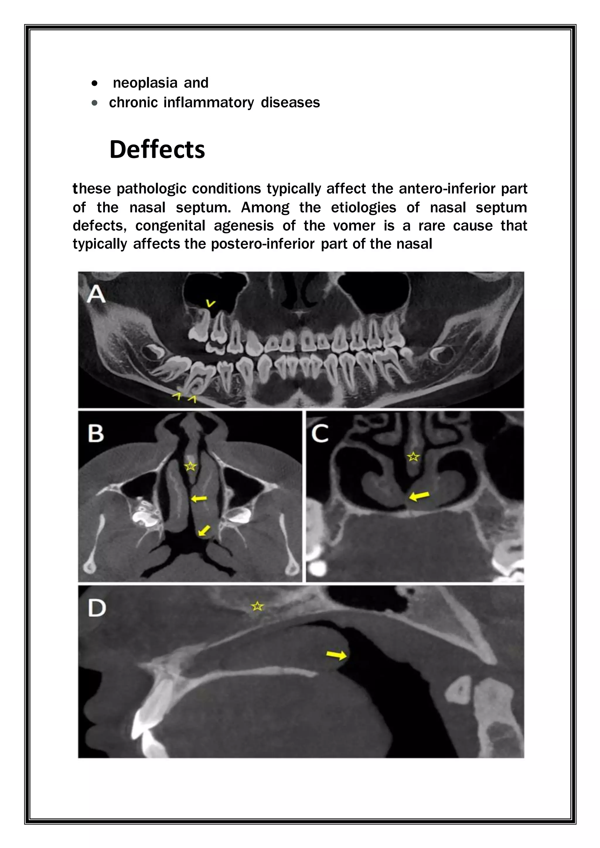
VOMER BONE
Contents
 Introduction
 Location
 structure
 Related structure
 Ossification
 Articulation
 Boarders
 foramens
 Nerve and blood supply
 Vomer fracture
Introduction
The vomer is a small, thin, plow-shaped, midline bone
that occupies and divides the nasal cavity. This is a single
bone which is one of the facial bones of the head. It is a
small flat bone that is approximately triangular in shape. It
is difficult to illustrate clearly in diagrams due it to its small
size.
It is usually described as resembling a ploughshare in
shape. Each lateral surface is covered with the thick mucous
membrane of the nasal sinus, and is traversed by a narrow
but well-marked groove, which lodges the naso- palatine
nerve from the spheno-palatine ganglion; hence it is
sometimes called the naso-palatine groove.
Location
The vomer is situated in the median plane,but its
anterior portion is frequently bent to one or other side. It is
thin, somewhat quadrilateral in shape, and forms the hinder
and lower part of the
nasalseptum.
Structure
The vomer is situated in the median plane, but its anterior
portion is frequently bent to one side.
It is thin, somewhat quadrilateral in shape, and forms the hinder
and lower part of the nasal septum; it has two surfaces and four
borders.
The surfaces are marked by small furrows for blood vessels, and on each is
the nasopalatine groove, which runs obliquelydownward and forward, and
lodges the nasopalatine nerve and vessels.
Related structure
The vomer is part of the nasal septum which follows the midline
of the viscerocranium and creates the division between the two
symmetrical sides of the nasal cavity. To be exact, the vomer
forms the posterior inferior aspect of the septum in between the
perpendicular plate of the ethmoid bone anterosuperiorly and
the palatine bone posteroinferiorly. In addition, the maxilla links
to the vomer anteriorly and inferiorly, the sphenoid
bone posteriorly and the nasal cartilage anteriorly.
Ossification
 The vomer is a membrane bone, and arises from a single
center deposited in the lower border of the perichondrium of
the ethmo-vomerine plate as early as the eighth week.
 From this single center a lamina of bone extends on each
side of the cartilage plate.
 For many weeks the vomer is a shallow bony trough.
 Gradually it presses upon and induces absorption of the
enclosed cartilage, and by degrees the laminae fuse, and
form a rectangular plate of bone.
 At birth the vomer presents an expanded lower border,
especially in cases of cleft palate.
ARTICULATION
1. The vomer articulates with six bones:
2. two of the cranium, the sphenoid and ethmoid.
3. four of the face, two maxillae; and two palatine
bones.
4. It also articulates with the septal cartilage of the nose
Boarders
 The vomer has 4 borders:
 superior: thickest border with laterally projecting alae which
articulate with the rostrum of the sphenoid bone body. The
alae also articulate with the:
o sphenoidal conchae
o vaginal processes of the medial pterygoid plates
o sphenoidal processes of the palatine bones
 inferior: articulates with the median nasal crests of
the maxilla and palatine bones, between the incisive canals
 anterior: longest border which articulates with:
o the perpendicular plate of the ethmoid bone along its
upper half
o the nasal septal cartilage along its lower half
 posterior: short concave border the is thicker (and bifid)
superiorly, which does not articulate with any bone, but
rather separates the choanae.
 The vomerovaginal canal is formed between the ala, body of
the sphenoid and the small medially directed vaginal
process arising from the sphenoid bone.
 The superior border, the thickest, presents a deep furrow,
bounded on either side by a horizontal projecting expansion of
bone – called the wing of vomer.
 the furrow receives the rostrum of the sphenoid, while the
margins of the alae articulate with the vaginal processes of
the medial pterygoid platesof the sphenoid behind, and with
the sphenoidal processes of the palatine bones in front.
 The inferior border articulates with the crest formed by the
maxillæ and palatine bones.
 The anterior border is the longest and slopes downward and
forward. Its upper half is fused with the perpendicular plate of
the ethmoid; its lower half is grooved for the inferior margin of
the septal cartilage of the nose.
 The posterior border is free of bony articulation, having no
muscle attachments. It is concave, separates the choanae, and
is thick and bifid above, thin below.
Function
 The vomeronasal organ, also called Jacobson's organ, .
 It is a chemoreceptor organ named for its closeness to the
vomer and nasal bones, .
 particularly developed in animals such as cats (who adopt a
characteristic pose called the Flagmen
reaction or flehming when making use of it),
 is thought to have to do with the perception of
certain pheromones.
Nerve supply and blood supply
The nose is innervated by three cranial nerves:
 Olfactionis provided by the olfactory nerve (CN I)
 General sensation is carried by the trigeminal nerve (CN V)
 Serous glands in the nasal mucosa which produce fluid that
constantly lubricates the nose walls are innervated by the
parasympathetic fibers of the facial nerve (CN VII).
Sympathetic innervation comes from T1 level of spinal cord
and is intended for regulation of blood flow through mucosa
VOMER BONE DISOREDR
Congenital vomer agenesis is an extremely rare condition
in which the vomer bone does not fully develop, which can
lead to septal perforation.vomer agenesis the defect is
normally located in the posteroinferior region of the septum.
CAUSES
Agenesis of the vomer bone is a rare condition that can lead to a
defect in the posterior of nasal septum, but more common
causes include
 Trauma
 irritation
 tuberculosis
 infection, irritation
 neoplasia and
 chronic inflammatory diseases
Deffects
these pathologic conditions typically affect the antero-inferior part
of the nasal septum. Among the etiologies of nasal septum
defects, congenital agenesis of the vomer is a rare cause that
typically affects the postero-inferior part of the nasal
Conclusion Of Vomer Agenesis
The etiology of nasal septum defect includes nasal surgery,
trauma, infection, chronic inflammatory disease, neoplasia and
drug abuse. As of today, only very few cases of isolated VA have
been reported in the literature. Since this anomaly is not
associated with any specific clinical symptoms, VA can be easily
overlooked or misinterpreted as septum perforation, especially if
found incidentally on CBCT. Knowledge of the pertinent imaging
features allows the correct diagnosis.
References
1. Lund V. Anatomy of the Nose and Sinuses. Butterworth Heinemann; Oxford, UK:
1997. [Google Scholar]
2. Jones N. The nose and paranasal sinuses physiology and anatomy. Adv. Drug
Deliv. Rev. 2001;51:5–19. doi: 10.1016/S0169-409X(01)00172-7. [PubMed]
[CrossRef] [Google Scholar]
3. Lanier B., Kai G., Marple B., Wall G.M. Pathophysiology and progression of nasal
septal perforation. Ann. Allergy Asthma Immunol. 2007;99:473–479. doi:
10.1016/S1081-1206(10)60373-0. [PubMed] [CrossRef] [Google Scholar]
4. Mohri M., Amatsu M. Congenital defects of the vomer. Ann. Otol. Rhinol.
Laryngol. 2000;109:497–499. doi: 10.1177/000348940010900510. [PubMed]
[CrossRef] [Google Scholar]

ANATOMY OF VOMER BONE

  • 1.
    VOMER BONE Contents  Introduction Location  structure  Related structure  Ossification  Articulation  Boarders  foramens  Nerve and blood supply  Vomer fracture
  • 2.
    Introduction The vomer isa small, thin, plow-shaped, midline bone that occupies and divides the nasal cavity. This is a single bone which is one of the facial bones of the head. It is a small flat bone that is approximately triangular in shape. It is difficult to illustrate clearly in diagrams due it to its small size. It is usually described as resembling a ploughshare in shape. Each lateral surface is covered with the thick mucous membrane of the nasal sinus, and is traversed by a narrow but well-marked groove, which lodges the naso- palatine nerve from the spheno-palatine ganglion; hence it is sometimes called the naso-palatine groove.
  • 3.
    Location The vomer issituated in the median plane,but its anterior portion is frequently bent to one or other side. It is thin, somewhat quadrilateral in shape, and forms the hinder and lower part of the nasalseptum.
  • 4.
    Structure The vomer issituated in the median plane, but its anterior portion is frequently bent to one side. It is thin, somewhat quadrilateral in shape, and forms the hinder and lower part of the nasal septum; it has two surfaces and four borders. The surfaces are marked by small furrows for blood vessels, and on each is the nasopalatine groove, which runs obliquelydownward and forward, and lodges the nasopalatine nerve and vessels. Related structure The vomer is part of the nasal septum which follows the midline of the viscerocranium and creates the division between the two symmetrical sides of the nasal cavity. To be exact, the vomer forms the posterior inferior aspect of the septum in between the perpendicular plate of the ethmoid bone anterosuperiorly and the palatine bone posteroinferiorly. In addition, the maxilla links to the vomer anteriorly and inferiorly, the sphenoid bone posteriorly and the nasal cartilage anteriorly.
  • 5.
    Ossification  The vomeris a membrane bone, and arises from a single center deposited in the lower border of the perichondrium of the ethmo-vomerine plate as early as the eighth week.  From this single center a lamina of bone extends on each side of the cartilage plate.  For many weeks the vomer is a shallow bony trough.  Gradually it presses upon and induces absorption of the enclosed cartilage, and by degrees the laminae fuse, and form a rectangular plate of bone.  At birth the vomer presents an expanded lower border, especially in cases of cleft palate. ARTICULATION 1. The vomer articulates with six bones: 2. two of the cranium, the sphenoid and ethmoid.
  • 6.
    3. four ofthe face, two maxillae; and two palatine bones. 4. It also articulates with the septal cartilage of the nose
  • 7.
    Boarders  The vomerhas 4 borders:  superior: thickest border with laterally projecting alae which articulate with the rostrum of the sphenoid bone body. The alae also articulate with the: o sphenoidal conchae o vaginal processes of the medial pterygoid plates o sphenoidal processes of the palatine bones  inferior: articulates with the median nasal crests of the maxilla and palatine bones, between the incisive canals  anterior: longest border which articulates with: o the perpendicular plate of the ethmoid bone along its upper half o the nasal septal cartilage along its lower half  posterior: short concave border the is thicker (and bifid) superiorly, which does not articulate with any bone, but rather separates the choanae.  The vomerovaginal canal is formed between the ala, body of the sphenoid and the small medially directed vaginal process arising from the sphenoid bone.
  • 8.
     The superiorborder, the thickest, presents a deep furrow, bounded on either side by a horizontal projecting expansion of bone – called the wing of vomer.  the furrow receives the rostrum of the sphenoid, while the margins of the alae articulate with the vaginal processes of the medial pterygoid platesof the sphenoid behind, and with the sphenoidal processes of the palatine bones in front.  The inferior border articulates with the crest formed by the maxillæ and palatine bones.  The anterior border is the longest and slopes downward and forward. Its upper half is fused with the perpendicular plate of the ethmoid; its lower half is grooved for the inferior margin of the septal cartilage of the nose.  The posterior border is free of bony articulation, having no muscle attachments. It is concave, separates the choanae, and is thick and bifid above, thin below.
  • 9.
    Function  The vomeronasalorgan, also called Jacobson's organ, .  It is a chemoreceptor organ named for its closeness to the vomer and nasal bones, .  particularly developed in animals such as cats (who adopt a characteristic pose called the Flagmen reaction or flehming when making use of it),  is thought to have to do with the perception of certain pheromones. Nerve supply and blood supply The nose is innervated by three cranial nerves:  Olfactionis provided by the olfactory nerve (CN I)  General sensation is carried by the trigeminal nerve (CN V)  Serous glands in the nasal mucosa which produce fluid that constantly lubricates the nose walls are innervated by the parasympathetic fibers of the facial nerve (CN VII). Sympathetic innervation comes from T1 level of spinal cord and is intended for regulation of blood flow through mucosa
  • 10.
    VOMER BONE DISOREDR Congenitalvomer agenesis is an extremely rare condition in which the vomer bone does not fully develop, which can lead to septal perforation.vomer agenesis the defect is normally located in the posteroinferior region of the septum. CAUSES Agenesis of the vomer bone is a rare condition that can lead to a defect in the posterior of nasal septum, but more common causes include  Trauma  irritation  tuberculosis  infection, irritation
  • 11.
     neoplasia and chronic inflammatory diseases Deffects these pathologic conditions typically affect the antero-inferior part of the nasal septum. Among the etiologies of nasal septum defects, congenital agenesis of the vomer is a rare cause that typically affects the postero-inferior part of the nasal
  • 12.
    Conclusion Of VomerAgenesis The etiology of nasal septum defect includes nasal surgery, trauma, infection, chronic inflammatory disease, neoplasia and drug abuse. As of today, only very few cases of isolated VA have been reported in the literature. Since this anomaly is not associated with any specific clinical symptoms, VA can be easily overlooked or misinterpreted as septum perforation, especially if found incidentally on CBCT. Knowledge of the pertinent imaging features allows the correct diagnosis. References 1. Lund V. Anatomy of the Nose and Sinuses. Butterworth Heinemann; Oxford, UK: 1997. [Google Scholar] 2. Jones N. The nose and paranasal sinuses physiology and anatomy. Adv. Drug Deliv. Rev. 2001;51:5–19. doi: 10.1016/S0169-409X(01)00172-7. [PubMed] [CrossRef] [Google Scholar] 3. Lanier B., Kai G., Marple B., Wall G.M. Pathophysiology and progression of nasal septal perforation. Ann. Allergy Asthma Immunol. 2007;99:473–479. doi: 10.1016/S1081-1206(10)60373-0. [PubMed] [CrossRef] [Google Scholar] 4. Mohri M., Amatsu M. Congenital defects of the vomer. Ann. Otol. Rhinol. Laryngol. 2000;109:497–499. doi: 10.1177/000348940010900510. [PubMed] [CrossRef] [Google Scholar]