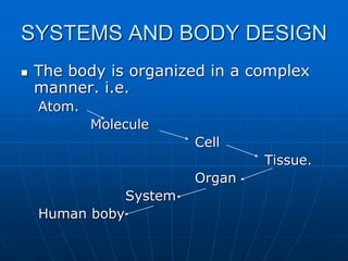
This document defines key terms related to anatomy, physiology, and cell biology. It provides an overview of the organization of the human body from atoms to organ systems. Key points include:
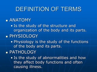
- Anatomy studies body structure, physiology studies functions, and pathology studies abnormalities.
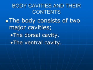
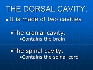
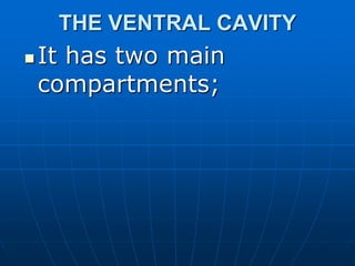
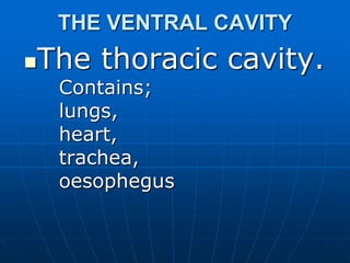
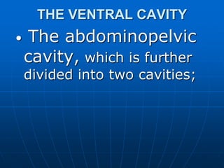
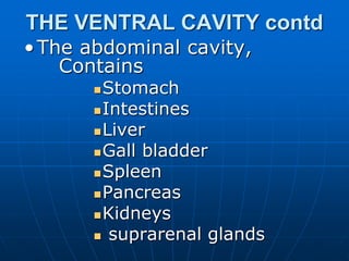
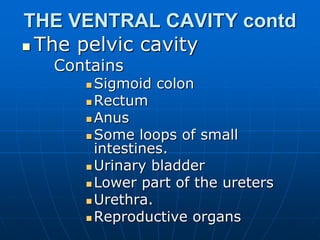
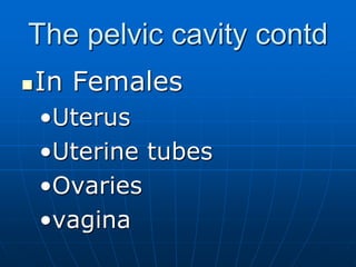
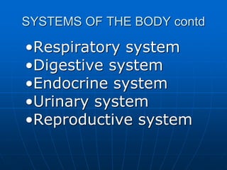
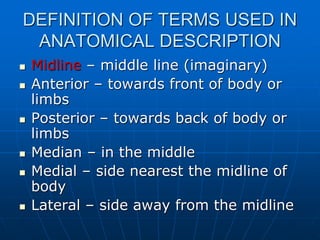
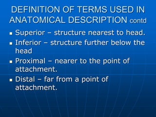
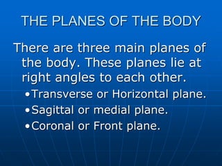
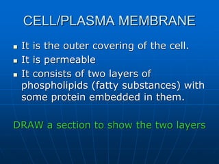
- The body is made of cells, tissues, organs, and systems organized into body cavities.
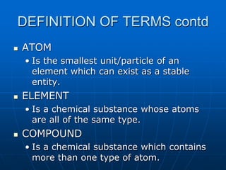
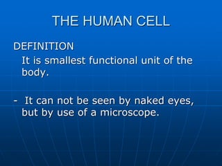
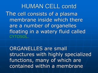
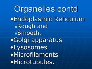
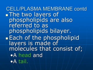
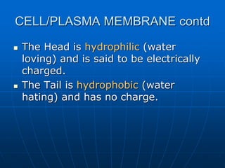
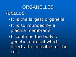
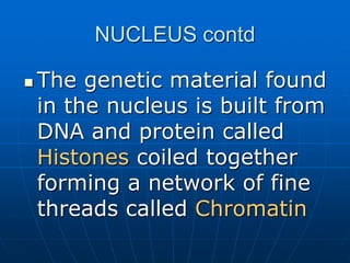
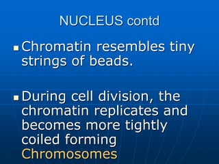
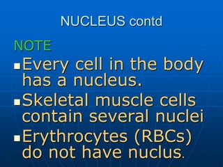
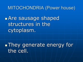
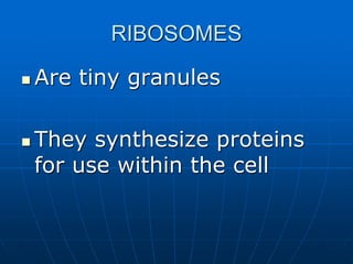
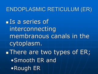
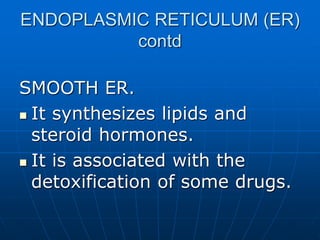
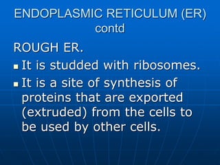
- The human cell contains organelles like the nucleus, mitochondria, and endoplasmic reticulum, surrounded by a plasma membrane.
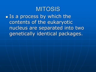
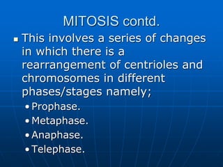
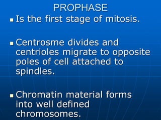
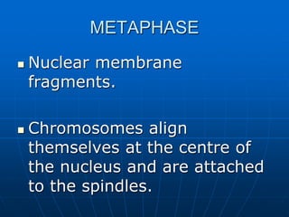
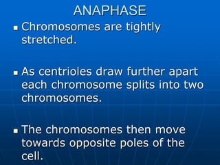
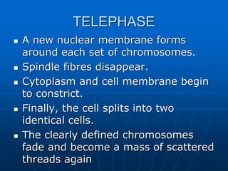
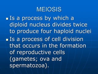
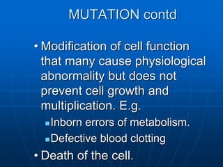
- Cells divide through mitosis and meiosis to produce genetically identical or variant cells.
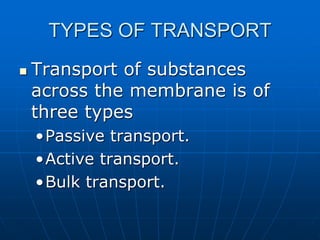
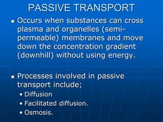
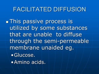
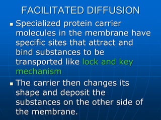
- Substances move across the cell membrane through passive, active, or bulk transport processes.
Human Anatomy and Physiology : Introduction
PCI Syllabus, B. Pharmacy, BP101T
Session III
Levels of Structural Organization.
Anatomical Nomenclature.
Anatomical terms and their meaning
lymphatic system, a subsystem of the circulatory system in the vertebrate body that consists of a complex network of vessels, tissues, and organs. The lymphatic system helps maintain fluid balance in the body by collecting excess fluid and particulate matter from tissues and depositing them in the bloodstream
HUMAN CYTOLOGY ( CELL AND TISSUE )
PRESENTED BY
BY SONU SHARMA. ROLL NO. 504/16
3RD YEAR B.SC RADIOGRAPHY & IMAGING TECHNOLOGY DEPARTMENT OF RADIO DIAGNOSIS
PANDIT BHAGWAT DAYAL SHARMA
UNIVERSITY OF MEDICAL SCIENCE AND RESEARCH P.G.I.M.S. ROHTAK,HARYANA.
Contact : - +91-7015728336
The skeletal system includes all of the bones and joints in the body. Each bone is a complex living organ that is made up of many cells, protein fibers, and minerals. The skeleton acts as a scaffold by providing support and protection for the soft tissues that make up the rest of the body. The skeletal system also provides attachment points for muscles to allow movements at the joints. New blood cells are produced by the red bone marrow inside of our bones.
Cartilage:
Cartilage is a specialized type of dense collective tissue designed to give support , bear weight and withstand tension , torsion , and bending.
General Features :
• Cartilage supports regions of body that requires flexibility.
• Non nervous structure
• Avascular
• Very poor regeneration power
• Usually surrounded by pericondrium (dense irregular connective tissue surrounding cartilage) except fibro cartilage.
Classification:
Hyaline cartilage
Elastic cartilage
Fibro cartilage
Fibro Cartilage :
White colored, tough cartilage containing dense connective tissue and collagen fibers often known as intervertebral discs is called fibro cartilage.
Structure :
Fibro cartilage consists of chondrocytes dispersed among bundles of type 1 collagen fibers.
Chondrocytes are present in lacunae (cavity).
The arrangement of cells is different from all other type of cartilages.
Chondrocytes are arranged in parallel rows of 2, 4 or 6 cells.
These rows of cells are called isogenous cell groups.
Chemical Compounds Present :
Proteoglycans rich in sulphated glucosaminoglycans especially
Chondroiton sulphate
Dermatan sulphate
Stain :
Due to the abundance of collagen type 1 fibers , the matrix of fibrocartilage stains intensely acidophilic/eosinophilic. (since collagen is basic in nature)
Stained by EOSIN which is pink in color.
Chondrocytes are stained in purple usually by HEMATOXYLIN and looks purple in color due to acidic nature of large centeral nucleus present.
Occurrence in body :
Intervertebral disc
Disc of pubic symphysis
Menisci of knee joint
Sternoclavicular joint
Temporomandibular joint
Ligamentum tere
Labrum glenoidale
Labrum acetabulare
Fibrocartilage is also found at places where tendons and ligaments attach to bones.
Disorders:
Degeneration of fibrocartilage is seen in degenerative disc disease.
A fibrocartilaginous embolism (FCE) is an unusual cause of spinal cord and cerebral ischemia (insufficient bloodsupply). Symptoms may include sudden, severe pain in the neck and/or back; progressive weakening reduced sensation and paralysis. It may be caused by the blocking of an artery interrupting vascular supply.
A herniated disk is a disk that ruptures. This allows the jelly-like center of the disk to leak, irritating the nearby nerves. This can cause sciatica or back pain.
References:
http://www.nlm.nih.gov/medlineplus/herniateddisk.html
http://www.ncbi.nlm.nih.gov/pubmed/3289246
histology by laiq hussaain
Cell: The cell is the ultimate structural and functional unit of the body.
The three principal constituents of the cell are:
1. Cell membrane
2. Cytoplasm and its organelles
3. Nucleus
This presentation file contains all about cell discovery, cell theory, organelles which are present within the cell and cell comparison between prokaryotic and Eukaryotic organisms.
Ozempic: Preoperative Management of Patients on GLP-1 Receptor Agonists Saeid Safari
Preoperative Management of Patients on GLP-1 Receptor Agonists like Ozempic and Semiglutide
ASA GUIDELINE
NYSORA Guideline
2 Case Reports of Gastric Ultrasound
Pulmonary Thromboembolism - etilogy, types, medical- Surgical and nursing man...VarunMahajani
Disruption of blood supply to lung alveoli due to blockage of one or more pulmonary blood vessels is called as Pulmonary thromboembolism. In this presentation we will discuss its causes, types and its management in depth.
Couples presenting to the infertility clinic- Do they really have infertility...Sujoy Dasgupta
Dr Sujoy Dasgupta presented the study on "Couples presenting to the infertility clinic- Do they really have infertility? – The unexplored stories of non-consummation" in the 13th Congress of the Asia Pacific Initiative on Reproduction (ASPIRE 2024) at Manila on 24 May, 2024.
Title: Sense of Taste
Presenter: Dr. Faiza, Assistant Professor of Physiology
Qualifications:
MBBS (Best Graduate, AIMC Lahore)
FCPS Physiology
ICMT, CHPE, DHPE (STMU)
MPH (GC University, Faisalabad)
MBA (Virtual University of Pakistan)
Learning Objectives:
Describe the structure and function of taste buds.
Describe the relationship between the taste threshold and taste index of common substances.
Explain the chemical basis and signal transduction of taste perception for each type of primary taste sensation.
Recognize different abnormalities of taste perception and their causes.
Key Topics:
Significance of Taste Sensation:
Differentiation between pleasant and harmful food
Influence on behavior
Selection of food based on metabolic needs
Receptors of Taste:
Taste buds on the tongue
Influence of sense of smell, texture of food, and pain stimulation (e.g., by pepper)
Primary and Secondary Taste Sensations:
Primary taste sensations: Sweet, Sour, Salty, Bitter, Umami
Chemical basis and signal transduction mechanisms for each taste
Taste Threshold and Index:
Taste threshold values for Sweet (sucrose), Salty (NaCl), Sour (HCl), and Bitter (Quinine)
Taste index relationship: Inversely proportional to taste threshold
Taste Blindness:
Inability to taste certain substances, particularly thiourea compounds
Example: Phenylthiocarbamide
Structure and Function of Taste Buds:
Composition: Epithelial cells, Sustentacular/Supporting cells, Taste cells, Basal cells
Features: Taste pores, Taste hairs/microvilli, and Taste nerve fibers
Location of Taste Buds:
Found in papillae of the tongue (Fungiform, Circumvallate, Foliate)
Also present on the palate, tonsillar pillars, epiglottis, and proximal esophagus
Mechanism of Taste Stimulation:
Interaction of taste substances with receptors on microvilli
Signal transduction pathways for Umami, Sweet, Bitter, Sour, and Salty tastes
Taste Sensitivity and Adaptation:
Decrease in sensitivity with age
Rapid adaptation of taste sensation
Role of Saliva in Taste:
Dissolution of tastants to reach receptors
Washing away the stimulus
Taste Preferences and Aversions:
Mechanisms behind taste preference and aversion
Influence of receptors and neural pathways
Impact of Sensory Nerve Damage:
Degeneration of taste buds if the sensory nerve fiber is cut
Abnormalities of Taste Detection:
Conditions: Ageusia, Hypogeusia, Dysgeusia (parageusia)
Causes: Nerve damage, neurological disorders, infections, poor oral hygiene, adverse drug effects, deficiencies, aging, tobacco use, altered neurotransmitter levels
Neurotransmitters and Taste Threshold:
Effects of serotonin (5-HT) and norepinephrine (NE) on taste sensitivity
Supertasters:
25% of the population with heightened sensitivity to taste, especially bitterness
Increased number of fungiform papillae
These lecture slides, by Dr Sidra Arshad, offer a quick overview of physiological basis of a normal electrocardiogram.
Learning objectives:
1. Define an electrocardiogram (ECG) and electrocardiography
2. Describe how dipoles generated by the heart produce the waveforms of the ECG
3. Describe the components of a normal electrocardiogram of a typical bipolar leads (limb II)
4. Differentiate between intervals and segments
5. Enlist some common indications for obtaining an ECG
Study Resources:
1. Chapter 11, Guyton and Hall Textbook of Medical Physiology, 14th edition
2. Chapter 9, Human Physiology - From Cells to Systems, Lauralee Sherwood, 9th edition
3. Chapter 29, Ganong’s Review of Medical Physiology, 26th edition
4. Electrocardiogram, StatPearls - https://www.ncbi.nlm.nih.gov/books/NBK549803/
5. ECG in Medical Practice by ABM Abdullah, 4th edition
6. ECG Basics, http://www.nataliescasebook.com/tag/e-c-g-basics
These simplified slides by Dr. Sidra Arshad present an overview of the non-respiratory functions of the respiratory tract.
Learning objectives:
1. Enlist the non-respiratory functions of the respiratory tract
2. Briefly explain how these functions are carried out
3. Discuss the significance of dead space
4. Differentiate between minute ventilation and alveolar ventilation
5. Describe the cough and sneeze reflexes
Study Resources:
1. Chapter 39, Guyton and Hall Textbook of Medical Physiology, 14th edition
2. Chapter 34, Ganong’s Review of Medical Physiology, 26th edition
3. Chapter 17, Human Physiology by Lauralee Sherwood, 9th edition
4. Non-respiratory functions of the lungs https://academic.oup.com/bjaed/article/13/3/98/278874
Report Back from SGO 2024: What’s the Latest in Cervical Cancer?bkling
Are you curious about what’s new in cervical cancer research or unsure what the findings mean? Join Dr. Emily Ko, a gynecologic oncologist at Penn Medicine, to learn about the latest updates from the Society of Gynecologic Oncology (SGO) 2024 Annual Meeting on Women’s Cancer. Dr. Ko will discuss what the research presented at the conference means for you and answer your questions about the new developments.
micro teaching on communication m.sc nursing.pdfAnurag Sharma
Microteaching is a unique model of practice teaching. It is a viable instrument for the. desired change in the teaching behavior or the behavior potential which, in specified types of real. classroom situations, tends to facilitate the achievement of specified types of objectives.
NVBDCP.pptx Nation vector borne disease control programSapna Thakur
NVBDCP was launched in 2003-2004 . Vector-Borne Disease: Disease that results from an infection transmitted to humans and other animals by blood-feeding arthropods, such as mosquitoes, ticks, and fleas. Examples of vector-borne diseases include Dengue fever, West Nile Virus, Lyme disease, and malaria.
The prostate is an exocrine gland of the male mammalian reproductive system
It is a walnut-sized gland that forms part of the male reproductive system and is located in front of the rectum and just below the urinary bladder
Function is to store and secrete a clear, slightly alkaline fluid that constitutes 10-30% of the volume of the seminal fluid that along with the spermatozoa, constitutes semen
A healthy human prostate measures (4cm-vertical, by 3cm-horizontal, 2cm ant-post ).
It surrounds the urethra just below the urinary bladder. It has anterior, median, posterior and two lateral lobes
It’s work is regulated by androgens which are responsible for male sex characteristics
Generalised disease of the prostate due to hormonal derangement which leads to non malignant enlargement of the gland (increase in the number of epithelial cells and stromal tissue)to cause compression of the urethra leading to symptoms (LUTS
Flu Vaccine Alert in Bangalore Karnatakaaddon Scans
As flu season approaches, health officials in Bangalore, Karnataka, are urging residents to get their flu vaccinations. The seasonal flu, while common, can lead to severe health complications, particularly for vulnerable populations such as young children, the elderly, and those with underlying health conditions.
Dr. Vidisha Kumari, a leading epidemiologist in Bangalore, emphasizes the importance of getting vaccinated. "The flu vaccine is our best defense against the influenza virus. It not only protects individuals but also helps prevent the spread of the virus in our communities," he says.
This year, the flu season is expected to coincide with a potential increase in other respiratory illnesses. The Karnataka Health Department has launched an awareness campaign highlighting the significance of flu vaccinations. They have set up multiple vaccination centers across Bangalore, making it convenient for residents to receive their shots.
To encourage widespread vaccination, the government is also collaborating with local schools, workplaces, and community centers to facilitate vaccination drives. Special attention is being given to ensuring that the vaccine is accessible to all, including marginalized communities who may have limited access to healthcare.
Residents are reminded that the flu vaccine is safe and effective. Common side effects are mild and may include soreness at the injection site, mild fever, or muscle aches. These side effects are generally short-lived and far less severe than the flu itself.
Healthcare providers are also stressing the importance of continuing COVID-19 precautions. Wearing masks, practicing good hand hygiene, and maintaining social distancing are still crucial, especially in crowded places.
Protect yourself and your loved ones by getting vaccinated. Together, we can help keep Bangalore healthy and safe this flu season. For more information on vaccination centers and schedules, residents can visit the Karnataka Health Department’s official website or follow their social media pages.
Stay informed, stay safe, and get your flu shot today!
2. DEFINITION OF TERMS
ANATOMY
• Is the study of the structure and
organization of the body and its parts.
PHYSIOLOGY
• Physiology is the study of the functions
of the body and its parts.
PATHOLOGY
• Is the study of abnormalities and how
they affect body functions and often
causing illness.
3. DEFINITION OF TERMS contd
ATOM
• Is the smallest unit/particle of an
element which can exist as a stable
entity.
ELEMENT
• Is a chemical substance whose atoms
are all of the same type.
COMPOUND
• Is a chemical substance which contains
more than one type of atom.
4. DEFINITION OF TERMS contd
MOLECULES
• Is the smallest unit of a compound that
contains two or more atoms chemically
combined.
CELL
• Is the smallest functional unit of the
body.
TISSIE
• Is a group of similar cells performing a
similar function.
5. DEFINITION OF TERMS contd
ORGAN
• Is a structure formed of two or
more tissues that performs
specific functions.
SYSTEM
• Is a combination of organs that
perform different functions.
HOMEOSTASIS
• Is the maintenance of a
dynamically stable internal
environment.
6. ORGANIZATION OF THE BODY
The body is organized in a
complex manner, its
structure and functions.
It operates as a single entity
and is made up of a number
of operational parts that work
independently.
7. ORGANIZATION OF THE BODY
contd
All the parts of the body
perform essential functions
for the well-being of an
individual.
The integrated working of the
body parts ensures the ability
of the individual to survive.
8. SYSTEMS AND BODY DESIGN
The body is organized in a complex
manner. i.e.
Atom.
Molecule
Cell
Tissue.
Organ
System
Human boby
9. ENVIRONMENT IN WHICH THE
BODY EXISTS.
The body maintains its
own internal environment
which is different from the
external environment in
which it exists.
10. ENVIRONMENT IN WHICH THE
BODY EXISTS contd.
EXTERNAL ENVIRONMENT
- It surrounds the body and
provides
- Oxygen and nutrients required
by all cells of the body.
- Waste products of cell activities
are excreted into this
environment.
11. ENVIRONMENT IN WHICH THE
BODY EXISTS contd.
INTERNAL ENVIRONMENT
- It is the water-based
medium in which body cells
exist.
12. BODY CAVITIES AND THEIR
CONTENTS
The body consists of two
major cavities;
•The dorsal cavity.
•The ventral cavity.
13. THE DORSAL CAVITY.
It is made of two cavities
•The cranial cavity.
•Contains the brain
•The spinal cavity.
•Contains the spinal cord
18. THE VENTRAL CAVITY contd
The pelvic cavity
Contains
Sigmoid colon
Rectum
Anus
Some loops of small
intestines.
Urinary bladder
Lower part of the ureters
Urethra.
Reproductive organs
20. The pelvic cavity contd
In Males
•Prostate gland
•Seminal vesicles
•Spermatic cord
•Vas deferens
•Ejaculatory duct
21. NOTE
CHIEF ORGANS OF THE BODY
The organs that are secured
in the body cavities and are
said to be chief organs of the
body include;
•The Brain.
•The lungs
•The heart
•Digestive organs
22. REGIONS OF THE ABDOMINAL
CAVITY
This region is divided into six
portions.
DRAW A DIAGRAM TO SHOW
23. SYSTEMS OF THE BODY
These include;
•The locomotor system
•The nervous system
•Cardiovascular system
•Skin and organs of special
senses
•Blood (haemopoietic
system)
24. SYSTEMS OF THE BODY contd
•Respiratory system
•Digestive system
•Endocrine system
•Urinary system
•Reproductive system
25. DEFINITION OF TERMS USED IN
ANATOMICAL DESCRIPTION
Midline – middle line (imaginary)
Anterior – towards front of body or
limbs
Posterior – towards back of body or
limbs
Median – in the middle
Medial – side nearest the midline of
body
Lateral – side away from the midline
26. DEFINITION OF TERMS USED IN
ANATOMICAL DESCRIPTION contd
Superior – structure nearest to head.
Inferior – structure further below the
head
Proximal – nearer to the point of
attachment.
Distal – far from a point of
attachment.
27. THE PLANES OF THE BODY
There are three main planes of
the body. These planes lie at
right angles to each other.
•Transverse or Horizontal plane.
•Sagittal or medial plane.
•Coronal or Front plane.
28. THE HUMAN CELL
DEFINITION
It is smallest functional unit of the
body.
- It can not be seen by naked eyes,
but by use of a microscope.
29. HUMAN CELL contd
The cell consists of a plasma
membrane inside which there
are a number of organelles
floating in a watery fluid called
CYTOSOL.
ORGANELLES are small
structures with highly specialized
functions, many of which are
contained within a membrane
30. HUMAN CELL contd
The organelles found
in the human cell
include the following;
•Nucleus
•Mitochondria
•Ribosomes
33. CELL/PLASMA MEMBRANE
It is the outer covering of the cell.
It is permeable
It consists of two layers of
phospholipids (fatty substances) with
some protein embedded in them.
DRAW a section to show the two layers
34. CELL/PLASMA MEMBRANE contd
The two layers of
phospholipids are also
referred to as
phospholipids bilayer.
Each of the phospholipid
layers is made of
molecules that consist of;
•A head and
•A tail.
35. CELL/PLASMA MEMBRANE contd
The Head is hydrophilic (water
loving) and is said to be electrically
charged.
The Tail is hydrophobic (water
hating) and has no charge.
36. ORGANELLES
NUCLEUS
•It is the largest organelle.
•It is surrounded by a
plasma membrane
•It contains the body’s
genetic material which
directs the activities of the
cell.
37. NUCLEUS contd
The genetic material found
in the nucleus is built from
DNA and protein called
Histones coiled together
forming a network of fine
threads called Chromatin
38. NUCLEUS contd
Chromatin resembles tiny
strings of beads.
During cell division, the
chromatin replicates and
becomes more tightly
coiled forming
Chromosomes
39. NUCLEUS contd
NOTE
Every cell in the body
has a nucleus.
Skeletal muscle cells
contain several nuclei
Erythrocytes (RBCs)
do not have nuclus.
40. MITOCHONDRIA (Power house)
Are sausage shaped
structures in the
cytoplasm.
They generate energy for
the cell.
41. RIBOSOMES
Are tiny granules
They synthesize proteins
for use within the cell
42. ENDOPLASMIC RETICULUM (ER)
Is a series of
interconnecting
membranous canals in the
cytoplasm.
There are two types of ER;
•Smooth ER and
•Rough ER
44. ENDOPLASMIC RETICULUM (ER)
contd
ROUGH ER.
It is studded with ribosomes.
It is a site of synthesis of
proteins that are exported
(extruded) from the cells to
be used by other cells.
45. GOLGI APPARATUS
It consists of stacks of closely
folded flattened membranous
sacs.
It is present in all cells.
It packages and stores
proteins synthesized in the
Rough ER into vesicles called
Secretory granules
46. LYSOSOMES
Are one type of secretory granules
from the golgi apparatus.
They contain a variety of enzymes
involved in breaking down fragments
of organelles and large molecules
(e.g. RNA, DNA, CHOs, proteins)
inside the cell into smaller particles.
These particles are either recycled or
extruded from the cell as waste
materials.
47. MICROFILAMENTS
Are tiny strands of proteins
They provide structural
support and maintain the
characteristic shape of the
cell.
48. MICROTUBULES
Are contractile protein
structures in the cytoplasm.
They are responsible for
•Movement of the cell
•Movement of organelles within
the cell
50. MITOSIS
Is a process by which the
contents of the eukaryotic
nucleus are separated into two
genetically identical packages.
51. MITOSIS contd.
This involves a series of changes
in which there is a
rearrangement of centrioles and
chromosomes in different
phases/stages namely;
• Prophase.
• Metaphase.
• Anaphase.
• Telephase.
52. PROPHASE
Is the first stage of mitosis.
Centrosme divides and
centrioles migrate to opposite
poles of cell attached to
spindles.
Chromatin material forms
into well defined
chromosomes.
54. ANAPHASE
Chromosomes are tightly
stretched.
As centrioles draw further apart
each chromosome splits into two
chromosomes.
The chromosomes then move
towards opposite poles of the
cell.
55. TELEPHASE
A new nuclear membrane forms
around each set of chromosomes.
Spindle fibres disappear.
Cytoplasm and cell membrane begin
to constrict.
Finally, the cell splits into two
identical cells.
The clearly defined chromosomes
fade and become a mass of scattered
threads again
56. MEIOSIS
Is a process by which a
diploid nucleus divides twice
to produce four haploid nuclei
Is a process of cell division
that occurs in the formation
of reproductive cells
(gametes; ova and
spermatozoa).
58. MEIOSIS contd.
Each daughter cell formed
has only 23 chromosomes.
This is referred to as haploid.
A full complement of 46
chromosomes that occurs
when ovum is fertilized is
referred to as diploid.
59. MUTATION
Change in the characteristics
of an organism when its
genetic make-up is altered.
61. MUTATION contd
• Modification of cell function
that many cause physiological
abnormality but does not
prevent cell growth and
multiplication. E.g.
Inborn errors of metabolism.
Defective blood clotting
• Death of the cell.
63. TYPES OF TRANSPORT
Transport of substances
across the membrane is of
three types
•Passive transport.
•Active transport.
•Bulk transport.
64. PASSIVE TRANSPORT
Occurs when substances can cross
plasma and organelles (semi-
permeable) membranes and move
down the concentration gradient
(downhill) without using energy.
Processes involved in passive
transport include;
• Diffusion
• Facilitated diffusion.
• Osmosis.
66. FACILITATED DIFFUSION
This passive process is
utilized by some substances
that are unable to diffuse
through the semi-permeable
membrane unaided eg.
•Glucose.
•Amino acids.
67. FACILITATED DIFFUSION
Specialized protein carrier
molecules in the membrane have
specific sites that attract and
bind substances to be
transported like lock and key
mechanism
The carrier then changes its
shape and deposit the
substances on the other side of
the membrane.
69. ACTIVE TRANSPORT
This is transport of
substances up their
concentration gradients
(uphill), i.e. from low to high
concentration.
70. ACTIVE TRANSPORT contd.
Chemical energy in form of
ATP drives specialized protein
carrier molecules that
transport substance across
the membrane in either
direction.
NB: Carrier sites are specific,
can only be used by one
substance.
71. BULK TRANSPORT
This involves transfer of
larger particles. e.g. cell
fragments, foreign
materials,microbes etc.
Transfer of particles to large
to cross cell membranes
occur by;
•phagocytosis
72. BULK TRANSPORT contd.
Particles are engulfed by
extensions of the cytoplasm
which enclose them, forming
a membrane-bound vacuole.
Extrusion of waste materials
by the reverse process
through the plasma
membrane is called
exocytosis