
Recommended
More Related Content
Similar to ANATOMY OF FEMA-WPS Office.pdf
Similar to ANATOMY OF FEMA-WPS Office.pdf (20)
More from PatrickMukoso
Recently uploaded
Recently uploaded (20)
ANATOMY OF FEMA-WPS Office.pdf
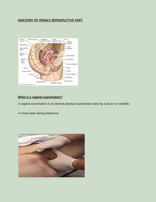
- 1. ANATOMY OF FEMALE REPRODUCTIVE PART What is a vaginal examination? A vaginal examination is an internal physical examination done by a doctor or midwife. In most cases during pregnancy
- 2. A bimanual vaginal examination may need to be performed in a number of different clinical scenarios including unexplained pelvic pain, irregular vaginal bleeding, abnormal vaginal discharge and as part of the assessment of a pelvic mass. . Gather the appropriate equipment: Gloves Lubricant Paper towels Introduction 1.Wash your hands and don PPE if appropriate. 2.Introduce yourself to the patient including your name and role. 3.Confirm the patient’s name and date of birth.
- 3. 4.Explain what the examination will involve using patient-friendly language: “Today I need to carry out a vaginal examination. This will involve me using one hand to feel your tummy and the other hand to place two fingers into your vagina. This will allow me to assess the vagina, womb and ovaries. It shouldn’t be painful, but it will feel a little uncomfortable. You can ask me to stop at any point.” 5.Explain the need for a chaperone: “One of the female ward staff members will be present throughout the examination, acting as a chaperone, would that be ok?” 6.Gain consent to proceed with the examination: “Do you understand everything I’ve said? Do you have any questions? Are you happy for me to carry out the examination?” 7.Ask the patient if they have any pain or if they think they may be pregnant before proceeding with the clinical examination. 8.Provide the patient with the opportunity to pass urine before the examination. 10.Explain to the patient that they’ll need to remove their underwear and lie on the
- 4. clinical examination couch, covering themselves with the sheet provided. Provide the patient with privacy to undress and check it is ok to re-enter the room before doing so. Vaginal examination 1.Position the patient supine in the modified lithotomy position: “Bring your heels towards your bottom and then let your knees fall to the sides.”. 2.Adequately expose the patient 3.Don a pair of non-sterile gloves. 4. Inspect the vulva for abnormalities: Ulcers: typically associated with genital herpes. Abnormal vaginal discharge: causes include candidiasis, bacterial vaginosis, chlamydia and gonorrhoea. Scarring: may relate to previous surgery (e.g. episiotomy) or (destructive scarring with associated adhesions). Vaginal atrophy: most commonly occurs in postmenopausal women. White lesions: may be patchy or in a figure of eight distribution around the
- 5. vulva and anus, associated with lichen sclerosus. Masses: causes include Bartholin’s cyst and vulval malignancy. Varicosities: varicose veins secondary to chronic venous disease or obstruction in the pelvis (e.g. pelvic malignancy). Female genital mutilation: total or partial removal of the clitoris and/or labia and/or narrowing of the vaginal introitus. 5. Inspect for evidence of vaginal prolapse (a bulge visible protruding from the vagina). Asking the patient to cough as you inspect can exacerbate the lump and help confirm the presence of prolapse. Bartholin’s cyst Bartholin’s glands are responsible for producing secretions which maintain vaginal moisture and are typically located at 4 and 8 o’clock in relation to the vaginal introitus. These glands can become blocked and/or infected, resulting in cyst formation. Typical findings on clinical examination include a unilateral, fluctuant mass, which may or may not be tender.
- 6. Lichen sclerosus Lichen sclerosus is a chronic inflammatory dermatological condition that can affect the anogenital region in women. It presents with pruritis and clinical examination typically reveals white thickened patches. Destructive scarring and adhesions develop causing distortion of the normal vaginal architecture (shrinking of the labia,
- 7. narrowing of the introitus, obscuration of the clitoris). Abnormal vaginal discharge There are several causes of abnormal vaginal discharge including: Bacterial vaginosis: typical findings include a thin, profuse fishy-smelling discharge without pruritis or inflammation. Candidiasis: typical findings include a curd-like, non-offensive discharge with associated pruritis and inflammation. Chlamydia and gonorrhoea (symptomatic): typical findings include purulent vaginal discharge Trichomoniasis: typical findings include offensive yellow, frothy vaginal discharge with associated pruritis and inflammation. Vaginal examination Warn the patient you are going to examine the vagina and ask if they’re still ok for you to do so. If the patient consents to the continuation of the examination:
- 8. 1. Lubricate the gloved index and middle fingers of your dominant hand. 2. Carefully separate the labia using the thumb and index finger of your non- dominant hand. 3. Gently insert the gloved index and middle finger of your dominant hand into the vagina. 4. Enter the vagina with your palm facing laterally and then rotate 90 degrees so that your palm is facing upwards. 5.Palpate the walls of the vagina for any irregularities or masses. 6.Examine the cervix to assess: Position (e.g. anterior or posterior)
- 9. Consistency (e.g. irregular, smooth) Cervical motion tenderness: involves severe pain on palpation of the cervix and may suggest pelvic inflammatory disease or ectopic pregnancy. Fornices The fornices are the superior portions of the vagina, extending into the recesses created by the vaginal portion of the cervix. Gently palpate lateral fornices for any masses. 7.Bimanually palpate the uterus: Place your non-dominant hand 4cm above the pubis symphysis. Place two of your dominant hand’s fingers into the posterior fornix. Push upwards with the internal fingers whilst simultaneously palpating the lower abdomen with your non-dominant hand. You should be able to feel the uterus between your hands. You should then assess the various characteristics
- 10. of the uterus: Size : the uterus should be approximately orange-sized in an average female. Shape: may be distorted by masses such as large fibroids. Position: the uterus may be anteverted or retroverted. Surface characteristics: note if the uterus feels smooth or nodular. Tenderness: may suggest inflammation (e.g. pelvic inflammatory disease, ectopic pregnancy). The position of the uterus can be described as: Anteverted: the uterus is orientated forwards towards the bladder. This is the most common position of the uterus. Retroverted: the uterus is orientated posteriorly, towards the spine. This is a less common uterine position present in approximately 1 in 5 women. Bimanually palpate the adnexa: The term adnexa refers to the area that includes the ovaries and fallopian tubes 1. Position your internal fingers in the left lateral fornix.
- 11. 2. Position your external hand onto the left iliac fossa. 3. Perform deep palpation of the left iliac fossa whilst moving your internal fingers upwards and laterally (towards the left). 4. Feel for any palpable masses, noting their size and shape (e.g. ovarian cyst, ovarian tumour, fibroid). 5. Repeat adnexal assessment on the right. 6. Withdraw your fingers and inspect the glove for blood or abnormal discharge. 7. Cover the patient with the sheet, explain that the examination is now complete and provide the patient with privacy so they can get dressed. Provide paper towels for the patient to clean themselves. 8. Dispose of the used equipment into a clinical waste bin.
- 12. To complete the examination… Thank the patient for their time. Dispose of PPE appropriately and wash your hands. Summarise your findings. Document the examination in the medical notes including the details of the chaperone. Example summary “Today I examined Mrs mukoso , a 28-year-old female. On general inspection, the patient appeared comfortable at rest. There were no objects or medical equipment around the bed of relevance.“ “Abdominal examination was unremarkable and there were no abnormalities noted on inspection of the vulva. Bimanual examination revealed an anteverted uterus of
- 13. normal size and shape. There were no masses palpated in the vaginal canal or adnexa.” “In summary, these findings are consistent with a normal vaginal examination.” “For completeness, I would like to perform the following further assessments and investigations.” Further assessments and investigations Urinalysis: including β-HCG to rule out pregnancy (including ectopic pregnancy). Speculum examination: to visualise the vaginal canal and cervix. Vaginal swabs/endocervical swabs: if there are concerns about infection (bacterial and viral). Ultrasound abdomen and pelvis: to better visualise any masses palpated and to assess endometrial thickness. Complete abdominal examination: if there are concerns about intraabdominal pathology (e.g. appendicitis).
- 14. ANATOMY OF FEMALE REPRODUCTIVE PART
- 15. What is a vaginal examination? A vaginal examination is an internal physical examination done by a doctor or midwife. In most cases during pregnancy A bimanual vaginal examination may need to be performed in a number of different
- 16. clinical scenarios including unexplained pelvic pain, irregular vaginal bleeding, abnormal vaginal discharge and as part of the assessment of a pelvic mass. . Gather the appropriate equipment: Gloves Lubricant Paper towels Introduction 1.Wash your hands and don PPE if appropriate. 2.Introduce yourself to the patient including your name and role. 3.Confirm the patient’s name and date of birth. 4.Explain what the examination will involve using patient-friendly language: “Today I need to carry out a vaginal examination. This will involve me using one hand to feel your tummy and the other hand to place two fingers into your vagina. This will allow
- 17. me to assess the vagina, womb and ovaries. It shouldn’t be painful, but it will feel a little uncomfortable. You can ask me to stop at any point.” 5.Explain the need for a chaperone: “One of the female ward staff members will be present throughout the examination, acting as a chaperone, would that be ok?” 6.Gain consent to proceed with the examination: “Do you understand everything I’ve said? Do you have any questions? Are you happy for me to carry out the examination?” 7.Ask the patient if they have any pain or if they think they may be pregnant before proceeding with the clinical examination. 8.Provide the patient with the opportunity to pass urine before the examination. 10.Explain to the patient that they’ll need to remove their underwear and lie on the clinical examination couch, covering themselves with the sheet provided. Provide the patient with privacy to undress and check it is ok to re-enter the room before doing so.
- 18. Vaginal examination 1.Position the patient supine in the modified lithotomy position: “Bring your heels towards your bottom and then let your knees fall to the sides.”. 2.Adequately expose the patient 3.Don a pair of non-sterile gloves. 4. Inspect the vulva for abnormalities: Ulcers: typically associated with genital herpes. Abnormal vaginal discharge: causes include candidiasis, bacterial vaginosis, chlamydia and gonorrhoea. Scarring: may relate to previous surgery (e.g. episiotomy) or (destructive scarring with associated adhesions). Vaginal atrophy: most commonly occurs in postmenopausal women. White lesions: may be patchy or in a figure of eight distribution around the vulva and anus, associated with lichen sclerosus. Masses: causes include Bartholin’s cyst and vulval malignancy. Varicosities: varicose veins secondary to chronic venous disease or obstruction
- 19. in the pelvis (e.g. pelvic malignancy). Female genital mutilation: total or partial removal of the clitoris and/or labia and/or narrowing of the vaginal introitus. 5. Inspect for evidence of vaginal prolapse (a bulge visible protruding from the vagina). Asking the patient to cough as you inspect can exacerbate the lump and help confirm the presence of prolapse. Bartholin’s cyst Bartholin’s glands are responsible for producing secretions which maintain vaginal moisture and are typically located at 4 and 8 o’clock in relation to the vaginal introitus. These glands can become blocked and/or infected, resulting in cyst formation. Typical findings on clinical examination include a unilateral, fluctuant mass, which may or may not be tender.
- 20. Lichen sclerosus Lichen sclerosus is a chronic inflammatory dermatological condition that can affect the anogenital region in women. It presents with pruritis and clinical examination typically reveals white thickened patches. Destructive scarring and adhesions develop causing distortion of the normal vaginal architecture (shrinking of the labia, narrowing of the introitus, obscuration of the clitoris).
- 21. Abnormal vaginal discharge There are several causes of abnormal vaginal discharge including: Bacterial vaginosis: typical findings include a thin, profuse fishy-smelling discharge without pruritis or inflammation. Candidiasis: typical findings include a curd-like, non-offensive discharge with associated pruritis and inflammation. Chlamydia and gonorrhoea (symptomatic): typical findings include purulent vaginal discharge Trichomoniasis: typical findings include offensive yellow, frothy vaginal discharge with associated pruritis and inflammation. Vaginal examination Warn the patient you are going to examine the vagina and ask if they’re still ok for you to do so. If the patient consents to the continuation of the examination:
- 22. 1. Lubricate the gloved index and middle fingers of your dominant hand. 2. Carefully separate the labia using the thumb and index finger of your non- dominant hand. 3. Gently insert the gloved index and middle finger of your dominant hand into the vagina. 4. Enter the vagina with your palm facing laterally and then rotate 90 degrees so that your palm is facing upwards. 5.Palpate the walls of the vagina for any irregularities or masses. 6.Examine the cervix to assess: Position (e.g. anterior or posterior) Consistency (e.g. irregular, smooth)
- 23. Cervical motion tenderness: involves severe pain on palpation of the cervix and may suggest pelvic inflammatory disease or ectopic pregnancy. Fornices The fornices are the superior portions of the vagina, extending into the recesses created by the vaginal portion of the cervix. Gently palpate lateral fornices for any masses. 7.Bimanually palpate the uterus: Place your non-dominant hand 4cm above the pubis symphysis. Place two of your dominant hand’s fingers into the posterior fornix. Push upwards with the internal fingers whilst simultaneously palpating the lower abdomen with your non-dominant hand. You should be able to feel the uterus between your hands. You should then assess the various characteristics of the uterus:
- 24. Size : the uterus should be approximately orange-sized in an average female. Shape: may be distorted by masses such as large fibroids. Position: the uterus may be anteverted or retroverted. Surface characteristics: note if the uterus feels smooth or nodular. Tenderness: may suggest inflammation (e.g. pelvic inflammatory disease, ectopic pregnancy). The position of the uterus can be described as: Anteverted: the uterus is orientated forwards towards the bladder. This is the most common position of the uterus. Retroverted: the uterus is orientated posteriorly, towards the spine. This is a less common uterine position present in approximately 1 in 5 women. Bimanually palpate the adnexa: The term adnexa refers to the area that includes the ovaries and fallopian tubes 1. Position your internal fingers in the left lateral fornix.
- 25. 2. Position your external hand onto the left iliac fossa. 3. Perform deep palpation of the left iliac fossa whilst moving your internal fingers upwards and laterally (towards the left). 4. Feel for any palpable masses, noting their size and shape (e.g. ovarian cyst, ovarian tumour, fibroid). 5. Repeat adnexal assessment on the right. 6. Withdraw your fingers and inspect the glove for blood or abnormal discharge. 7. Cover the patient with the sheet, explain that the examination is now complete and provide the patient with privacy so they can get dressed. Provide paper towels for the patient to clean themselves. 8. Dispose of the used equipment into a clinical waste bin.
- 26. To complete the examination… Thank the patient for their time. Dispose of PPE appropriately and wash your hands. Summarise your findings. Document the examination in the medical notes including the details of the chaperone. Example summary “Today I examined Mrs mukoso , a 28-year-old female. On general inspection, the patient appeared comfortable at rest. There were no objects or medical equipment around the bed of relevance.“ “Abdominal examination was unremarkable and there were no abnormalities noted on inspection of the vulva. Bimanual examination revealed an anteverted uterus of normal size and shape. There were no masses palpated in the vaginal canal or
- 27. adnexa.” “In summary, these findings are consistent with a normal vaginal examination.” “For completeness, I would like to perform the following further assessments and investigations.” Further assessments and investigations Urinalysis: including β-HCG to rule out pregnancy (including ectopic pregnancy). Speculum examination: to visualise the vaginal canal and cervix. Vaginal swabs/endocervical swabs: if there are concerns about infection (bacterial and viral). Ultrasound abdomen and pelvis: to better visualise any masses palpated and to assess endometrial thickness. Complete abdominal examination: if there are concerns about intraabdominal pathology (e.g. appendicitis).