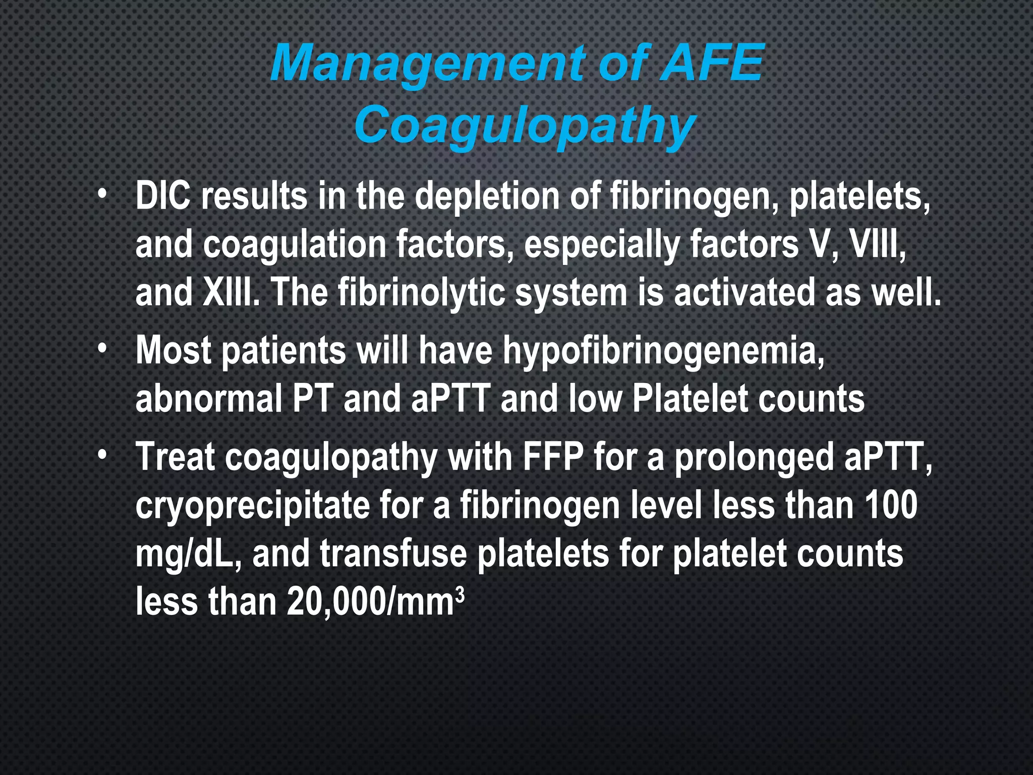
Amniotic fluid embolism (AFE) is a life threatening obstetric emergency characterized by sudden cardiorespiratory collapse and disseminated intravascular coagulation.
Steiner and Luschbaugh first described AFE in 1941, after they found fetal debris in the pulmonary circulation of women who died during labor. Data from the National Amniotic Fluid Embolus Registry (USA) suggest that the process is more similar to anaphylaxis than to embolism, and the term anaphylactoid syndrome of pregnancy has been suggested because fetal tissue or amniotic fluid components are not universally found in women who present with signs and symptoms attributable to AFE.
The diagnosis of AFE has traditionally been made at autopsy when fetal squamous cells are found in the maternal pulmonary circulation; however, fetal squamous cells are commonly found in the circulation of laboring patients who do not develop the syndrome. The diagnosis is essentially one of exclusion based on clinical presentation. Other causes of hemodynamic instability should not be neglected.
When fetal head is delivered, but shoulders are stuck and cannot be delivered it is known as shoulder dystocia.
The anterior shoulder becomes trapped behind on the symphysis pubis, whilst the posterior shoulder may be in the hollow of the sacrum or high above the sacral promontory.
Cord prolapse is a frightening and life-threatening event that occurs in labor. Rapid identification and immediate appropriate response may well save the life of a neonate. Therefore, clinicians should be knowledgeable in its recognition and management.
This topic contains definition, meaning, classification, pathophysiology, clinical menifestations, metabolic and general changes, management of obstetrical shock
Uterine Rupture
Deepa Mishra
Assistant Professor (OBG)
Introduction
Uterine rupture is when the muscular wall of the uterus tears during pregnancy or childbirth
Symptoms while classically including increased pain, vaginal bleeding, or a change in contractions are not always present.
Disability or death of the mother or baby may result.
Definition
Uterine rupture is giving way of gravid uterus or dissolution in the continuity of uterine wall anytime after 28 weeks of gestation with or without expulsion of the fetus.
Incidence
Rates of uterine rupture during vaginal birth following one previous C-section, done by the typical technique, are estimated at 0.9%
Rates are greater among those who have had multiple prior C-sections or an atypical type of C-section.
In those who do have uterine scarring, the risk during a vaginal birth is about 1 per 12,000
Risk of death of the baby is about 6%
Etiology
Risk Factors
Previous cesarean section
Myomectomy
Dysfunctional labor
Labor augmentation by oxytocin or prostaglandins
High parity
First pregnancy- very rare
Types of uterine rupture
Complete Rupture
All the layers including peritoneum are torn and the uterine contents escape into the peritoneal cavity.
Usually results in death
Incomplete Rupture
Visceral peritoneum is intact and usually the fetus remains in the uterine cavity
Sign & Symptoms
Uterine dehiscence and abdominal pain and vaginal bleeding
Deterioration of fetal heart rate
Loss of fetal station on manual vaginal exam
Hypovolemic shock due to intrabdominal bleeding
Chest pain between the scapulae, pain during inspiration due to irritation of blood below the perineum
Cessation of uterine contractions
Palpation of fetus outside the uterus
Signs of abdominal pregnancy
Post term pregnancy
Diagnosis
Signs of obstructed labor with dehydration, exhaustion, tachycardia raised temperature tonic contraction , pathological retraction ring
Absent fetal heart sound
On PV hot, dry vagina with a large caput over the presenting part
Prevention
Early diagnosis and management of CPD mal presentation and obstructed labor
Proper selection of cases for vaginal delivery
Carefull monitoring of oxytocin infusion specially in multipara
Avoid intra uterine manipulation no version in single fetus
Instrumental delivery after cervical dilatation
Immediate CS in obstructed labor
Hospital delivery for high risk cases
ECV should be avoided during general anaesthesia
Careful manual removal of placenta
Treatment
Resuscitation with adequate hydration and blood transfusion
Laprotomy
Hysterectomy
Repair
Complication
Rupture uterus with haemorrhage, shock and sepsis
Fetal loss is high in spontaneous and traumatic rupture
Mortality is low in LSCS scar rupture
CONCLUSIONS:
- Cardiologist, obstetrician and anestesiologist should cooperate to each other
- The advantage of regional anesthesia is patients can communicate if symptoms occur
- If palpitations, chest pain and shortness of breath happened, immediate action should be performed
- RA should be given using lower dose of local anesthetics opioids and slow induction
- GA : standard technique “rapid sequence induction”
The increased cardiac output related to pregnancy can lead to heart failure, and the increased heart rate in the third trimester can lead to ischemic events. The potential obstetrical complications include preeclampsia or other hypertensive related disorders, premature birth, and small-for-gestational-age births.
This is an acute terrifying emergency following delivery in which patient suddenly develops dyspnoea, apnoea, cyanosis with or without seizure and shock among other features. It usually occurs secondary to several catastrophic events and may be a tragic experience to the patient and the physician
a clinical syndrome that results from inadequate tissue perfusion.
Hypovolemic shock - Blood or fluid loss, both leading to a decreased circulating blood volume, diastolic filling pressure, and volume.
Cardiogenic shock - due to cardiac pump failure related to loss of myocardial contractility/functional myocardium or structural/mechanical failure of the cardiac anatomy and characterized by elevations of diastolic filling pressures and volumes
Extra-cardiac/obstructive shock - due to obstruction to flow in the cardiovascular circuit and characterized by either impairment of diastolic filling or excessive afterload
Distributive shock - caused by loss of vasomotor control resulting in arteriolar/venular dilatation leading to a decrease in preload, with decreased, normal, or elevated cardiac output, depending on the presence of myocardial depression.
The primary treatment goals for patients with hepatitis B (HBV) infection are to prevent progression of the disease, particularly to cirrhosis, liver failure, and hepatocellular carcinoma (HCC).
Risk factors for progression of chronic HBV include the following :
Persistently elevated levels of HBV DNA and, in some patients, alanine aminotransferase (ALT), as well as the presence of core and precore mutations seen most commonly in HBV genotype C and D infections
Male sex
Older age
Family history of HCC
Alcohol use
Elevated alpha-fetoprotein (AFP)
Coinfection with hepatitis D (delta) virus (HDV), hepatitis C virus (HCV), or human immunodeficiency virus (HIV)
A synergistic approach of suppressing viral load and boosting the patient’s immune response with immunotherapeutic interventions is needed for the best prognosis. The prevention of HCC often includes the use of antiviral treatment using pegylated interferon (PEG-IFN) or nucleos(t)ide analogues.
HBV infection can be self-limited or chronic. No specific therapy is available for persons with acute hepatitis B; treatment is supportive.
Patients with acute hepatitis C virus (HCV) infection appear to have an excellent chance of responding to 6 months of standard therapy with interferon (IFN). Because spontaneous resolution is common, no definitive timing of therapy initiation can be recommended; however, waiting 2-4 months after the onset of illness seems reasonable.
Treatment for chronic HCV is based on guidelines from the Infectious Diseases Society of America (IDSA) and the American Associations for the Study of Liver Diseases (AASLD), in collaboration with the International Antiviral Society-USA (IAS-USA). These guidelines are constantly being updated. For more information, see HCV Guidance: Recommendations for Testing, Managing, and Treating Hepatitis C.
The guidelines propose that because all patients cannot receive treatment immediately upon the approval of new agents, priority should be given to those with the most urgent need.
The recommendations include the following :
Patients with advanced fibrosis, those with compensated cirrhosis, liver transplant recipients, and those with severe extraheptic hepatitis are to be given the highest priority for treatment
Based on available resources, patients at high risk for liver-related complications and severe extrahepatic hepatitis C complications should be given high priority for treatment
Treatment decisions should balance the anticipated reduction in transmission versus the likelihood of reinfection in patients whose risk of HCV transmission is high and in whom HCV treatment may result in a reduction in transmission (eg, men who have high-risk sex with men, active injection drug users, incarcerated persons, and those on hemodialysis)
Interstitial Lung Diseases [ILD] Approach to ManagementArun Vasireddy
Diffuse (interstitial) lung disease includes a wide variety of relatively uncommon conditions presenting with characteristic clusters of clinical features and marked by an immune response. There are over 200 specific diffuse lung diseases, many of unknown etiology. The combined incidence is 50 per 100,000, or 1 in 2000 people. Because these conditions cause aberrant lung function, morbidity and mortality due to lung injury and fibrosis are not uncommon. Both environmental and genetic factors are believed to contribute to the development of diffuse lung disease. Antigen processing and presentation are important in the development of the immune response seen in the disease, and it is thought that the likely candidate genes predisposing patients to this category of disease are those of the major histocompatibility complex. Genes that affect the immune, inflammatory, and fibrotic processes may also influence who develops the disease. If we can identify the genes that cause diseases characterized by lung injury and fibrosis, we can eventually develop genetic interventional approaches to treatment.
Approach to Management of Upper Gastrointestinal (GI) BleedingArun Vasireddy
Upper gastrointestinal bleeding is gastrointestinal bleeding in the upper gastrointestinal tract, commonly defined as bleeding arising from the esophagus, stomach, or duodenum. Blood may be observed in vomit (hematemesis) or in altered form in the stool (melena). Depending on the severity of the blood loss, there may be symptoms of insufficient circulating blood volume and shock. As a result, upper gastrointestinal bleeding is considered a medical emergency and typically requires hospital care for urgent diagnosis and treatment. Upper gastrointestinal bleeding can be caused by peptic ulcers, gastric erosions, esophageal varices, and some rarer causes such as gastric cancer.
The initial assessment includes measurement of the blood pressure and heart rate, as well as blood tests to determine hemoglobin concentration. In significant bleeding, fluid replacement is often required, as well as blood transfusion, before the source of bleeding can be determined by endoscopy of the upper digestive tract with an esophagogastroduodenoscopy. Depending on the source, endoscopic therapy can be applied to reduce rebleeding risk. Specific medical treatments (such as proton pump inhibitors for peptic ulcer disease) or procedures (such as TIPS for variceal hemorrhage) may be used. Recurrent or refractory bleeding may lead to need for surgery, although this has become uncommon as a result of improved endoscopic and medical treatment.
Tachy Arrhythmias - Approach to ManagementArun Vasireddy
Tachyarrhythmias are disorders of heart rhythm which may present with a tachycardia i.e. a heart rate >100 bpm.
This article provides an overview of tachyarrhythmias in general and goes on to cover the most common tachyarrhythmias in more detail. The acute management of tachyarrhythmias, in an emergency setting, will be covered in the 'Acute' section of the fastbleep website.
Tachyarrhythmias are clinically important as they can precipitate cardiac arrest, cardiac failure, thromboembolic disease and syncopal events. As such, they crop up time and time again in exam papers and on the wards.
Tachyarrhythmias are classified based on whether they have broad or narrow QRS complexes on the ECG. Broad is defined as >0.12s (or more than 3 small squares on the standard ECG). Narrow is equal to or less than 0.12s. Broad QRS complexes are slower ventricular depolarisations that arise from the ventricles. Narrow complexes are ventricular depolarisations initiated from above the ventricles (known as supraventricular). One important exception is when there is a supraventricular depolarisation conducted through a diseased AV node. This will produce wide QRS complexes despite the rhythm being supraventricular in origin.
Scrub typhus is a mite-borne disease caused by Orientia tsutsugamushi (formerly Rickettsia tsutsugamushi). Symptoms are fever, a primary lesion, a macular rash, and lymphadenopathy. (See also Overview of Rickettsial and Related Infections.) Scrub typhus is related to rickettsial diseases.
Pulmonary edema is often caused by congestive heart failure. When the heart is not able to pump efficiently, blood can back up into the veins that take blood through the lungs. As the pressure in these blood vessels increases, fluid is pushed into the air spaces (alveoli) in the lungs.
The jugular venous pressure (JVP, sometimes referred to as jugular venous pulse) is the indirectly observed pressure over the venous system via visualization of the internal jugular vein. It can be useful in the differentiation of different forms of heart and lung disease.
A treadmill exercise stress test is used to determine the effects of exercise on the heart. Exercise allows doctors to detect abnormal heart rhythms (arrhythmias) and diagnose the presence or absence of coronary artery disease.
This test involves walking in place on a treadmill while monitoring the electrical activity of your heart. Throughout the test, the speed and incline of the treadmill increase. The results show how well your heart responds to the stress of different levels of exercise.
Description
A technologist will explain the test to you, take a brief medical history, and answer any questions you may have. Your blood pressure, heart rate, and electrocardiogram (ECG) will be monitored before, during, and after the test.
You will be asked to sign a consent form. This form is required before the test can proceed.
You will be asked to remove all upper body clothing, and to put on a gown with the opening to the front.
Adhesive electrodes will be put onto your chest to capture an ECG. The sites where the electrodes are placed will be cleaned with alcohol and shaved if necessary. A mild abrasion may also be used to ensure a good quality ECG recording.
Your resting blood pressure, heart rate, and ECG will be recorded.
You will be asked to walk on a treadmill. The walk starts off slowly, then the speed and incline increases at set times. It is very important that you walk as long as possible because the test is effort-dependent.
You will be monitored throughout the test. If a problem occurs, the technologist will stop the test right away. It is very important for you to tell the technologist if you experience any symptoms, such as chest pain, dizziness, unusual shortness of breath, or extreme fatigue.
Following the test, you will be asked to lie down. Your blood pressure, heart rate, and ECG will be monitored for three to five minutes after exercise.
The data will be reviewed by a cardiologist after the test is completed. A report will be sent to the doctor(s) involved in your care.
A mosquito-borne viral disease occurring in tropical and subtropical areas.
Spreads by animals or insects
Requires a medical diagnosis
Lab tests or imaging often required
Short-term: resolves within days to weeks
Those who become infected with the virus a second time are at a significantly greater risk of developing severe disease.
Symptoms include high fever, headache, rash and muscle and joint pain. In severe cases there is serious bleeding and shock, which can be life threatening.
Treatment includes fluids and pain relievers. Severe cases require hospital care.
Adrenal gland & Cushing's Disease - Seminar August 2015Arun Vasireddy
A condition that occurs from exposure to high cortisol levels for a long time.
Fewer than 1 million cases per year (India)
Treatable by a medical professional
Requires a medical diagnosis
Lab tests or imaging always required
Chronic: can last for years or be lifelong
The most common cause is the use of steroid drugs, but it can also occur from overproduction of cortisol by the adrenal glands.
Signs are a fatty hump between the shoulders, a rounded face and pink or purple stretch marks.
Treatment options include reducing steroid use, surgery, radiation and medication.
Osteoporosis is a progressive systemic skeletal disease characterized by low bone mass and microarchitecture deterioration of bone tissue, leading to enhanced bone fragility and a consequent increase in fracture risk.
The prostate is an exocrine gland of the male mammalian reproductive system
It is a walnut-sized gland that forms part of the male reproductive system and is located in front of the rectum and just below the urinary bladder
Function is to store and secrete a clear, slightly alkaline fluid that constitutes 10-30% of the volume of the seminal fluid that along with the spermatozoa, constitutes semen
A healthy human prostate measures (4cm-vertical, by 3cm-horizontal, 2cm ant-post ).
It surrounds the urethra just below the urinary bladder. It has anterior, median, posterior and two lateral lobes
It’s work is regulated by androgens which are responsible for male sex characteristics
Generalised disease of the prostate due to hormonal derangement which leads to non malignant enlargement of the gland (increase in the number of epithelial cells and stromal tissue)to cause compression of the urethra leading to symptoms (LUTS
These simplified slides by Dr. Sidra Arshad present an overview of the non-respiratory functions of the respiratory tract.
Learning objectives:
1. Enlist the non-respiratory functions of the respiratory tract
2. Briefly explain how these functions are carried out
3. Discuss the significance of dead space
4. Differentiate between minute ventilation and alveolar ventilation
5. Describe the cough and sneeze reflexes
Study Resources:
1. Chapter 39, Guyton and Hall Textbook of Medical Physiology, 14th edition
2. Chapter 34, Ganong’s Review of Medical Physiology, 26th edition
3. Chapter 17, Human Physiology by Lauralee Sherwood, 9th edition
4. Non-respiratory functions of the lungs https://academic.oup.com/bjaed/article/13/3/98/278874
MANAGEMENT OF ATRIOVENTRICULAR CONDUCTION BLOCK.pdfJim Jacob Roy
Cardiac conduction defects can occur due to various causes.
Atrioventricular conduction blocks ( AV blocks ) are classified into 3 types.
This document describes the acute management of AV block.
Explore natural remedies for syphilis treatment in Singapore. Discover alternative therapies, herbal remedies, and lifestyle changes that may complement conventional treatments. Learn about holistic approaches to managing syphilis symptoms and supporting overall health.
Factory Supply Best Quality Pmk Oil CAS 28578–16–7 PMK Powder in Stockrebeccabio
Factory Supply Best Quality Pmk Oil CAS 28578–16–7 PMK Powder in Stock
Telegram: bmksupplier
signal: +85264872720
threema: TUD4A6YC
You can contact me on Telegram or Threema
Communicate promptly and reply
Free of customs clearance, Double Clearance 100% pass delivery to USA, Canada, Spain, Germany, Netherland, Poland, Italy, Sweden, UK, Czech Republic, Australia, Mexico, Russia, Ukraine, Kazakhstan.Door to door service
Hot Selling Organic intermediates
Ethanol (CH3CH2OH), or beverage alcohol, is a two-carbon alcohol
that is rapidly distributed in the body and brain. Ethanol alters many
neurochemical systems and has rewarding and addictive properties. It
is the oldest recreational drug and likely contributes to more morbidity,
mortality, and public health costs than all illicit drugs combined. The
5th edition of the Diagnostic and Statistical Manual of Mental Disorders
(DSM-5) integrates alcohol abuse and alcohol dependence into a single
disorder called alcohol use disorder (AUD), with mild, moderate,
and severe subclassifications (American Psychiatric Association, 2013).
In the DSM-5, all types of substance abuse and dependence have been
combined into a single substance use disorder (SUD) on a continuum
from mild to severe. A diagnosis of AUD requires that at least two of
the 11 DSM-5 behaviors be present within a 12-month period (mild
AUD: 2–3 criteria; moderate AUD: 4–5 criteria; severe AUD: 6–11 criteria).
The four main behavioral effects of AUD are impaired control over
drinking, negative social consequences, risky use, and altered physiological
effects (tolerance, withdrawal). This chapter presents an overview
of the prevalence and harmful consequences of AUD in the U.S.,
the systemic nature of the disease, neurocircuitry and stages of AUD,
comorbidities, fetal alcohol spectrum disorders, genetic risk factors, and
pharmacotherapies for AUD.
- Video recording of this lecture in English language: https://youtu.be/lK81BzxMqdo
- Video recording of this lecture in Arabic language: https://youtu.be/Ve4P0COk9OI
- Link to download the book free: https://nephrotube.blogspot.com/p/nephrotube-nephrology-books.html
- Link to NephroTube website: www.NephroTube.com
- Link to NephroTube social media accounts: https://nephrotube.blogspot.com/p/join-nephrotube-on-social-media.html
Pulmonary Thromboembolism - etilogy, types, medical- Surgical and nursing man...VarunMahajani
Disruption of blood supply to lung alveoli due to blockage of one or more pulmonary blood vessels is called as Pulmonary thromboembolism. In this presentation we will discuss its causes, types and its management in depth.
Ozempic: Preoperative Management of Patients on GLP-1 Receptor Agonists Saeid Safari
Preoperative Management of Patients on GLP-1 Receptor Agonists like Ozempic and Semiglutide
ASA GUIDELINE
NYSORA Guideline
2 Case Reports of Gastric Ultrasound
micro teaching on communication m.sc nursing.pdfAnurag Sharma
Microteaching is a unique model of practice teaching. It is a viable instrument for the. desired change in the teaching behavior or the behavior potential which, in specified types of real. classroom situations, tends to facilitate the achievement of specified types of objectives.
Anti ulcer drugs and their Advance pharmacology ||
Anti-ulcer drugs are medications used to prevent and treat ulcers in the stomach and upper part of the small intestine (duodenal ulcers). These ulcers are often caused by an imbalance between stomach acid and the mucosal lining, which protects the stomach lining.
||Scope: Overview of various classes of anti-ulcer drugs, their mechanisms of action, indications, side effects, and clinical considerations.
HOT NEW PRODUCT! BIG SALES FAST SHIPPING NOW FROM CHINA!! EU KU DB BK substit...GL Anaacs
Contact us if you are interested:
Email / Skype : kefaya1771@gmail.com
Threema: PXHY5PDH
New BATCH Ku !!! MUCH IN DEMAND FAST SALE EVERY BATCH HAPPY GOOD EFFECT BIG BATCH !
Contact me on Threema or skype to start big business!!
Hot-sale products:
NEW HOT EUTYLONE WHITE CRYSTAL!!
5cl-adba precursor (semi finished )
5cl-adba raw materials
ADBB precursor (semi finished )
ADBB raw materials
APVP powder
5fadb/4f-adb
Jwh018 / Jwh210
Eutylone crystal
Protonitazene (hydrochloride) CAS: 119276-01-6
Flubrotizolam CAS: 57801-95-3
Metonitazene CAS: 14680-51-4
Payment terms: Western Union,MoneyGram,Bitcoin or USDT.
Deliver Time: Usually 7-15days
Shipping method: FedEx, TNT, DHL,UPS etc.Our deliveries are 100% safe, fast, reliable and discreet.
Samples will be sent for your evaluation!If you are interested in, please contact me, let's talk details.
We specializes in exporting high quality Research chemical, medical intermediate, Pharmaceutical chemicals and so on. Products are exported to USA, Canada, France, Korea, Japan,Russia, Southeast Asia and other countries.
Triangles of Neck and Clinical Correlation by Dr. RIG.pptx
Amniotic Fluid Embolism [AFE] Approach to Management
1.
2.
3.
4.
5. Nevertheless, these and other frequently cited risk factors
are not consistently observed and at the present time
Experts agree that this condition is not preventable.
20. Management of AFE
Coagulopathy
• DIC results in the depletion of fibrinogen, platelets,
and coagulation factors, especially factors V, VIII,
and XIII. The fibrinolytic system is activated as well.
• Most patients will have hypofibrinogenemia,
abnormal PT and aPTT and low Platelet counts
• Treat coagulopathy with FFP for a prolonged aPTT,
cryoprecipitate for a fibrinogen level less than 100
mg/dL, and transfuse platelets for platelet counts
less than 20,000/mm3
21.
22. Sympathomimetic Vasopressor agent
• Dopamine increases myocardial contractility and systolic BP
with little increase in diastolic BP. Also dilates the renal
vasculature, increasing renal blood flow and GFR.
• DOSE: 2-5 mcg/kg/min IV; titrate to BP and cardiac output.
• Contraindications: ventricular fibrillation, hypovolemia,
pheochromocytoma.
• Precautions: Monitor urine flow, cardiac output, pulmonary
wedge pressure, and BP during infusion; prior to infusion,
correct hypovolemia with either whole blood or plasma, as
indicated; monitoring central venous pressure or left
ventricular filling pressure may be helpful
23. Maternal Mortality in AFE
• Maternal death usually occurs in one of three ways: (1)
sudden cardiac arrest, (2) hemorrhage due to coagulopathy,
or (3) initial survival with death due to acute respiratory
distress syndrome (ARDS) and multiple organ failure
• For women diagnosed as having AFE, mortality rates
ranging from 26% to as high as 86% have been reported.
• The variance in these numbers is explained by dissimilar
case definitions and possibly improvements in intensive
care management of affected patients.
24.
25. SUMMARY
• AFE is a sudden and unexpected rare but life
threatining complication of pregnancy.
• It has a complex pathogenesis and serious
implications for both mother and infant
• Associated with high rates of mortality and
morbidity.
• Diagnosis of exclusion.
• Suspect AFE when confronted with any pregnant
patient who has sudden onset of respiratory
distress, cardiac collapse, seizures, unexplained
fetal distress, and abnormal bleeding
• Obstetricians should be alert to the symptoms of
AFE and strive for prompt and aggressive treatment.
Editor's Notes
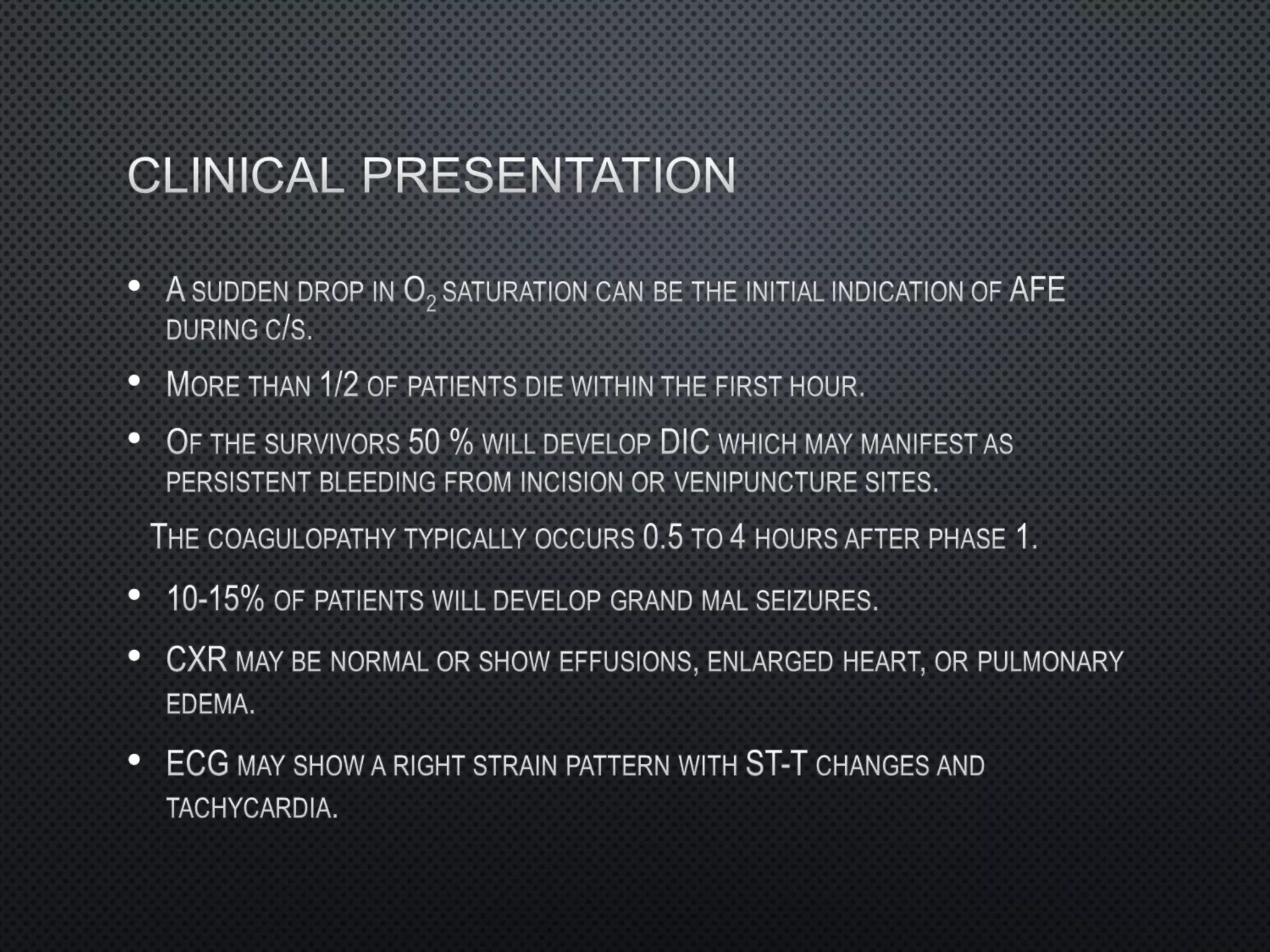
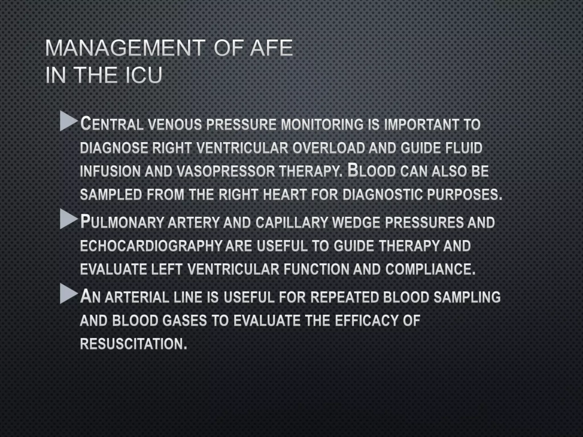
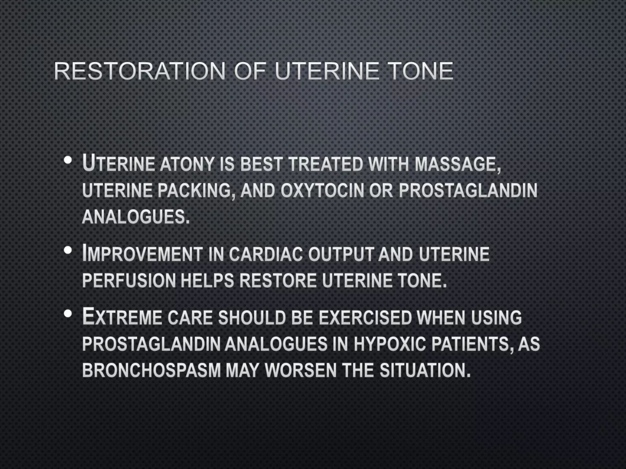
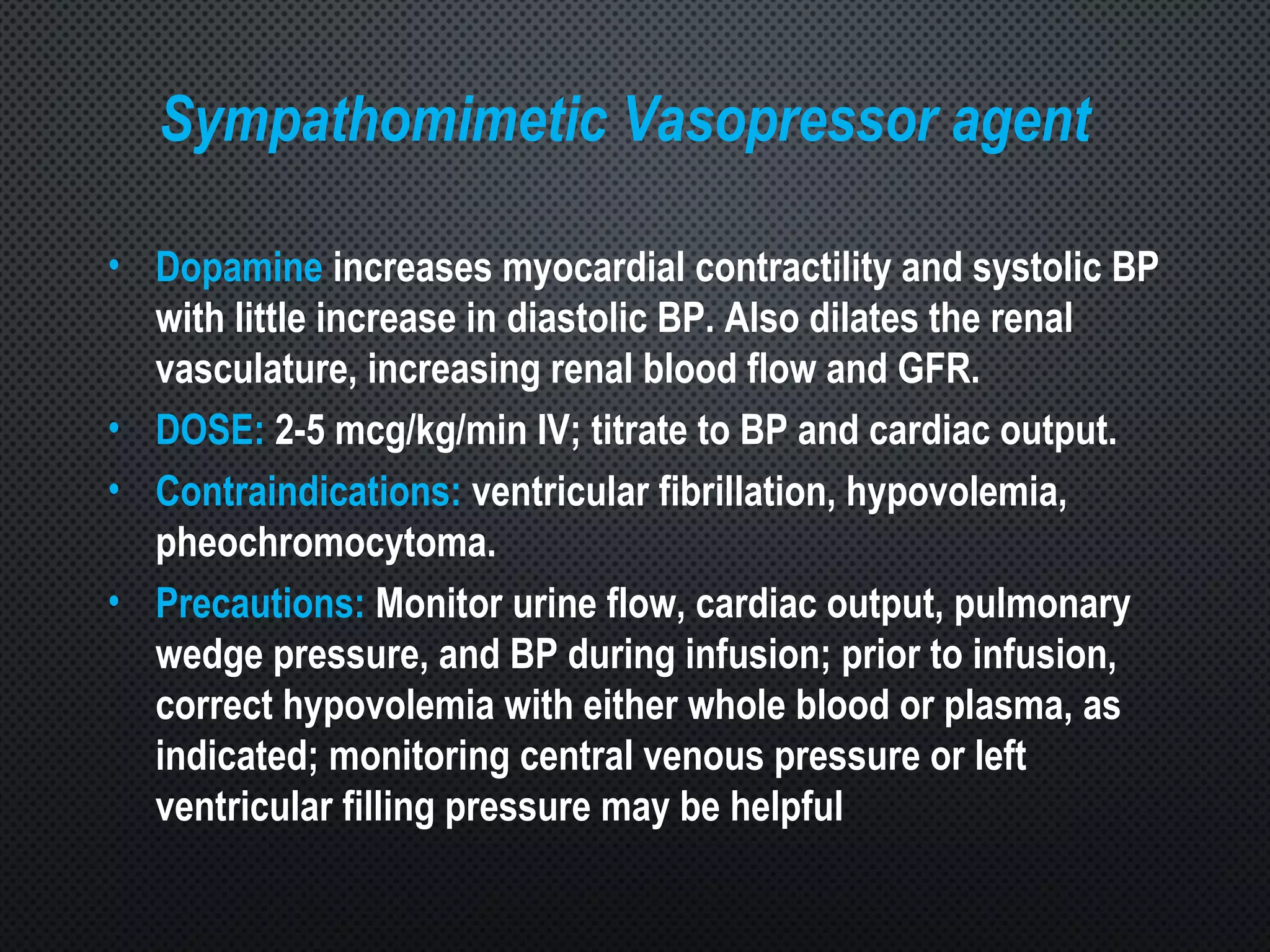
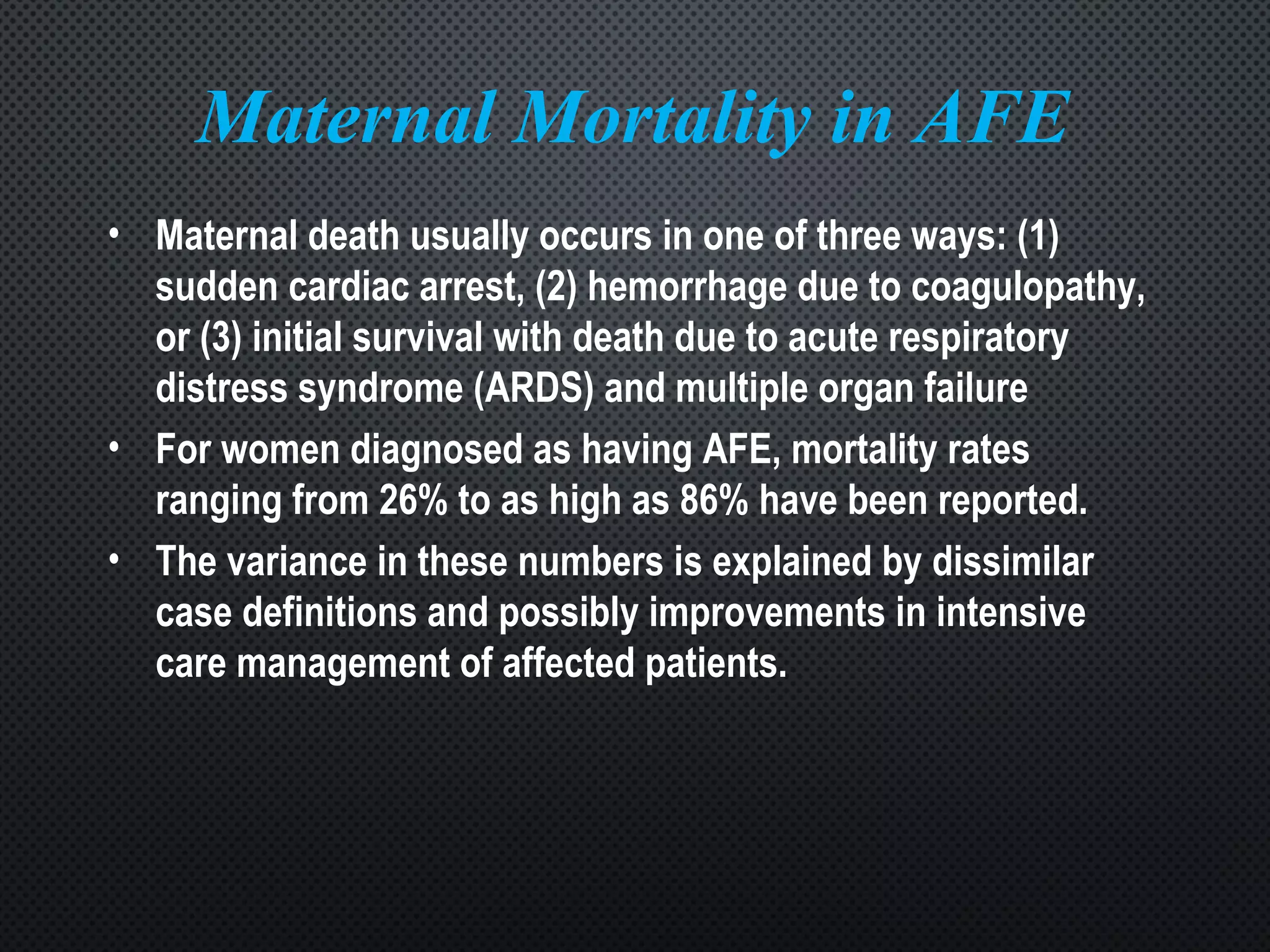
Amniotic fluid embolism (AFE) is a rare obstetric emergency in which it is postulated that amniotic fluid, fetal cells, hair, or other debris enter the maternal circulation, causing cardiorespiratory collapse.
In 1941, Steiner and Luschbaugh described AFE for the first time after they found fetal debris in the pulmonary circulation of women who died during labor.
Current data from the National Amniotic Fluid Embolus Registry suggest that the process is more similar to anaphylaxis than to embolism, and the term anaphylactoid syndrome of pregnancy has been suggested because fetal tissue or amniotic fluid components are not universally found in women who present with signs and symptoms attributable to AFE.[1]
The diagnosis of AFE has traditionally been made at autopsy when fetal squamous cells are found in the maternal pulmonary circulation; however, fetal squamous cells are commonly found in the circulation of laboring patients who do not develop the syndrome. In a patient who is critically ill, a sample obtained by aspiration of the distal port of a pulmonary artery catheter that contains fetal squamous cells is considered suggestive of but not diagnostic of AFE syndrome.[2] The diagnosis is essentially one of exclusion based on clinical presentation. Other causes of hemodynamic instability should not be neglected.
The pathophysiology of AFE is poorly understood. Based on the original description, it was theorized that amniotic fluid and fetal cells enter the maternal circulation, possibly triggering an anaphylactic reaction to fetal antigens. However, fetal material is not always found in the maternal circulation in patients with AFE, and material of fetal origin is often found in women who do not develop AFE.
Benson et al[3] tested 2 hypotheses concerning the pathophysiology of AFE: (1) Clinical symptoms result from mast cell degranulation with the release of histamine and tryptase, or (2) Clinical symptoms result from activation of the complement pathway. Nine women with AFE were compared with 22 women with normal labors. Serum from patients with AFE was collected within 14 hours of symptom onset and frozen. Urine was collected within 12-24 hours after symptom onset. Control patients had complement levels measured on admission, during labor, and the day after delivery.
Six of the 9 women with AFE died, and all 9 required blood transfusions for disseminated intravascular coagulation (DIC). Seven women had no evidence of mast cell degranulation (ie, either urinary histamine or serum tryptase). Compared with postpartum control patients, complement levels in the AFE group were severely depressed. C3 in the AFE group was 44 compared with 117.2 in the postpartum group. C4 was 10.7 in the AFE group versus 29.4 in the postpartum group. These differences were statistically significant. This suggests that complement activation may play an important role in the pathophysiology of AFE.
Farrar and Gherman[4] reported the case of a 40-year-old multipara in active labor with acute onset of facial erythema, seizures, hypoxia, cardiac arrest, DIC, and ultimately death. Fetal squames and fibrin thrombi were found in the pulmonary tree at autopsy. Blood drawn 2 hours after symptom onset had a serum tryptase level of 4.7 ng/mL (normal < 1 ng/mL).
A case reported by Marcus et al[5] , in which AFE developed after a spontaneous rupture of membranes, demonstrated no increase in mast cells or degranulation in lung tissue as shown by Giemsa staining. Serum tryptase levels were 11.4 ng/mL (normal < 11.4 ng/mL).
The initiating event is poorly understood. However, usually during labor or other procedure, amniotic fluid and debris, or some as yet unidentified substance, enters the maternal circulation; this may trigger a massive anaphylactic reaction, activation of the complement cascade, or both. Progression usually occurs in 2 phases. In phase I, pulmonary artery vasospasm with pulmonary hypertension and elevated right ventricular pressure cause hypoxia. Hypoxia causes myocardial capillary damage and pulmonary capillary damage, left heart failure, and acute respiratory distress syndrome. Women who survive these events may enter phase II. This is a hemorrhagic phase characterized by massive hemorrhage with uterine atony and DIC; however, fatal consumptive coagulopathy may be the initial presentation.
AFE is considered an unpredictable and unpreventable event with an unknown cause. In the national registry, 41% of patients had a history of allergies.
Reported risk factors for development of AFE include multiparity, advanced maternal age, male fetus, and trauma. In a retrospective review of a 12-year period encompassing 180 cases of AFE, of which 24 were fatal, medical induction of labor increased the risk of AFE.[10] In the same study, AFE was positively associated with multiparity, cesarean section or operative vaginal delivery, abruption, placenta previa, and cervical laceration or uterine rupture
Amniotic fluid embolism (AFE) usually occurs during labor but has occurred during abortion, after abdominal trauma, and during amnioinfusion.
A woman in the late stages of labor becomes acutely dyspneic with hypotension; she may experience seizures quickly followed by cardiac arrest. Massive DIC-associated hemorrhage follows and then death. Most patients die within an hour of onset.
Currently no definitive diagnostic test exists. The United States and United Kingdom AFE registries recommend the following 4 criteria, all of which must be present to make the diagnosis of AFE.[7, 1, 9]
Acute hypotension or cardiac arrest
Acute hypoxia
Coagulopathy or severe hemorrhage in the absence of other explanations
All of these occurring during labor, cesarean delivery, dilation and evacuation, or within 30 minutes postpartum with no other explanation of findings
In case reports, patients are described as developing acute shortness of breath, sometimes with a cough, followed by severe hypotension. The following signs and symptoms are indicative of possible AFE:
Hypotension: Blood pressure may drop significantly with loss of diastolic measurement.
Dyspnea: Labored breathing and tachypnea may occur.
Seizure: Tonic clonic seizures are seen in 50% of patients.
Cough: This is usually a manifestation of dyspnea.
Cyanosis: As hypoxia/hypoxemia progresses, circumoral and peripheral cyanosis and changes in mucous membranes may manifest.
Fetal bradycardia: In response to the hypoxic insult, fetal heart rate may drop to less than 110 beats per minute (bpm). If this drop lasts for 10 minutes or more, it is a bradycardia. A rate of 60 bpm or less over 3-5 minutes may indicate a terminal bradycardia.
Pulmonary edema: This is usually identified on chest radiograph.
Cardiac arrest
Uterine atony: Uterine atony usually results in excessive bleeding after delivery. Failure of the uterus to become firm with bimanual massage is diagnostic.
Coagulopathy or severe hemorrhage in absence of other explanation (DIC occurs in 83% of patients.)[9]
Altered mental status/confusion/agitation